Search
- Page Path
- HOME > Search
Case Study
- Histopathological characteristics of Epstein-Barr virus (EBV)–associated encephalitis and colitis in chronic active EBV infection
- Betty A Kasimo, James J Yahaya, Sun Och Yoon, Se Hoon Kim, Minsun Jung
- J Pathol Transl Med. 2025;59(3):188-194. Published online April 16, 2025
- DOI: https://doi.org/10.4132/jptm.2025.02.21
- 4,242 View
- 168 Download
-
 Abstract
 PDF
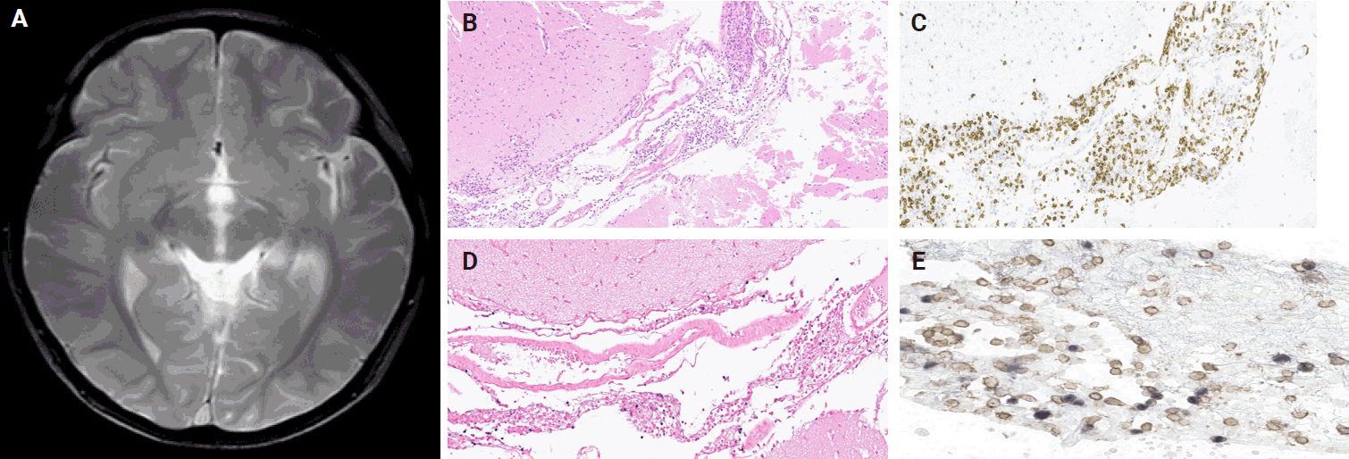
PDF - Chronic active Epstein-Barr virus (CAEBV) can induce complications in various organs, including the brain and gastrointestinal tract. A 3-year-old boy was referred to the hospital with a history of fever and seizures for 15 days. A diagnosis of encephalitis based on computed tomography (CT) and magnetic resonance imaging findings and clinical correlation was made. Laboratory tests showed positive serology for Epstein-Barr virus (EBV) and negative for Rotavirus antigen and IgG and IgM antibodies for cytomegalovirus, herpes simplex virus, and varicella zoster virus, respectively. Abdominal CT showed diffuse wall thickening with fluid distension of small bowel loops, lower abdomen wall thickening, and a small amount of ascites. The biopsy demonstrated positive Epstein-Barr encoding region in situ hybridization in cells within the crypts and lamina propria. The patient was managed with steroids and hematopoietic stem cell transplantation (HSCT). This case showed histopathological characteristics of concurrent EBV-associated encephalitis and colitis in CAEBV infection. The three-step strategy of immunosuppressive therapy, chemotherapy, and allogeneic HSCT should be always be considered for prevention of disease progression.
Original Article
- Genetic Analysis of Epstein-Barr Virus Latent Membrane Protein 1 and Immunohistochemical Expression of Transforming Growth Factor (TGF)-beta1, TGF-betaRII, p21, p16, E2F1, Thymidylate Synthase, and NF-kappaB in Epstein-Barr Virus Encoded RNA-positive Gastric Adenocarcinoma
- Mee Yon Cho, Minseob Eom, Kwang Hwa Park, Mee Dong Kim, Seung Hoon Sung, Myoung Soo Kim, Dae Sung Kim, Sun Ju Choi
- Korean J Pathol. 2006;40(3):176-184.
- 2,113 View
- 22 Download
-
 Abstract
 PDF
PDF - BACKGROUND
:Although clinicopathologic differences have been described between Epstein-Barr virus (EBV)-positive and negative gastric adenocarcinomas, the pathogenetic basis for these differences remains unclear. In this study, efforts were made to confirm that expression of EBV-latent membrane protein (LMP1) and immunohistochemical characteristics of EBVpositive gastric adenocarcinomas.
METHODS
We investigated genomic deletion, and RNA & protein expression of the EBV-LMP1, as well as immunohistochemical protein expression of transforming growth factor (TGF)-beta1, TGF-bata RII, p21, p16, E2F1, thymidylate synthase, and NF-kappaB in relation to EBV positive gastric adenocarcinoma.
RESULTS
A total of 38 Epstein-Barr Virus Encoded RNA-positive and 80 negative gastric carcinomas were examined. A 30 bp DNA deletion in the EBV-LMP1 gene, initiating at codon 342, was detected in 94.4% of EBVpositive cases. By RT-PCR and western blotting, EBV-LMP1 mRNA and protein expressions were absent in all cases, re-gardless of DNA deletion. No significant differences in TGF-bata1, TGF-betaRII, p21, NF-kappaB, E2F1, or thymidylate synthase expression were identified. However, the decreased expression of p16 was found in 84.2% of EBV-positive carcinomas, relative to only 57.5% of EBV-negative tumors (p=0.024).
CONCLUSION
EBV-LMP1 DNA deletion, mRNA and protein losses are highly prevalent in EBV-positive gastric adenocarcinoma among Korean patients, along with decreased p16 expression.
Case Reports
- Cytologic Findings of Infectious Mononucleosis Lymphadenitis: A Report of Four Cases .
- Jin Hee Sohn, Eun Ha Jung, Hye Rim Park
- J Pathol Transl Med. 1998;9(2):227-232.
- 4,981 View
- 282 Download
-
 Abstract
 PDF
PDF - Infectious mononucleosis(IM) is an acute self-limiting lymphoproliferative disorder associated with infection by the Epstein-Barr Virus(EBV), with the characteristic triad of fever, sore throat, and cervical or generalized lymphadenopathy. And also there are atypical lymphocytes in the peripheral blood. Cytological findings of IM lymphadenitis are characterized by a florid immunoblastic and atypical lymphoid cell proliferation. However, the small number of cases were studied by fineneedle aspiration cytology(FNAC) even though there was a complexity of lymph node pathology. It is important to recognize the reactive pattern of IM that would initiate EBV study and to avoid unnecessary biopsy. We studied findings of lymph node FNAC from 4 patients with EBV infection confirmed by EBV-specific serologic studies. All of the cases were positive for viral capsid antigen(VCA) and one case was positive for anti-EBV nuclear antigen(EBNA). Cytologically, all of the cases exhibited high cellularity and atypia with great numbers of large immunoblastic lymphocytes.
- Nasal Type NK/T Cell Lymphoma with Pseudoepitheliomatous Hyperplasia: A Case Report.
- Hoon Kyu Oh, Jeong Kyu Kim, Kwan Kyu Park
- Korean J Pathol. 2006;40(3):250-254.
- 2,080 View
- 31 Download
-
 Abstract
 PDF
PDF - A case of prominent pseudoepitheliomatous hyperplasia (PEH), that was misdiagnosed as squamous cell carcinoma (SCC) on the frozen section occured in the nasal mucosa of a patient suffering with nasal type NK/T cell lymphoma. To prevent misdiagnosis of this lesion, pathologists should be aware that NK/T cell lymphoma may be associated with overlying mucosal PEH, and so the physician must adhere to strict diagnostic criteria for making the diagnosis of SCC. The pathogenesis of PEH associated with NK/T cell lymphoma is not still clear, but it may be related to the production of growth factors, especially epidermal growth factor and transforming growth factor, by the underlying tumors.
Original Articles
- Epstein-Barr Virus and p53 in Laryngeal and Nasopharyngeal Carcinomas.
- Eun Sook Nam, Duck Hwan Kim, Hyung Sik Shin, Young Euy Park, Young Sik Kim, Insun Kim
- Korean J Pathol. 1998;32(8):551-562.
- 2,144 View
- 10 Download
-
 Abstract
- To investigate the correlation between EBV infection and p53 overexpression in laryngeal carcinomas (LC) and nasopharyngeal carcinomas (NPC) in Korea, we analyzed 37 laryngeal squamous cell carcinomas and 33 nasopharyngeal (11 squamous cell and 22 undifferentiated) carcinomas. We used the immunohistochemistry and polymerase chain reaction-single stranded conformational polymorphism (PCR-SSCP) for p53 overexpression and p53 gene mutation, respectively, and EBER-1 in situ hybridization and PCR using primer for EBNA-1 and EBNA-2 type 1 and 2 for prevalence and the subtype of EBV. The results were as follows; 1) The p53 expression was found in 43.2% of squamous cell LCs, in 54.6% of squamous cell NPCs and in 22.7% of undifferentiated NPCs. The p53 gene mutation was detected in 6 of 23 squamous cell LCs and 3 of 14 undifferentiated NPCs. 2) EBV was detected more frequently in undifferentiated NPCs (95.5%) than in squamous cell NPCs (63.6%) and squamous cell LCs (37.0%). Only type 1 was found in squamous cell LCs and NPCs, whereas both type 1 and type 2 were detected in undifferentiated NPCs. 3) There was no difference according to EBV infection (EBV+ ; 7 cases, EBV- ; 7 cases) in the cases with p53 protein overexpression but mutaion. From the above results, it can be concluded that squamous cell LCs and NPCs are associated with both p53 and EBV, whereas undifferentiated NPCs are more closely associated with EBV than p53. In Korea, both type 1 and 2 are detected in undifferentiated NPCs. Also, our result suggests that EBV infection does not seem to contribute to p53 overexpression. The interrelationship between EBV infection and p53 remains to be further defined.
- Correlation between Expression of p53 and Bcl-2 Protein and Epstein-Barr Virus Detection in Gastric Adenocarcinoma.
- Ki Jung Yun, Weon Cheol Han, Hyung Bae Moon, Sang Woo Juhng
- Korean J Pathol. 1998;32(8):574-580.
- 1,848 View
- 10 Download
-
 Abstract
- Epstein-Barr virus (EBV) has been known to be associated with a wide variety of neoplastic conditions including nasopharyngeal carcinoma, Hodgkin's disease, and non-Hodgkin's lymphoma. Recent studies reveal the presence of EBV in certain subtypes of gastric carcinoma in which EBV appears to be pathogenetically related. To evaluate the relationship between EBV and gastric adenocarcinoma, we examined EBV DNA using direct in situ polymerase chain reaction, and expression of p53 protein and bcl-2 protein using immunohistochemical staining method on paraffin embedded tissues. The materials consisted of one hundred twenty-eight gastric adenocarcinomas and twenty benign peptic ulcers. EBV DNA was detected in 14 of 128 gastric adenocarcinomas (10.9%). p53 protein was positive in 10 of 14 EBV positive adenocarcinomas (71.4%) and in 61 of 114 EBV negative adenocarcinomas (53.5%). Bcl-2 protein was positive in 2 of 14 EBV positive adenocarcinomas (14.3%) and in 19 of 114 EBV negative adenocarcinomas (16.7%). The above results indicate that EBV is associated with gastric adenocarcinoma, and p53 protein may play a role in carcinogenesis of EBV in gastric adenocarcinoma.
- Genotype of Epstein-Barr Virus and Comparative Genomic Hybridization Analysis of NK/T Cell Lymphoma.
- Keying Eun Choi, Young Hyeh Ko
- Korean J Pathol. 2000;34(8):541-549.
- 1,957 View
- 12 Download
-
 Abstract
 PDF
PDF - NK/T cell lymphoma is a distinct clinicopathologic entity which is more prevalent in Asia than in America and Europe and is highly associated with Epstein-Barr virus (EBV) infection. Although the clinicopathologic features of the tumor have been clearly defined, genetic changes and roles of virus associated with the development and progression of tumor have not been well studied. In this study, we carried out polymerase chain reaction (PCR) for EBNA-3B, EBNA-3C, and LMP-1 30 bp deletion to investigate EBV subtype and variants in tumor tissue and performed comparative genomic hybridization (CGH) to screen chromosomal imbalances using frozen tissues from 7 patients with nasal-type NK/T cell lymphoma and 1 patient with blastic NK cell lymphoma. Of 6 cases infected with EBV, there were EBV type 1 in six, LMP-1 30 bp deletion variant in four, and LMP-1 40 bp deletion variant in one. Frequent chromosomal imbalances included deletions at 1p31-pter (4), 12q23-q24 (3), and 17p (4), and gains at 2q (5), 10q (3), and 13q34-qter (4). Blastic NK cell lymphoma displayed deletions of 9q, 7q, and 6q, similar to that of nasal-type NK/T-cell lymphoma. With these results, we assumed that candidate genes in these imbalanced chromosomal loci would provide the clue for further molecular studies to identify putative tumor suppressor genes or proto-oncogenes associated with pathogenesis of this neoplasm.
- Epstein-Barr Virus in Korean Malignant Lymphomas.
- Young Hyeh Ko, Jung Dal Lee
- Korean J Pathol. 1996;30(11):1011-1017.
- 1,889 View
- 19 Download
-
 Abstract
 PDF
PDF - To determine the prevalence of Epstein-Barr virus infection in lymphoid neoplasms of various histology and location, the paraffin tissues of 74 non-Hodgkin's lymphomas and 13 Hodgkin's diseases were studied by EBER and BHLF RNA in situ hybridization as well as immunostaining using LMP-1, EBNA-2, and ZEBRA. As a control, non-neoplastic lymphoid tissues from the nasal cavity(10), lymph node(38) and Waldeyer's ring(12) were investigated. In non-neoplastic control, EBV genome was detected in none of 10 nasal mucosa, 6 of 38 lymph node, and 1 of 12 Waldeyer's ring. EBV-positive non-neoplastic lymphocytes expressed CD45RO in 2 cases and CD20 in 4 cases. Non-Hodgkin's lymphoma was positive for EBV in 37.8% of the cases in which T-cell lymphoma showed higher rate(56%) than B-cell lymphoma(15%), especially in nasal lymphoma(80%) and angiocentric lymphoma(63.6%). Hodgkin's disease was EBV positive in 38.4% of the cases. EBV genome in tumor tissue existed in latent form as well as in lytic form. LMP-1 was positive in 80% of Hodgkin's disease and 39% of non-Hodgkin's lymphoma in which EBV genome was detected. EBNA-2 was expressed in 3 cases of non-Hodgkin's lymphoma. On the basis of protein expression, most lymphomas belonged to type II latency. These results support that EBV is associated with pathogenesis of malignant lymphoma although its mechanism still awaits to be clarified.

 E-submission

 First
First Prev
Prev


