 E-submission
Search
- Page Path
- HOME > Search
Review Article
- The evolving role of TRPS1 in dermatopathology: insights from the past 4 years
- Mokhtar H. Abdelhammed, Woo Cheal Cho
- J Pathol Transl Med. 2026;60(2):129-143. Published online January 29, 2026
- DOI: https://doi.org/10.4132/jptm.2025.11.25
- 3,411 View
- 230 Download
-
 Abstract
 PDF
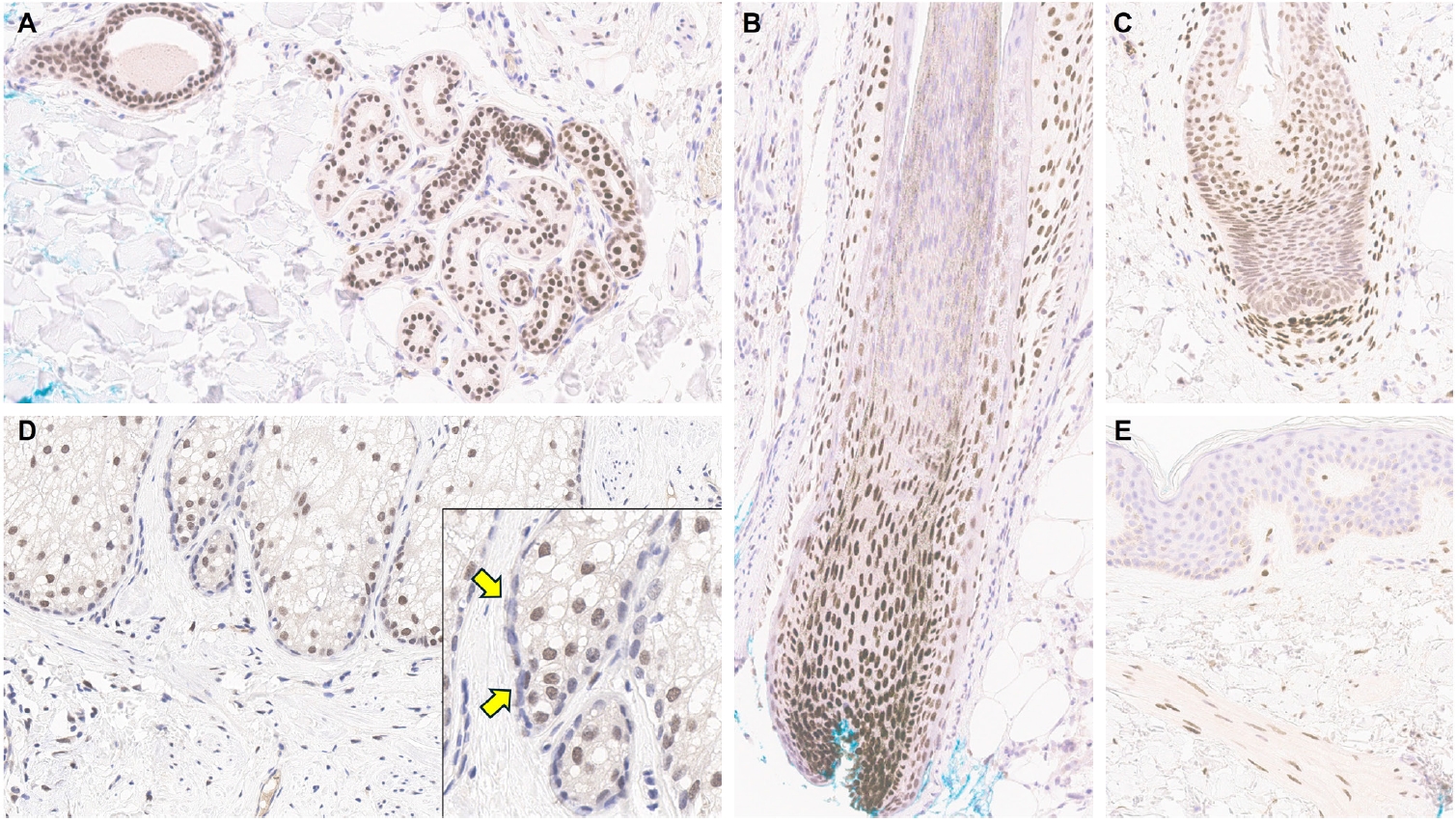
PDF - Over the past 4 years, trichorhinophalangeal syndrome type 1 (TRPS1) has rapidly gained attention among practicing pathologists, with numerous studies emerging that both support and question its diagnostic utility. Initially regarded as a highly specific marker for tumors of mammary origin, TRPS1 is now recognized to have broader expression patterns, including in a variety of cutaneous neoplasms. This is likely due to embryologic parallels between breast tissue and skin adnexal structures, an overlap that was underappreciated in early investigations. Although TRPS1 lacks absolute specificity—even among cutaneous neoplasms—it can still offer meaningful diagnostic value when interpreted alongside conventional immunohistochemical markers and within the appropriate morphologic context. Noteworthy diagnostic applications include mammary Paget disease, primary extramammary Paget disease, rare adnexal neoplasms such as endocrine mucin-producing sweat gland carcinoma and primary cutaneous NUT adnexal carcinoma, and cutaneous metastases from breast carcinoma. In this review, we present the most comprehensive and up-to-date evaluation of the utility and limitations of TRPS1 immunohistochemistry in dermatopathology. Our aim is to deepen understanding of this emerging marker and provide practical guidance on its optimal integration with established immunohistochemical panels to enhance diagnostic accuracy in routine practice.
Case Report
- Invasive Extramammary Paget Disease: A Report of 2 Cases with Immunohistochemical and Ultrastructural Findings.
- Kyu Rae Kim, Chong Woo You, Jeong Ho Han, Young Hyeh Ko
- Korean J Pathol. 1996;30(9):858-864.
- 2,421 View
- 42 Download
-
Abstract
PDF
- We present 2 cases of invasive extramammary Paget disease occuring in the vulva area of a 60 year old female, and in the scrotal and penile area of a 63 year old male patient. The histologically typical Paget cells were not only seen in the surface epithelium but were also involved in the outer root sheath of the hair follicles. Stromal infiltration of tumor cells into the upper dermis were present in both cases, however, no underlying primary sweat gland carcinoma was present. Metastatic foci of inguinal lymph nodes showed apocrine-type epithelium with abundant eosinophilic granular cytoplasm, which were positive for anti-CEA and GCDFP-15, as well as eccrine-type epithelium containing mucinous secretory materials in the lumen and the cytoplasm. Ultrastructural findings showed interdigitating plasma membranes with prominent desmosomes between the Paget cells, intracytoplasmic tonofibrils, intracellular tubules, lipid vacuoles, and enlarged mitochondria. Histological, immunohistochemical, and ultrastructural findings suggested that Paget cells showed both eccrine and apocrine differentiation.
 First
First Prev
Prev


