 E-submission
Search
- Page Path
- HOME > Search
Review Article
- A comprehensive review of ossifying fibromyxoid tumor: insights into its clinical, pathological, and molecular landscape
- Kyriakos Chatzopoulos, Antonia Syrnioti, Mohamed Yakoub, Konstantinos Linos
- J Pathol Transl Med. 2026;60(1):6-19. Published online January 14, 2026
- DOI: https://doi.org/10.4132/jptm.2025.10.02
- 2,477 View
- 131 Download
-
 Abstract
 PDF
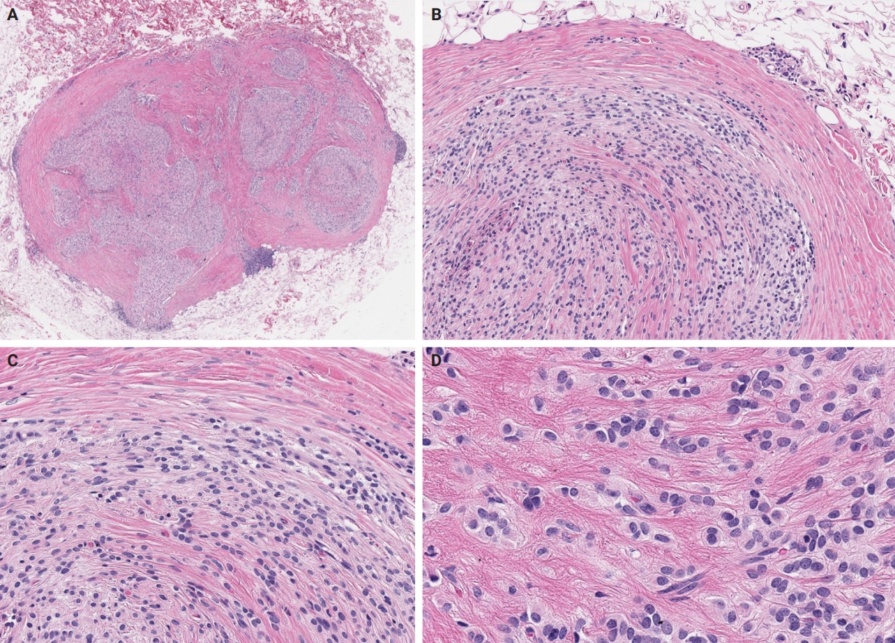
PDF - Ossifying fibromyxoid tumor (OFMT) is a rare mesenchymal neoplasm first described in 1989. It typically arises in the superficial soft tissues of the extremities as a slow-growing, painless mass. Histologically, it is commonly characterized by a multilobular architecture composed of uniform epithelioid cells embedded in a fibromyxoid matrix, often surrounded by a rim of metaplastic bone. While classic cases are readily identifiable, the tumor's histopathological heterogeneity can mimic a range of benign and malignant neoplasms, posing significant diagnostic challenges. Molecularly, most OFMTs harbor PHF1 rearrangements, commonly involving fusion partners such as EP400, MEAF6, or TFE3. This review underscores the importance of an integrated diagnostic approach- incorporating histopathological, immunohistochemical, and molecular data- to accurately classify OFMT and distinguish it from its mimics. Expanding awareness of its morphologic and molecular spectrum is essential for precise diagnosis, optimal patient management, and a deeper understanding of this enigmatic neoplasm.
Case Report
- Ossifying Fibromyxoid Tumor of Soft Parts.
- Seok Hoon Jeon, Seung Sam Paik, Eun Kyung Hong, Moon Hyang Park, Jung Dal Lee
- Korean J Pathol. 1997;31(2):174-178.
- 2,018 View
- 18 Download
-
Abstract
PDF
- An ossifying fibromyxoid tumor of soft parts is a rare, recently described, fibro-osseous neoplasm of uncertain histogenesis. It occurs most frequently within the subcutis or skeletal muscle of the extremities. Its biologic behavior is generally regarded as benign with at worst a locally aggressive clinical course. But, atypical and malignant variants have been recently reported. Herein we report a case of a benign ossifying fibromyxoid tumor which occurred in the left upper back of 41-year-old man. The tumor is composed of uniformly round or polygonal cells arranged in cords or nests which are separated by myxoid and hyalinzed fibrous matrix and associated with irregular bony trabeculae. The tumor cells are strong positive for vimentin. Ultrastructural findings and a review of literatures are added.
 First
First Prev
Prev


