 E-submission
Search
- Page Path
- HOME > Search
Original Articles
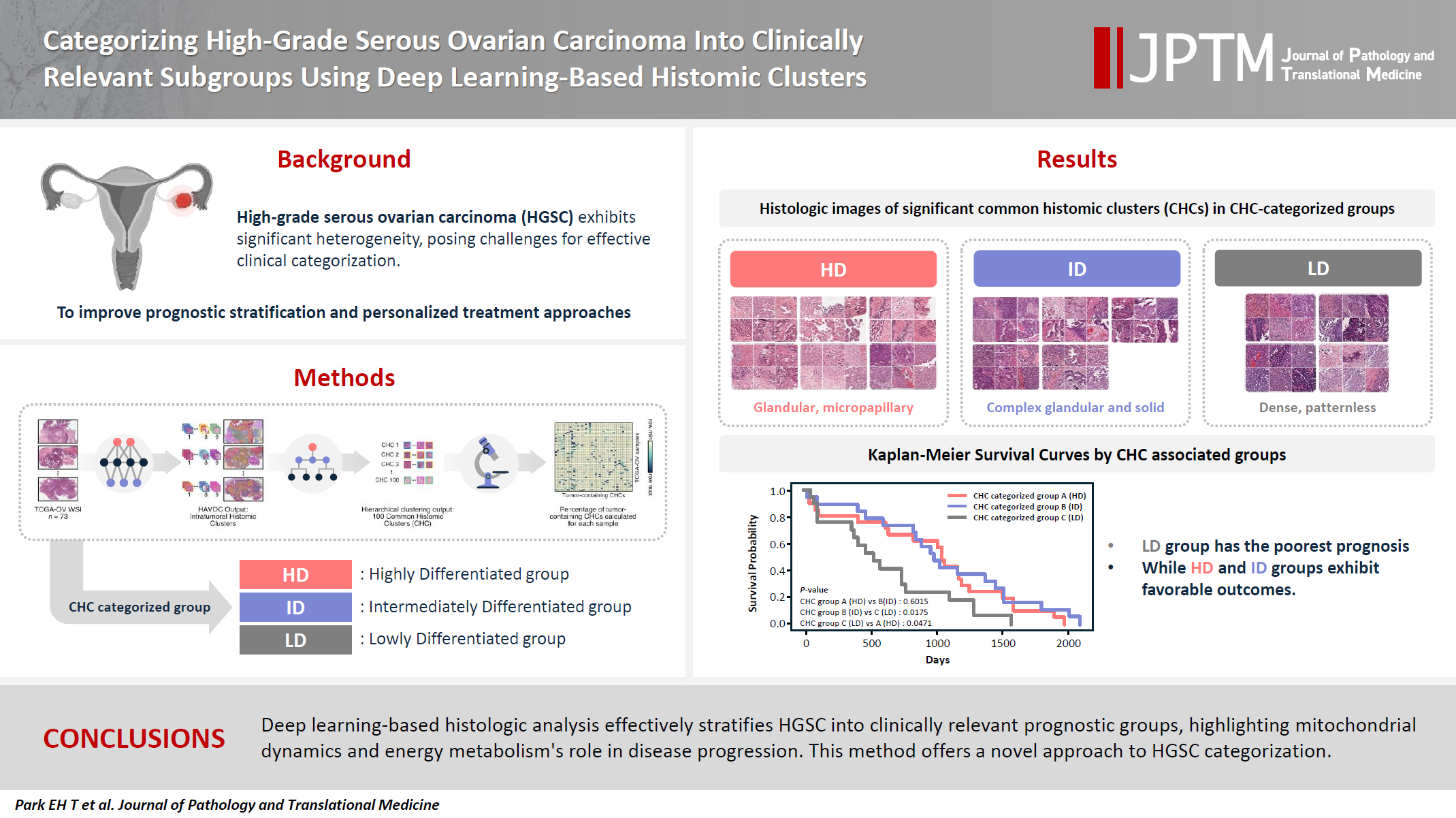
- Categorizing high-grade serous ovarian carcinoma into clinically relevant subgroups using deep learning–based histomic clusters
- Byungsoo Ahn, Eunhyang Park
- J Pathol Transl Med. 2025;59(2):91-104. Published online February 18, 2025
- DOI: https://doi.org/10.4132/jptm.2024.10.23
- 7,984 View
- 276 Download
- 1 Web of Science
- 2 Crossref
-
 Abstract
 PDF
PDF Supplementary Material
 - Background
High-grade serous ovarian carcinoma (HGSC) exhibits significant heterogeneity, posing challenges for effective clinical categorization. Understanding the histomorphological diversity within HGSC could lead to improved prognostic stratification and personalized treatment approaches. Methods: We applied the Histomic Atlases of Variation Of Cancers model to whole slide images from The Cancer Genome Atlas dataset for ovarian cancer. Histologically distinct tumor clones were grouped into common histomic clusters. Principal component analysis and K-means clustering classified HGSC samples into three groups: highly differentiated (HD), intermediately differentiated (ID), and lowly differentiated (LD). Results: HD tumors showed diverse patterns, lower densities, and stronger eosin staining. ID tumors had intermediate densities and balanced staining, while LD tumors were dense, patternless, and strongly hematoxylin-stained. RNA sequencing revealed distinct patterns in mitochondrial oxidative phosphorylation and energy metabolism, with upregulation in the HD, downregulation in the LD, and the ID positioned in between. Survival analysis showed significantly lower overall survival for the LD compared to the HD and ID, underscoring the critical role of mitochondrial dynamics and energy metabolism in HGSC progression. Conclusions: Deep learning-based histologic analysis effectively stratifies HGSC into clinically relevant prognostic groups, highlighting the role of mitochondrial dynamics and energy metabolism in disease progression. This method offers a novel approach to HGSC categorization. -
Citations
Citations to this article as recorded by
- Ovarian Cancer: Epidemiology, Disease Mechanisms, New Diagnosis and Treatment Strategies, and Research Directions
Zunera Khalid, Weirong Fan, Farah Nazir, Yixiang Xing, Tengchuan Jin
iNew Medicine.2026;[Epub] CrossRef - Learning Disabilities in the 21st Century: Integrating Neuroscience, Education, and Technology for Better Outcomes
Syed Mohammed Basheeruddin Asdaq, Ahmad H. Alhowail, Syed Imam Rabbani, Naira Nayeem, Syed Mohammed Emaduddin Asdaq, Faiqa Nausheen
SAGE Open.2025;[Epub] CrossRef
- Ovarian Cancer: Epidemiology, Disease Mechanisms, New Diagnosis and Treatment Strategies, and Research Directions
- Detection of the c-m c Oncogene Amplification in Ovarian Carcinomas by Differential Polymerase Chain Reaction.
- Geun Shin Lyu, Chan Kum Park, Chun Geun Lee, Youl Hee Cho, Youn Yeoung Hwang, Jung Dal Lee
- Korean J Pathol. 1997;31(7):644-654.
- 2,252 View
- 13 Download
-
Abstract
PDF
- The amplification of c-myc oncogene was evaluated in 42 cases of ovarian carcinomas to correlate with clinical parameters. Using oligonucleotide primers, sequences from the c-myc exon-3 gene and from a control gene, tissue plasminogen activator (tPA), were amplified simultaneously by polymerase chain reaction (PCR). After the products of differential PCR (d-PCR) were electrophoresed, slot blot hybridization was performed, and hybridized with P32 dATP-labeled myc and tPA oligonucleotide probes and then autoradiographed. The signal intensities of the two products were quantitated by densitometry and the ratios of two products (c-myc/tPA) were measured. The ovarian carcinomas showed significantly increased amplification of c-myc oncogene Oligonucleoti compared to normal control group (p<0.05). 15 of 42 cases (35.7%) showed various degrees of the MYC gene amplification up to 27 folds in various histologic types of ovarian carcinomas. No significant differences of the MYC gene amplification according to histologic subtypes, tumor action) grades and clinical stages of ovarian carcinomas were present.
- Tumor Angiogenesis and Stage in Ovarian Carcinoma.
- Eun Sook Chang, Hyun Chang Joo, Tae Sung Lee
- Korean J Pathol. 1999;33(8):596-602.
- 2,152 View
- 13 Download
-
Abstract
PDF
- Tumor angiogenesis has been found to have prognostic significance in many tumor types for predicting an increased risk of metastasis. We assessed tumor vascularity in 28 cases of borderline malignancy and 71 cases of carcinoma of the ovary which had been resected and diagnosed, using the highly specific endothelial cell marker CD34. The numbers of microvessels were counted in 200 magnification in three highly vascularised areas. The numbers of microvessels in carcinomas were higher than that in the borderline malignancy of serous and mucinous tumors. The number of microvessels of mucinous carcinomas was significantly higher than that of serous carcinomas. There were neither significant differences in the number of microvessels according to histological tumor types (p=0.075) nor significant differences in the number of microvessels according to FIGO stages (p=0.072). But in serous carcinomas, the number of microvessels was higher in the FIGO III-IV stage than in the FIGO I-II stage (p=0.017). This study showed higher neovascularization in malignant tumor than borderline malignancy, and in the advanced stage (FIGO III-IV) than less advanced stage (FIGO I-II) of serous carcinomas.
- Anticancer Effect and Apoptosis of All-trans-retinoic Acid on the Human Ovarian Epithelial Carcinoma Cell Lines.
- Jee Young Han, Woo Hee Jung, Tai Seung Kim
- Korean J Pathol. 2000;34(3):225-234.
- 2,108 View
- 18 Download
-
Abstract
PDF
- Ovarian carcinoma is a serious disease in women. Some reports revealed all-trans-retinoic acid (tRA) inhibited the proliferation of ovarian carcinoma cell lines and induced apoptosis. The aim of this study was to evaluate the anticancer and apoptotic effects of tRA and the expression of the retinoic acid receptor (RAR) alpha, beta, gamma, p53, bcl-2, and c-myc genes on the ovarian carcinoma cell lines, NIH OVCAR3 and SKOV3. In both cell lines, the proliferation of tumor cells was inhibited and characteristic morphologic patterns of apoptosis were shown after treatment of tRA. The number of apoptotic cells and the percentage of apoptosis were increased after treatment of tRA. The gel electrophoresis revealed the DNA ladder pattern in the NIH OVCAR3. Gene expressions were observed using northern blotting. There was no RARalpha expression in both cell lines. In NIH OVCAR3, there was no changes in the expression of RARbeta and bcl-2 gene. The RARgamma gene expression of tRA treated group was slightly increased, but p53 gene expression was decreased, and c-myc gene was not expressed. In SKOV3, the expressions of RARbeta, gamma, and p53 genes were increased and that of bcl-2 was decreased in the tRA treated group. There was no change in c-myc gene expression. These results suggest tRA has anticancer and apoptotic effect on both ovarian carcinoma cell lines. RARbeta, RARgamma, bcl-2, and p53 gene expressions were correlated with these effects of tRA on SKOV3 but not on NIH OVCAR3.
- A Study on the DNA Ploidy and Expression of c-erbB-2 Oncogen in the Ovarian Carcinomas.
- Jong Jae Jung, Chang Soo Park, Sang Woo Juhng
- Korean J Pathol. 1997;31(1):15-22.
- 1,990 View
- 15 Download
-
Abstract
PDF
- To evaluate the relationships among the c-erbB-2 oncogene expression, DNA ploidy and other prognostic factors, an immunohistochemical study of the c-erbB-2 oncogene product and flow cytometric analysis of DNA ploidy were performed in paraffin sections of 42 cases of ovarian carcinomas. The results were as follows: 1) The positive reaction for c-erbB-2 oncogene product was observed mainly along the cytoplasmic membrane, and occasionally within the cytoplasm of the tumor cells. 2) Overall the positivity of c-erbB-2 oncogene expression was 45.2% of the ovarian carcinomas. By the histological types, the positivity was 35.7% in serous carcinoma, 80.0% in mucinous carcinoma, and 45.2% in endometrioid carcinoma; by the degree of differentiation, 57.1% in well differentiated carcinoma, 40.0% in moderately differentiated, and 27.3% in poorly differentiated; by the nuclear grading, 58.3% in grade I, 52.6% in grade II, and 18.2 % in grade III; and by the clinical staging, 57.1% in stage I, 42.8% in stage II, and 35.0% in stage III. The expression of the c-erbB-2 oncogene in the ovarian carcinomas was higher in the tumors of good differentiation, of the lower nuclear grade and of the lower clinical stage. 3) The incidence of DNA aneuploidy in the cases positive for the c-erbB-2 oncogene expression(47.3%) was higher than that in the negative cases(31.4%). From the above results, therefore, it is suggested that the c-erbB-2 oncogene may be involved in the early stage of ovarian carcinogenesis. Also suggested is that ovarian carcinomas positive for the c-erbB-2 oncogene in the early stages may have higher probability of having a DNA aneuploid cell line during the progress of the tumors.
 First
First Prev
Prev


