
 E-submission
Search
- Page Path
- HOME > Search
Review Article
- Sentinel lymph node biopsy in melanoma: pathologic evaluation and diagnostic considerations
- Dre Barnachea, Bonnie Lee
- J Pathol Transl Med. 2026;60(5):493-501. Published online September 1, 2026
- DOI: https://doi.org/10.4132/jptm.2026.07.01
- 2,283 View
- 64 Download
-
 Abstract
 PDF
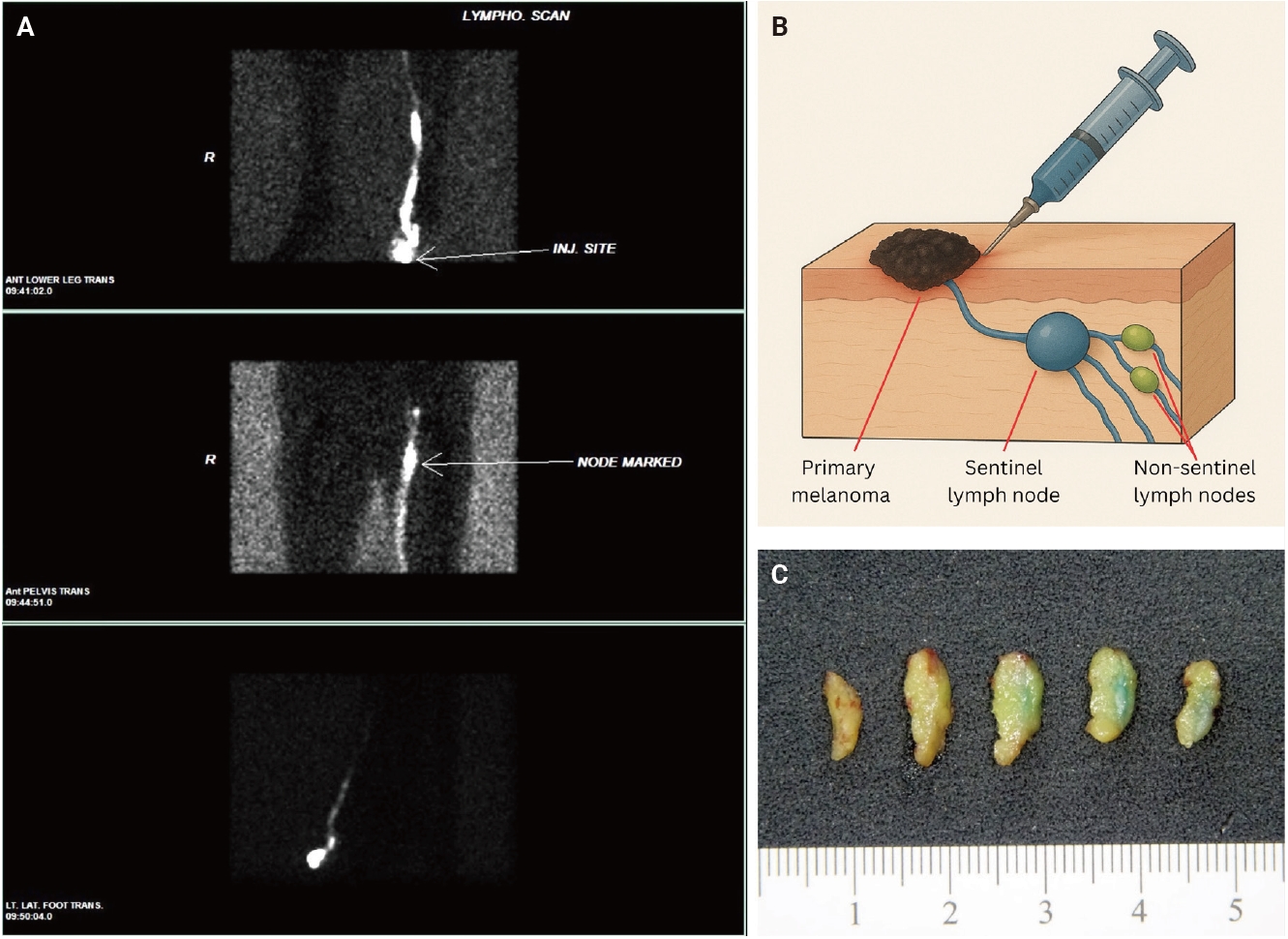
PDF - Sentinel lymph node biopsy (SLNB) is a widely used staging procedure in melanoma that provides important prognostic information and guides clinical management. The sentinel lymph node (SLN), defined as the first lymph node in the lymphatic drainage pathway from a primary tumor, represents the most likely site of early regional metastasis. Accordingly, identification of metastatic melanoma within SLNs has significant implications for staging and risk stratification and is associated with worse clinical outcomes. The SLNB procedure involves preoperative and intraoperative lymphatic mapping techniques, including radiotracer localization with or without blue dye injection, which allow identification of SLNs. Histopathologic evaluation includes careful gross examination, serial sectioning, and immunohistochemical analysis using melanocytic markers such as SOX10, Melan-A, HMB45, and occasionally PRAME to detect metastatic disease. In addition, prognostic features such as tumor burden, extranodal extension, and microanatomic location of metastases within the lymph node further refine risk assessment. This review provides an overview of SLNB in melanoma, with emphasis on clinical indications, histopathologic evaluation, and key diagnostic and prognostic considerations.
Case Report
- Pseudometastasis in Sentinel Lymph Nodes with Cytokeratin Debris-containing Histiocytes in Breast Cancer Patient: A Case Report.
- Keum Ha Choi, Eun Jung Cha, Ha Na Choi, Woo Sung Moon
- Korean J Pathol. 2007;41(6):427-429.
- 3,044 View
- 53 Download
-
Abstract
PDF
- Immunohistochemical staining for cytokeratins can detect false negative nodes in patients with breast carcinoma. We report on a patient with breast carcinoma and pseudometastasis detected by immunohistochemical staining within a negative sentinel lymph node. A 66-year-old woman underwent a simple mastectomy and sentinel lymph node biopsy. Immunohistochemical staining of the sentinel nodes for cytokeratin in permanent sections showed cells with intense cytoplasmic staining in the subcapsular sinus. The cells were negative for epithelial membrane antigen staining, but positive for CD68. In combination with morphologic findings and immunohistochemistry, cytokeratin-positive cells were confirmed as histiocytes with phagocytized cytokeratin debris. Careful correlation with histology and additional IHC could help avoid a misinterpretation of this type of pseudometastasis.
 First
First Prev
Prev



