 E-submission
Search
- Page Path
- HOME > Search
- Diagnostic value of cytology in detecting human papillomavirus–independent cervical malignancies: a nation-wide study in Korea
- Hye-Ra Jung, Junyoung Shin, Chong Woo Yoo, Eun Na Kim, Cheol Lee, Kyeongmin Kim, Ho-chang Lee, Yonghee Lee, Ji Hye Kim, Soo Jin Jung, Yumin Chung, Joo Yeon Kim, Hye Eun Park, Tae Hoen Kim, Wonae Lee, Min-Sun Cho, Ran Hong, Yoon Jung Choi, Younghee Choi, Young Sub Lee, Sang-Ryung Lee, Myunghee Kang, Young Jin Seo, Seung-Sook Lee, Yoon-Jung Hwang, Hyun-Jung Kim
- J Pathol Transl Med. 2025;59(6):444-452. Published online November 11, 2025
- DOI: https://doi.org/10.4132/jptm.2025.10.21

- 4,233 View
- 140 Download
-
 Abstract
 PDF
PDF - Background
Human papillomavirus (HPV) independent cervical malignancies (HPV-IDCMs) have recently been classified by the World Health Organization (WHO) 5th edition. These malignancies have historically received limited attention due to their rarity and the potential for evasion of HPV-based screening.
Methods
We retrospectively reviewed 5,854 biopsy-confirmed cervical malignancies from 22 institutions over 3 years (July 2020–June 2023). Histologic classification followed the WHO guidelines. HPV independence was confirmed by dual negativity for p16 and HPV; discordant cases (p16-positive/HPV-negative) underwent additional HPV testing using paraffin-embedded tissue. Cytological results were matched sequentially to histological confirmation.
Results
The prevalence of HPV-IDCM was 4.4% (257/5,854) overall and was 3.6% (208/5,805 cases) among primary cervical malignancy. Patient age of HPV-IDCM was 29 to 89 years (median, 57.79). Its histologic subtypes included primary adenocarcinoma (n = 116), endometrial adenocarcinoma (n = 35), squamous cell carcinoma (n = 72), metastatic carcinoma (n = 14), carcinoma, not otherwise specified (n = 10), neuroendocrine carcinoma (n = 3), and others (n = 7). Among 155 cytology-histological matched cases, the overall and primary Pap test detection rates were 85.2% (132/155) and 83.2% (104/125), respectively. The interval between cytology and histologic confirmation extended up to 38 months.
Conclusions
HPV-IDCMs comprised 3.6% of primary cervical malignancies with a high detection rate via cytology (83.2%). These findings affirm the value of cytological screening, particularly in patients with limited screening history or at risk for HPV-independent lesions, and may guide future screening protocols.
- National quality assurance program using digital cytopathology: a 5-year digital transformation experience by the Korean Society for Cytopathology
- Yosep Chong, Hyeong Ju Kwon, Soon Auck Hong, Sung Soon Kim, Bo-Sung Kim, Younghee Choi, Yoon Jung Choi, Jung-Soo Pyo, Ji Yun Jeong, Soo Jin Jung, Hoon Kyu Oh, Seung-Sook Lee
- J Pathol Transl Med. 2025;59(5):320-333. Published online September 15, 2025
- DOI: https://doi.org/10.4132/jptm.2025.06.27
- 4,097 View
- 107 Download
- 2 Web of Science
- 2 Crossref
-
Abstract
PDF
 Supplementary Material
 - Background
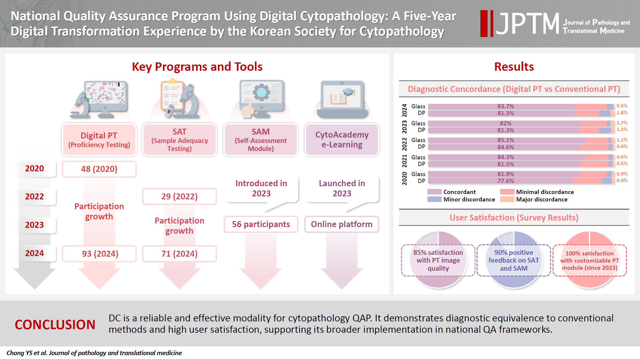
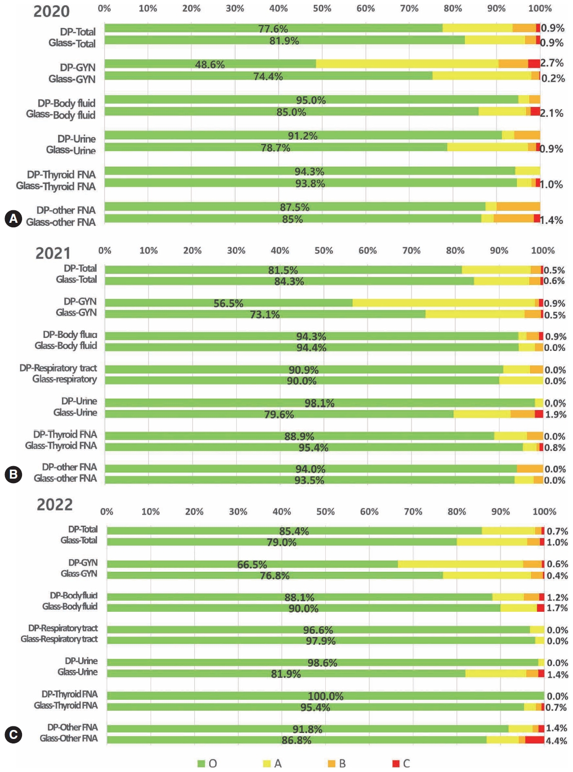
Digital cytopathology (DC) is emerging as a transformative approach in quality assurance programs (QAP), though its comprehensive evaluation remains limited. Since 2020, the Korean Society for Cytopathology has progressively incorporated DC into its national QAP, including digital proficiency testing (PT), sample adequacy testing (SAT), a customizable PT module, and a self-assessment module (SAM), aiming for full digital implementation by 2026. Methods: This 5-year study assessed diagnostic concordance between conventional and digital PT formats and analyzed participant feedback on service quality and digital image usability across PT, SAT, and SAM. Parallel testing was conducted during the transitional phase, and satisfaction was measured through structured surveys. Results: Participation in digital PT increased from 48 institutions in 2020 to 93 in 2024, while digital SAT participation rose from 29 to 71 between 2022 and 2024. In 2023, 56 institutions joined SAM. Diagnostic concordance rates were comparable between digital and conventional PTs (78.6%–84.6% vs. 82.0%–85.1%), including similar category C (major discordance) rates. Satisfaction with digital PT services and image quality exceeded 85%, and over 90% of institutions reported positive feedback on SAT and SAM. Over 80% were satisfied with the customizable PT module. Conclusions: DC is a reliable and effective modality for cytopathology QAP. It demonstrates diagnostic equivalence to conventional methods and high user satisfaction, supporting its broader implementation in national quality assurance frameworks. -
Citations
Citations to this article as recorded by
- Practice of Cytopathology in Korea: A 40‐Year Evolution Through Standardization, Digital Transformation, and Global Partnership
Yosep Chong, Ran Hong, Hyeong Ju Kwon, Haeryoung Kim, Lucia Kim, Soon Jae Kim, Yoon Jung Choi
Diagnostic Cytopathology.2026; 54(2): 146. CrossRef - Validation of Digital Cytology for Primary Diagnosis Across a Range of Specimen Types
Talisa Mistry, Harriet Hunter, Dahmane Oukrif, Sabine Pomplun, Reena Khiroya, Mary Falzon, Tanya Alan, Manuel Rodriguez‐Justo, Adam P. Levine
Cytopathology.2026;[Epub] CrossRef
- Practice of Cytopathology in Korea: A 40‐Year Evolution Through Standardization, Digital Transformation, and Global Partnership
- Diagnostic proficiency test using digital cytopathology and comparative assessment of whole slide images of cytologic samples for quality assurance program in Korea
- Yosep Chong, Soon Auck Hong, Hoon Kyu Oh, Soo Jin Jung, Bo-Sung Kim, Ji Yun Jeong, Ho-Chang Lee, Gyungyub Gong
- J Pathol Transl Med. 2023;57(5):251-264. Published online August 24, 2023
- DOI: https://doi.org/10.4132/jptm.2023.07.17
- 8,310 View
- 344 Download
- 9 Web of Science
- 9 Crossref
-
Abstract
PDFSupplementary Material
- Background
The Korean Society for Cytopathology introduced a digital proficiency test (PT) in 2021. However, many doubtful opinions remain on whether digitally scanned images can satisfactorily present subtle differences in the nuclear features and chromatin patterns of cytological samples.
Methods
We prepared 30 whole-slide images (WSIs) from the conventional PT archive by a selection process for digital PT. Digital and conventional PT were performed in parallel for volunteer institutes, and the results were compared using feedback. To assess the quality of cytological assessment WSIs, 12 slides were collected and scanned using five different scanners, with four cytopathologists evaluating image quality through a questionnaire.
Results
Among the 215 institutes, 108 and 107 participated in glass and digital PT, respectively. No significant difference was noted in category C (major discordance), although the number of discordant cases was slightly higher in the digital PT group. Leica, 3DHistech Pannoramic 250 Flash, and Hamamatsu NanoZoomer 360 systems showed comparable results in terms of image quality, feature presentation, and error rates for most cytological samples. Overall satisfaction was observed with the general convenience and image quality of digital PT.
Conclusions
As three-dimensional clusters are common and nuclear/chromatin features are critical for cytological interpretation, careful selection of scanners and optimal conditions are mandatory for the successful establishment of digital quality assurance programs in cytology. -
Citations
Citations to this article as recorded by- Practice of Cytopathology in Korea: A 40‐Year Evolution Through Standardization, Digital Transformation, and Global Partnership
Yosep Chong, Ran Hong, Hyeong Ju Kwon, Haeryoung Kim, Lucia Kim, Soon Jae Kim, Yoon Jung Choi
Diagnostic Cytopathology.2026; 54(2): 146. CrossRef - ThinPrep® Whole-Slide Digital Images versus Conventional Microscopy in Negative for Intraepithelial Lesion or Malignancy, Atypical Squamous Cells of Undetermined Significance, and Low-Grade Squamous Intraepithelial Lesion Cervical Lesions: European Federa
Ester Puntonen, Sara Bazzon, Massimo Bongiovanni, Rosario Granados, Ines Krivak Bolanca, Maria Nasioutziki, Maurizio Pinamonti, Danijela Vrdoljak-Mozetic, Arrigo Capitanio, Beatrix Cochand-Priollet, Ambrogio Fassina, Giovanni Negri, Laura Ventura, Ivana K
Pathobiology.2026; : 1. CrossRef - Sensitivity, Specificity, and Cost–Benefit Effect Between Primary Human Papillomavirus Testing, Primary Liquid‐Based Cytology, and Co‐Testing Algorithms for Cervical Lesions
Chang Gok Woo, Seung‐Myoung Son, Hye‐Kyung Hwang, Jung‐Sil Bae, Ok‐Jun Lee, Ho‐Chang Lee
Diagnostic Cytopathology.2025; 53(1): 35. CrossRef - Integration of AI‐Assisted in Digital Cervical Cytology Training: A Comparative Study
Yihui Yang, Dongyi Xian, Lihua Yu, Yanqing Kong, Huaisheng Lv, Liujing Huang, Kai Liu, Hao Zhang, Weiwei Wei, Hongping Tang
Cytopathology.2025; 36(2): 156. CrossRef - National quality assurance program using digital cytopathology: a 5-year digital transformation experience by the Korean Society for Cytopathology
Yosep Chong, Hyeong Ju Kwon, Soon Auck Hong, Sung Soon Kim, Bo-Sung Kim, Younghee Choi, Yoon Jung Choi, Jung-Soo Pyo, Ji Yun Jeong, Soo Jin Jung, Hoon Kyu Oh, Seung-Sook Lee
Journal of Pathology and Translational Medicine.2025; 59(5): 320. CrossRef - Integration of Digital Cytology in Quality Assurance Programs for Cytopathology
Yosep Chong, Maria Jesús Fernández Aceñero, Zaibo Li, Andrey Bychkov
Acta Cytologica.2025; 70(1): 126. CrossRef - Quantitative Assessment of Focus Quality in Whole-Slide Imaging of Thyroid Liquid-Based Cytology Using Laplacian Variance
Chan Kwon Jung, Chankyung Kim, Sora Jeon, Andrey Bychkov
Endocrine Pathology.2025;[Epub] CrossRef - Validation of digital image slides for diagnosis in cervico-vaginal cytology
Francisco Tresserra, Gemma Fabra, Olga Luque, Miriam Castélla, Carla Gómez, Carmen Fernández-Cid, Ignacio Rodríguez
Revista Española de Patología.2024; 57(3): 182. CrossRef - Improved Diagnostic Accuracy of Thyroid Fine-Needle Aspiration Cytology with Artificial Intelligence Technology
Yujin Lee, Mohammad Rizwan Alam, Hongsik Park, Kwangil Yim, Kyung Jin Seo, Gisu Hwang, Dahyeon Kim, Yeonsoo Chung, Gyungyub Gong, Nam Hoon Cho, Chong Woo Yoo, Yosep Chong, Hyun Joo Choi
Thyroid®.2024; 34(6): 723. CrossRef
- Practice of Cytopathology in Korea: A 40‐Year Evolution Through Standardization, Digital Transformation, and Global Partnership
- Sebaceous Carcinoma Arising in Mature Cystic Teratoma of Ovary
- Hyo Jeong An, Yong Han Jung, Hye Kyoung Yoon, Soo Jin Jung
- Korean J Pathol. 2013;47(4):383-387. Published online August 26, 2013
- DOI: https://doi.org/10.4132/KoreanJPathol.2013.47.4.383
- 9,292 View
- 69 Download
- 11 Crossref
-
Abstract
PDF
Roughly 1% of mature cystic teratomas undergo malignant transformation. In particular, cutaneous-type adnexal neoplasms may occur in mature cystic teratomas. Sebaceous carcinomas, which arise from mature cystic teratomas, have rarely been observed, with only seven cases previously reported. Here, we present a case of a 69-year-old female who had pelvic pain for two weeks and who subsequently underwent bilateral salpingo-oophorectomy and hysterectomy. Her left ovary showed a unilocular cyst, measuring 22.0 cm in diameter, filled with sebaceous material and a few hairs. A luminally-protruding solid mass measuring 4.0 cm in diameter was also noted. Microscopic findings revealed lobular or diffusely arranged basophilic, atypical sebaceous cells connected to a typical mature cystic teratoma. Tumor cells demonstrated positive immunoreactivity for high molecular weight cytokeratin, cytokeratin 7, cytokeratin 19, epithelial membrane antigen, and carcinoembryonic antigen. Here, we present a case of sebaceous carcinoma arising from a mature cystic teratoma along with a review of previously published reports.
-
Citations
Citations to this article as recorded by- How can we best manage ovarian sebaceous carcinomas arising from mature cystic teratomas?
Hong Min Shaye Peng, Sung Hock Chew, Yang Huang Grace Ng, Felicia Hui Xian Chin
BMJ Case Reports.2025; 18(2): e264651. CrossRef - Genetic Profiling of Sebaceous Carcinoma Arising from an Ovarian Mature Teratoma: A Case Report
Sumika Zaitsu, Yoko Aoyagi, Haruto Nishida, Kohei Nakamura, Mitsutake Yano, Eiji Kobayashi
International Journal of Molecular Sciences.2024; 25(12): 6351. CrossRef - Extraocular sebaceous carcinoma arising in a mature cystic teratoma of ovary: A case report and review of literature
Sara Pakbaz, Tanya Chawla, Marcus Q Bernardini, Liat Hogen, Marjan Rouzbahman
Human Pathology Reports.2022; 27: 300592. CrossRef - Sebaceous adenoma occurring within an intracranial dermoid cyst
Takashi Minamisaka, Johji Imura, Keitaro Shiraishi, Kohji Takagi, Takahiko Tomia, Sinichi Tanaka, Akira Noguchi, Takuya Akai, Kyo Noguchi, Satoshi Kuroda
Neuropathology.2022; 42(4): 289. CrossRef - Malignant transformation of mature cystic teratoma of the ovary
Doaa Atwi, Maria Kamal, Michael Quinton, Lewis A. Hassell
Journal of Obstetrics and Gynaecology Research.2022; 48(12): 3068. CrossRef - Sebaceous Carcinoma Arising in Ovarian Teratoma: First Report Associated With Germline Mismatch Repair Gene Mutation
Jacinta Murray, Patrick McIlwaine, Patrick J. Morrison, W. Glenn McCluggage
International Journal of Gynecological Pathology.2022; 41(6): 608. CrossRef - Impact of surgery and adjuvant treatment on the outcome of extraocular sebaceous carcinoma: a systematic review and individual patient's data analysis of 206 cases
Prashanth Giridhar, Lakhan Kashyap, Supriya Mallick, Ashish Dutt Upadhyay, Goura K. Rath
International Journal of Dermatology.2020; 59(4): 494. CrossRef - Mismatch repair deficiency is implicated in carcinoma arising from ovarian teratoma
Alvin Ho-Kwan Cheung, Chit Chow, Mei-Yung Yu, Wendy Wai-Tak Law, Peggy Pui-Ying Law, Paul Cheung-Lung Choi, Wei Kang, Ka-Fai To
Pathology.2019; 51(1): 67. CrossRef - Malignant transformation of an ovary mature cystic teratoma: case report and review of the literature
Elkin Fabián Dorado-Roncancio, Oscar Joel Carrillo-Garibaldi
Obstetrics & Gynecology International Journal.2019;[Epub] CrossRef - A case of ovarian clear cell carcinoma arising from ovarian mature cystic teratoma
Kazuya Maeda, Yoshito Terai, Shinichi Terada, Hiroshi Maruoka, Yuhei Kogata, Keisuke Ashihara, Yoshimichi Tanaka, Tomohito Tanaka, Hiroshi Sasaki, Satoshi Tsunetoh, Takashi Yamada, Masahide Ohmichi
Journal of Ovarian Research.2018;[Epub] CrossRef - Sebaceous carcinoma arising within an ovarian mature cystic teratoma: A case report with discussion of clinical management and genetic evaluation
Alyssa Wield, Melissa Hodeib, Mohammad Khan, Lindsay Gubernick, Andrew J. Li, Shivani Kandukuri
Gynecologic Oncology Reports.2018; 26: 37. CrossRef
- How can we best manage ovarian sebaceous carcinomas arising from mature cystic teratomas?
- Crush Cytologic Findings of Myxopapillary Ependymoma in Spinal Cord: A Case Report.
- Soo Jin Jung, Young Il Yang
- J Pathol Transl Med. 1999;10(1):73-78.
- 2,208 View
- 32 Download
-
Abstract
PDF
- Myxopapillary ependymoma generally arise in the conus medullaris and filum terminale of adult spinal cord. These tumors are readily recognized due to unique histopathologic features, however, their cytologic features are not well described. When only a tiny sample is obtained, cytologic examination using crush preparation may be a useful diagnostic tool to help appropriate intraoperative diagnosis. We present the crush cytologic features of myxopapillary ependymoma arising in thoracic and lumbar spinal cord of a 13-year-old boy. The patient had complained of paraparesis and back pain for 1 month. The MRI image revealed a relatively well demarcated intramedullary mass in T11-L1 levels. Crush preparation for cytology were performed by biopsy material. Crush cytologic findings revealed high cellularity and small sized branching papillary clusters on fibrillary or mucinous background. The tumor cells had uniform round or elongated nuclei. The cytoplasmic process of tumor cells were attached to the vascular wall. Between the tumor cells and vascular walls, the perivascular collar of globoid acellular stroma with metachromatic reaction on toluidin blue stain was noted. The crush preparation of myxopapillary ependymoma is considered as a simple and highly accurate diagnostic tool for differentiation from other intramedullary neoplasms of central nervous system.
- The Difference of Cathepsin D Expression between Invasive Ductal Carcinoma and Ductal Carcinoma In Situ of the Breast.
- Hye Kyoung Yoon, Soo Jin Jung
- Korean J Pathol. 2004;38(6):408-414.
- 2,133 View
- 15 Download
-
Abstract
PDF
- BACKGROUND
It is known that cathepsin D expression in host stromal cells is associated with a more aggressive tumor behavior in breast cancers.
METHODS
Cathepsin D expression was examined in 222 cases of invasive ductal carcinoma (CA) and 25 cases of ductal carcinoma in situ (DCIS) by the immunohistochemical staining. Cathepsin D expression was evaluated according to the expression site, either in the tumor cells (CD-T) or in the stromal cells (CD-S), and graded according to the immunopositivity. The differences of CD-T and CD-S in each case were evaluated according to the pathologic parameters and estrogen receptor (ER)/progesterone receptor (PR) status.
RESULTS
The rate of CD-S was significantly higher in the CA than in the DCIS (p<0.0001). In the CA, the rate of CD-S was higher than that of CD-T, while in the DCIS, the rate of CD-T was higher than that of CD-S. In the CA, the rate of CD-S and the tumor grade showed a positive relationship (p=0.0281). There were positive correlations between the ER positivity and CD-S (p=0.0236), and between the PR positivity and CD-T (p=0.0246). For the DCIS, no significant relationships were noted between the pathologic parameters including ER/PR status and CD-T/CD-S.
CONCLUSION
Cathepsin D expression in the stromal cells seems to be related to the invasiveness and aggressive biological behavior in breast cancers. In addition, there might be some relationship betweeen the ER positivity and CD-S, and between the PR positivity and CD-T.
- Immunohistochemical Findings in 10 Cases of Inflammatory Myofibroblastic Tumor.
- Soo Jin Jung, Mi Seon Kang, Chang Hoon Lee, Sook Hee Hong, Hye Kyoung Yoon
- Korean J Pathol. 1999;33(9):717-722.
- 2,462 View
- 22 Download
-
Abstract
PDF
- A wide range of denomination has been used for inflammatory myofibroblastic tumor (IMT). IMT is not entirely homogeneous, even though it shows some overlapping histologic features such as haphazard proliferation of spindle cell and polymorphic chronic inflammatory cell infiltraion. The spindle cell is considered to be of myofibroblastic origin but follicular dendritic cell origin was reported recently. IMT is known as nonneoplastic, aberrant inflammatory response. However, IMT could show local invasion, recurrence, vascular invasion, and malignant transformation, and clonal characteristics and aneuploidy of IMT support the hypothesis that IMT may be a neoplastic process. In order to define the nature of spindle cell of IMT, immunohistochemical stains for smooth muscle actin (SMA), vimentin (VMT), lysozyme, S-100 protein, cytokeratin, CD21 were done. Additional immunohistochemical stains for MIB-1 for proliferating activity and LMP (latent membrane protein) for Epstein-Barr virus (EBV) were done. IMTs were composed of each 2 cases from lung, liver and lymph node and one case from common bile duct, maxillary sinus, bladder and thigh, and were histologically subclassified according to Coffin et al. Nine cases (90%) were positive for SMA and VMT, but no correlation between SMA and VMT immunoreactivity and histologic types was identified. Five cases (50%) were positive for lysozyme and S-100 protein, and histologic type III was negative for lysozyme and S-100 protein, and immunoreactivity for S-100 protein was different according to the histologic subtypes. All 11 cases were negative for CD21 and EBV LMP. MIB-1 labelling index was less than 1% in all cases. In summary, the spindle cell is regarded as myofibroblastic origin rather than follicular dendritic cell origin. Relationship with EBV is not clear, and negligible MIB-1 reaction suggests that IMT might have a good prognosis.
- Cytologic Findings of a Plasmacytoid Variant of Urothelial Carcinoma of the Urinary Bladder in Voided Urine.
- Soo Jin Jung, Joo Yeon Song, Hye Kyoung Yoon, Sung Hyup Choi
- J Pathol Transl Med. 2006;17(1):51-55.
- 2,323 View
- 26 Download
-
Abstract
PDF
- The plasmacytoid variant is an extremely rare form of urothelial carcinoma in which the malignant cells resemble those of plasmacytoma. We report the cytologic features of 3 cases of this disorder. All 3 patients were male and presented with painless macroscopic hematuria. The voided urine cytology revealed a few scattered clusters of tumor cells in a bloody background. Each tumor cell had an abundant amount of cytoplasm that was clear or densely stained and characterized by eccentrically located nuclei. A histological examination of tissue obtained from a radical cystectomy confirmed the cytologic diagnosis in each 3 case, revealing a diffusely infiltrating tumor composed of round, noncohesive tumor cells demonstrating a high nuclear grade. These cells had infiltrated the tunica propria in 2 cases, but were limited to the submucosa in 1 case. The tumor cells were plasmacytoid in appearance, each demonstrating an eccentric nucleus and dense cytoplasm, as seen in the cytologic findings. All of the tumors were immunoreactive for pancytokeratin, CK7, CK20; negative for epithelial membrane antigen (EMA), leukocyte common antigen (LCA), kappa, lambda, and CD79a. Thus, it is important to consider the plasmacytoid variant of urothelial carcinoma in addition to plasmacytoma or lymphoma as a diagnosis when encountering plasmacytoid tumor cells in a voided urine sample.
- Pulmonary Endometriosis: A case report.
- Soo Jin Jung, Young Ju Kim, Hye Kyoung Yoon
- Korean J Pathol. 1998;32(5):382-384.
- 2,123 View
- 10 Download
-
Abstract
- Pulmonary endometriosis is a rare disease which is characterized by hemoptysis during menstruation (catamenial hemoptysis). We report a case of pulmonary endometriosis in a 33-year-old housewife. She has had regular menses with moderate flow and minimal dysmenorrhea. She had undergone curettage in May 1995 for artificial abortion. In July 1995, she experienced the first episode of hemoptysis. A chest CT scan revealed a 2.0 1.0 cm sized ill-defined soft tissue density in the periphery of anterior segment of the left upper lobe with a surrounding irregular ground-glass opacity. A left upper lobectomy was done under the diagnosis of pulmonary endometriosis. Cut section of the resected lung showed a round red-brownish solid lesion, measuring 2.0x1.0cm in cross. Microscopically a focus of the endometrial tissue, which was composed of endometrial glands and stroma, was found in the lung parenchyme and many hemosiderin laden macrophages were seen in the surrounding alveoli. The postoperative course was favorable with no further episodes of hemoptysis.
- Comparison of Fine Needle Aspiration Cytologic Diagnoses and Histologic Diagnoses in 256 Breast Lesions.
- Mi Sun Kang, Soo Jin Jung, Hye Kyoung Yoon
- J Pathol Transl Med. 1997;8(2):120-128.
- 1,886 View
- 16 Download
-
Abstract
PDF
- PURPOSE: Henoch-Schonlein purpura nephritis(HSPN) accompanied by nephrotic syndrome(NS) is known to have a poor prognosis and effective treatment is still controversial, even though both corticosteroids and immunosuppresant have been used for therapy. Cyclosporine A(CsA) is a well known immunosuppresant and widely used in renal transplantation and glomerular diseases especially steroid resistant. The aims of this study was to evaluate the therapeutic effect of CsA and to compare CsA with previously reported our data of rifampin(RFP) and azathioprine(AZA) in children with HSPN accompanied by NS.
METHODS
37 HSPN patients with NS confirmed by renal biopsy were selected. Of these, 17 patients were treated with CsA(5 mg/kg/day) for 6-8 months, 7 children were treated with RFP(10-20 mg/kg/day) for 9-12 months and 13 patients were treated with AZA(2 mg/kg/day) for 8 months. Along with these regimens, low dose oral prednisolone(0.5-1 mg/kg, qod) was also used. Sequential renal biopsy was done in all patients 1 month after termination of treatment. RESULTS: Complete remission rate of nephrotic syndrome was 5S.8% in CsA, 57.1% in RFP and 38.4% in AZA group after 17, 22, 11 months of mean follow-up period. Overall remission rate including partial remission was 88.2% in CsA, 85.7% in RFP and 84.6% in AZA group. Disappearance rate of hematuria was 58.8% in CsA, 57.1% in RFP and 46.2% in AZA group. Improvement of grade of clinical status was observed in 17 out of 17 CsA, 7 out of 7 RFP and 10 out of 13 AZA group. Improvement of pathologic class on sequencial renal biopsy was shown in 5 CsA(29.4%), none RFP(0%) and 2 AZA group(12.4%). Improvement on histologic immune-deposition was seen in 15 CsA(88.2%), 6 RFP(85.9%) and 4 AZA group(30.8%). CONCLUSION: In conclusion, Both CsA and RFP treated groups showed better result in complete remission rate of nephrotic syndrome and significant inprovement of histologic immune-deposition compared with AZA treated group(p=0.004). So, we recommend CsA and RFP rather than AZA for immunosuppresant treatment in HSPN with nephrotic syndrome.
- Fine Needle Aspiration Cytology of Adenomyoepithelioma of the Breast: Comparison with Typical Fibroadenoma.
- Hye Kyoung Yoon, Soo Jin Jung, Mi Seon Kang
- J Pathol Transl Med. 1998;9(1):105-110.
- 2,206 View
- 38 Download
-
Abstract
PDF
- Adenomyoepithelioma is an uncommon benign tumor of the breast. We present the fine needle aspiration cytologic features of adenomyoepithelioma in a 23 year-old Korean women, initially diagnosed as fibroadenoma. Aspiration cytologic findings of the left breast mass revealed high cellularity, small to medium sized, less cohesive epithelial clusters, rich naked cells and amorphous materials on background. The epithelial cells were round and uniform with no cytologic atypia or mitosis. Myoepithelial cells were conspicuous with peripheral rimming along the epithelial clusters. Small amount of fibrotic stromal tissues were observed. Distinguishing features from typical fibroadenoma are less tight epithelial clusters, dyscohesive epithelial cell aggregates, more abundant naked cells and scant stromal tissue fragments.
 First
First Prev
Prev


