 E-submission
Search
- Page Path
- HOME > Search
Original Articles
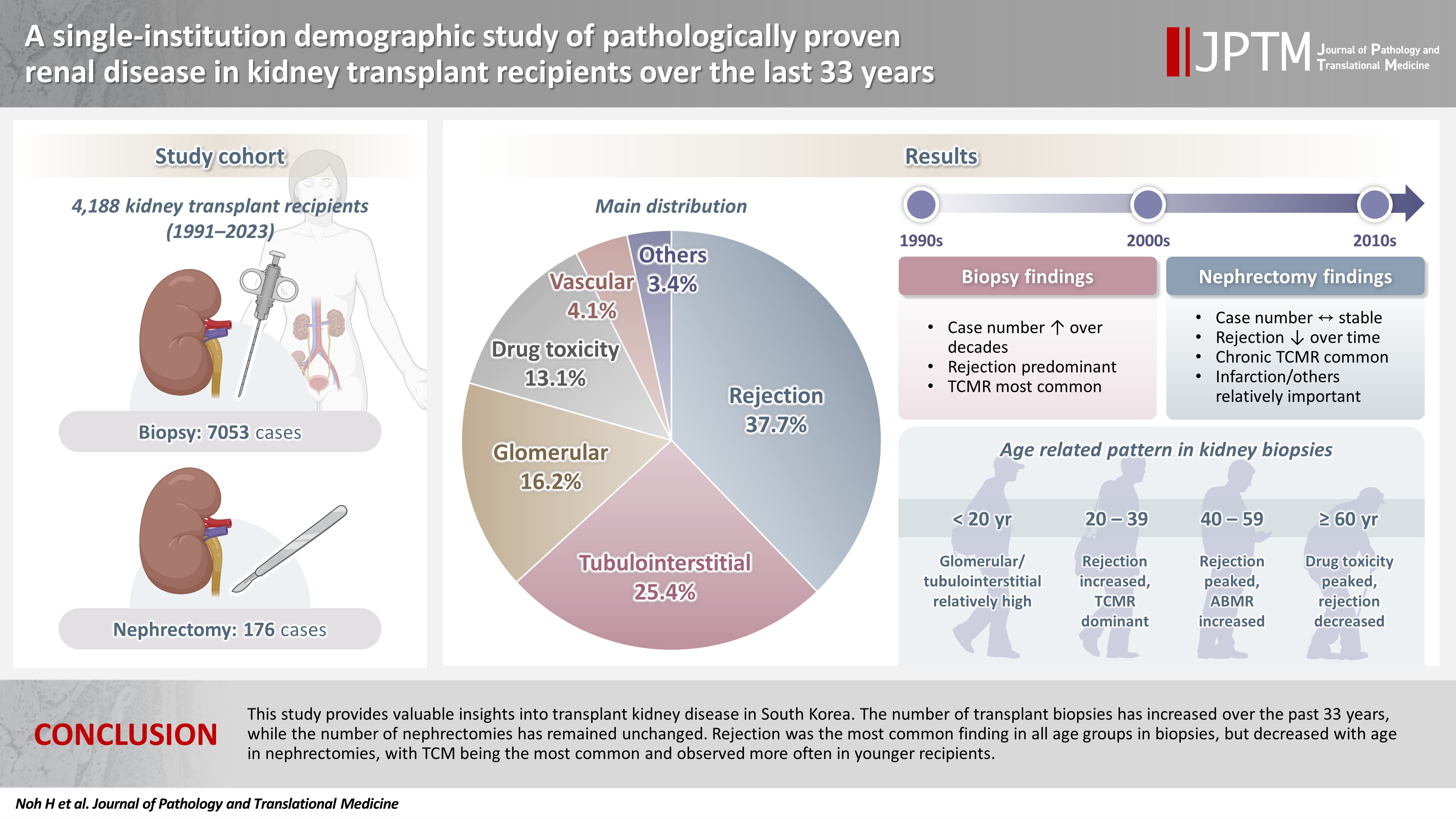
- A single-institution demographic study of pathologically proven renal disease in kidney transplant recipients over the last 33 years
- Hyejin Noh, Jiyeon Kim, Yeong Jin Choi
- J Pathol Transl Med. 2026;60(4):398-412. Published online May 26, 2026
- DOI: https://doi.org/10.4132/jptm.2026.03.28
- 1,244 View
- 20 Download
-
 Abstract
 PDF
PDF Supplementary Material
 - Background
While the number of kidney transplants for end-stage renal disease (ESRD) is increasing, studies examining the long-term demographic analyses based on pathological diagnosis of transplant kidney remain limited. Methods: We conducted a retrospective analysis of 4,188 transplant recipients who underwent either biopsy or nephrectomy from 1991 to 2023 at Seoul St. Mary’s Hospital. Results: Among 7,229 pathologically confirmed cases, rejection was the most prevalent (37.7%), followed by tubulointerstitial (25.4%), glomerular, drug toxicity, and vascular diseases. In 7,053 transplant biopsies, rejection was predominant across all age groups, with T-cell mediated (TCM) category being the most common (60.1%), followed by antibody-mediated and mixed. Drug toxicity increased with age (p = .047), while glomerular and tubulointerstitial diseases were highest in recipients under 20 (p < .001). Among glomerular diseases, IgA-related glomerulonephritis (45.2%) was the most common. In 176 transplant nephrectomies, the most common diagnosis was rejection (33.5%), followed by renal infarction (19.9%), tubulointerstitial, vascular, glomerular disease, and drug toxicity. “Others” included infarction, ESRD, and lymphangiectasia, which increased with age (p = .011). In nephrectomy cases, rejection decreased over time, with chronic TCM rejection (40.7%) being the most frequent. Conclusions: This study provides valuable insights into transplant kidney disease in South Korea. The number of transplant biopsies has increased over the past 33 years, while the number of nephrectomies has remained unchanged. Rejection was the most common finding in all age groups in biopsies, but decreased with age in nephrectomies, with TCM being the most common and observed more often in younger recipients.
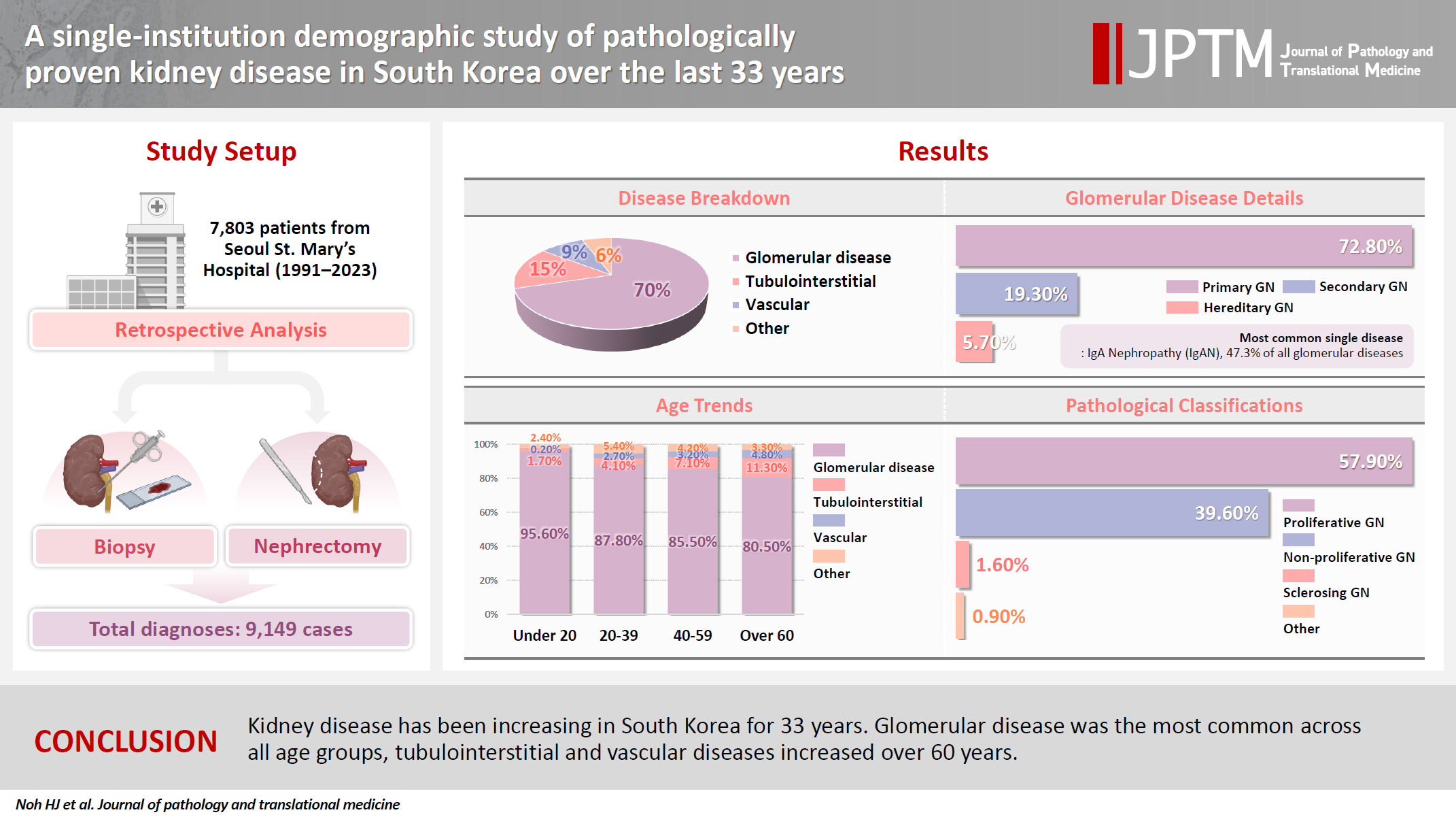
- A single-institution demographic study of pathologically proven kidney disease in South Korea over the last 33 years
- Hyejin Noh, Jiyeon Kim, Yeong Jin Choi
- J Pathol Transl Med. 2025;59(5):306-319. Published online September 10, 2025
- DOI: https://doi.org/10.4132/jptm.2025.06.18
- 3,647 View
- 100 Download
- 1 Web of Science
- 1 Crossref
-
Abstract
PDFSupplementary Material
- Background
To date, epidemiological studies on the entire spectrum of kidney disease based on pathology have been rarely reported. Methods: A retrospective study was conducted on patients diagnosed with kidney disease at Seoul St. Mary's Hospital between 1991 and 2023. Results: Among 7,803 patients with native kidney disease, glomerular disease (70.3%) was the most common, followed by tubulointerstitial (15.1%) and vascular disease (8.8%). In kidney biopsy, glomerular disease (77.8%) showed the highest frequency, particularly in those under 20s (95.6%) (p = .013). Primary glomerulonephritis (GN) (72.8%) was the predominant glomerular disease, with IgA nephropathy (IgAN) (47.3%) being the most common one. Tubulointerstitial and vascular diseases increased with age, showing the highest prevalence in those over 60 years (p = .008 and p = .032, respectively). Glomerular disease was diagnosed at a younger age (39.7 ± 16.7 years) than tubulointerstitial (49.1 ± 16.2) and vascular (48.1 ± 15.3) diseases (p < .001). When glomerular diseases were classified morphologically, proliferative GN (57.9%) was the most common, followed by non-proliferative (39.6%) and sclerosing (1.6%). When classified by etiology, primary GN accounted for the most (72.8%), followed by secondary (19.3%) and hereditary GN (5.7%). In nephrectomy, tubulointerstitial disease (64.6%) was the most common. Those with a tubulointerstitial disease had a higher mean age than those with a glomerular disease (p < .001). In cases where nephrectomy was performed for glomerular diseases, IgAN (34.1%) was the most common diagnosis. Conclusions: Kidney disease has been increasing in South Korea for 33 years. Glomerular disease was the most common across all age groups, tubulointerstitial and vascular diseases increased over 60 years. -
Citations
Citations to this article as recorded by
- A single-institution demographic study of pathologically proven renal disease in kidney transplant recipients over the last 33 years
Hyejin Noh, Jiyeon Kim, Yeong Jin Choi
Journal of Pathology and Translational Medicine.2026; 60(4): 398. CrossRef
- A single-institution demographic study of pathologically proven renal disease in kidney transplant recipients over the last 33 years
Review
- Professional biobanking education in Korea based on ISO 20387
- Jong Ok Kim, Chungyeul Kim, Sangyong Song, Eunah Shin, Ji-Sun Song, Mee Sook Roh, Dong-chul Kim, Han-Kyeom Kim, Joon Mee Kim, Yeong Jin Choi
- J Pathol Transl Med. 2025;59(1):11-25. Published online January 15, 2025
- DOI: https://doi.org/10.4132/jptm.2024.11.04

- 8,742 View
- 204 Download
- 4 Web of Science
- 4 Crossref
-
Abstract
PDF
- To ensure high-quality bioresources and standardize biobanks, there is an urgent need to develop and disseminate educational training programs in accordance with ISO 20387, which was developed in 2018. The standardization of biobank education programs is also required to train biobank experts. The subdivision of categories and levels of education is necessary for jobs such as operations manager (bank president), quality manager, practitioner, and administrator. Essential training includes programs tailored for beginner, intermediate, and advanced practitioners, along with customized training for operations managers. We reviewed and studied ways to develop an appropriate range of education and training opportunities for standard biobanking education and the training of experts based on KS J ISO 20387. We propose more systematic and professional biobanking training programs in accordance with ISO 20387, in addition to the certification programs of the National Biobank and the Korean Laboratory Accreditation System. We suggest various training programs appropriate to a student’s affiliation or work, such as university biobanking specialized education, short-term job training at unit biobanks, biobank research institute symposiums by the Korean Society of Pathologists, and education programs for biobankers and researchers. Through these various education programs, we expect that Korean biobanks will satisfy global standards, meet the needs of users and researchers, and contribute to the advancement of science.
-
Citations
Citations to this article as recorded by- Establishing and Managing a Biobank at an Academic Institution in a Resource-Limited Setting: A Case Study from Ecuador
Alexander Maldonado, Andrés Herrera-Yela, Evaluna Chicango, Micaela Gómez, Gabriela Naranjo, Camila Maldonado, Paula Echeverría
Biopreservation and Biobanking.2026;[Epub] CrossRef - Biobanking for intelligent medicine: assessment and evaluation with the SHARE principle
Yin Yang, Amin Ullah, Yingbo Zhang, Hui Zong, Xingyun Liu, Chi Zhang, Shanshan Hu, Jiakun Li, Bairong Shen
Journal of the American Medical Informatics Association.2026; 33(7): 1333. CrossRef - Development of a big data platform for collecting and utilizing clinical information from the Korea Biobank Network
Yun Seon Im, Seol Whan Oh, Ki Hoon Kim, Wona Choi, In Young Choi
BMC Medical Informatics and Decision Making.2025;[Epub] CrossRef - Frozen section histopathology and preanalytical factors affecting nucleic acid integrity in biobanked fresh-frozen human cancer tissues
Soungeun Kim, Jaewon Kang, Boyeon Kim, Yoonjin Kwak, Hye Seung Lee
Journal of Pathology and Translational Medicine.2025; 59(6): 398. CrossRef
- Establishing and Managing a Biobank at an Academic Institution in a Resource-Limited Setting: A Case Study from Ecuador
Original Articles
- KRAS Mutation Test in Korean Patients with Colorectal Carcinomas: A Methodological Comparison between Sanger Sequencing and a Real-Time PCR-Based Assay
- Sung Hak Lee, Arthur Minwoo Chung, Ahwon Lee, Woo Jin Oh, Yeong Jin Choi, Youn-Soo Lee, Eun Sun Jung
- J Pathol Transl Med. 2017;51(1):24-31. Published online December 25, 2016
- DOI: https://doi.org/10.4132/jptm.2016.10.03
- 14,095 View
- 174 Download
- 5 Web of Science
- 5 Crossref
-
Abstract
PDFSupplementary Material
- Background
Mutations in the KRAS gene have been identified in approximately 50% of colorectal cancers (CRCs). KRAS mutations are well established biomarkers in anti–epidermal growth factor receptor therapy. Therefore, assessment of KRAS mutations is needed in CRC patients to ensure appropriate treatment.
Methods
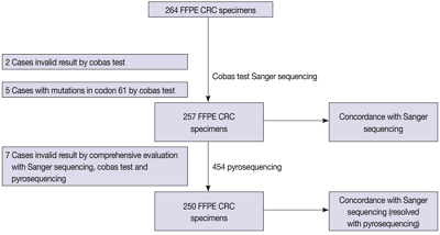
We compared the analytical performance of the cobas test to Sanger sequencing in 264 CRC cases. In addition, discordant specimens were evaluated by 454 pyrosequencing.
Results
KRAS mutations for codons 12/13 were detected in 43.2% of cases (114/264) by Sanger sequencing. Of 257 evaluable specimens for comparison, KRAS mutations were detected in 112 cases (43.6%) by Sanger sequencing and 118 cases (45.9%) by the cobas test. Concordance between the cobas test and Sanger sequencing for each lot was 93.8% positive percent agreement (PPA) and 91.0% negative percent agreement (NPA) for codons 12/13. Results from the cobas test and Sanger sequencing were discordant for 20 cases (7.8%). Twenty discrepant cases were subsequently subjected to 454 pyrosequencing. After comprehensive analysis of the results from combined Sanger sequencing–454 pyrosequencing and the cobas test, PPA was 97.5% and NPA was 100%.
Conclusions
The cobas test is an accurate and sensitive test for detecting KRAS-activating mutations and has analytical power equivalent to Sanger sequencing. Prescreening using the cobas test with subsequent application of Sanger sequencing is the best strategy for routine detection of KRAS mutations in CRC. -
Citations
Citations to this article as recorded by- Single-center study on clinicopathological and typical molecular pathologic features of metastatic brain tumor
Su Hwa Kim, Young Suk Lee, Sung Hak Lee, Yeoun Eun Sung, Ahwon Lee, Jun Kang, Jae-Sung Park, Sin Soo Jeun, Youn Soo Lee
Journal of Pathology and Translational Medicine.2023; 57(4): 217. CrossRef - Assessment of KRAS and NRAS status in metastatic colorectal cancer: Experience of the National Institute of Oncology in Rabat Morocco
Chaimaa Mounjid, Hajar El Agouri, Youssef Mahdi, Abdelilah Laraqui, En-nacer Chtati, Soumaya Ech-charif, Mouna Khmou, Youssef Bakri, Amine Souadka, Basma El Khannoussi
Annals of Cancer Research and Therapy.2022; 30(2): 80. CrossRef - The current understanding on the impact of KRAS on colorectal cancer
Mingjing Meng, Keying Zhong, Ting Jiang, Zhongqiu Liu, Hiu Yee Kwan, Tao Su
Biomedicine & Pharmacotherapy.2021; 140: 111717. CrossRef - Droplet digital PCR revealed high concordance between primary tumors and lymph node metastases in multiplex screening of KRAS mutations in colorectal cancer
Barbora Vanova, Michal Kalman, Karin Jasek, Ivana Kasubova, Tatiana Burjanivova, Anna Farkasova, Peter Kruzliak, Dietrich Busselberg, Lukas Plank, Zora Lasabova
Clinical and Experimental Medicine.2019; 19(2): 219. CrossRef - CRISPR Technology for Breast Cancer: Diagnostics, Modeling, and Therapy
Rachel L. Mintz, Madeleine A. Gao, Kahmun Lo, Yeh‐Hsing Lao, Mingqiang Li, Kam W. Leong
Advanced Biosystems.2018;[Epub] CrossRef
- Single-center study on clinicopathological and typical molecular pathologic features of metastatic brain tumor
- Differential Immunohistochemical Profiles for Distinguishing Prostate Carcinoma and Urothelial Carcinoma
- Woo Jin Oh, Arthur Minwoo Chung, Jee Soon Kim, Ji Heun Han, Sung Hoo Hong, Ji Yeol Lee, Yeong Jin Choi
- J Pathol Transl Med. 2016;50(5):345-354. Published online August 7, 2016
- DOI: https://doi.org/10.4132/jptm.2016.06.14
- 17,142 View
- 362 Download
- 34 Web of Science
- 37 Crossref
-
Abstract
PDF
- Background
The pathologic distinction between high-grade prostate adenocarcinoma (PAC) involving the urinary bladder and high-grade urothelial carcinoma (UC) infiltrating the prostate can be difficult. However, making this distinction is clinically important because of the different treatment modalities for these two entities.
Methods
A total of 249 patient cases (PAC, 111 cases; UC, 138 cases) collected between June 1995 and July 2009 at Seoul St. Mary’s Hospital were studied. An immunohistochemical evaluation of prostatic markers (prostate-specific antigen [PSA], prostate-specific membrane antigen [PSMA], prostate acid phosphatase [PAP], P501s, NKX3.1, and α-methylacyl coenzyme A racemase [AMACR]) and urothelial markers (CK34βE12, p63, thrombomodulin, S100P, and GATA binding protein 3 [GATA3]) was performed using tissue microarrays from each tumor.
Results
The sensitivities of prostatic markers in PAC were 100% for PSA, 83.8% for PSMA, 91.9% for PAP, 93.7% for P501s, 88.3% for NKX 3.1, and 66.7% for AMACR. However, the urothelial markers CK34βE12, p63, thrombomodulin, S100P, and GATA3 were also positive in 1.8%, 0%, 0%, 3.6%, and 0% of PAC, respectively. The sensitivities of urothelial markers in UC were 75.4% for CK34βE12, 73.9% for p63, 45.7% for thrombomodulin, 22.5% for S100P, and 84.8% for GATA3. Conversely, the prostatic markers PSA, PSMA, PAP, P501s, NKX3.1, and AMACR were also positive in 9.4%, 0.7%, 18.8%, 0.7%, 0%, and 8.7% of UCs, respectively.
Conclusions
Prostatic and urothelial markers, including PSA, NKX3.1, p63, thrombomodulin, and GATA3 are very useful for differentiating PAC from UC. The optimal combination of prostatic and urothelial markers could improve the ability to differentiate PAC from UC pathologically. -
Citations
Citations to this article as recorded by- Comparative histologic survey and transcriptomic investigation into canine prostate carcinoma
Nathan K. Hoggard, Said M. Elshafae, Nigel A. Daniels, Jonathan A. Young, Chris Premanandan, John B. Echols, Darshan S. Chandrashekar, Blake E. Hildreth, Michael C. Haffner, Thomas J. Rosol
Research in Veterinary Science.2026; 198: 105981. CrossRef - Plasmacytoid Urothelial Carcinoma with Initial Presentation as a Secondary
Prostatic Tumor: Diagnostic Pitfalls
and Literature Review
丰锦 李
Advances in Clinical Medicine.2026; 16(02): 1264. CrossRef - The molecular pathology of prostate cancer: an update for practising pathologists
Fernanda Caramella Pereira, Angelo M De Marzo
Histopathology.2026; 89(1): 3. CrossRef - High-Grade Urothelial Carcinoma with Divergent Prostatic Differentiation Mimicking a Collision Tumor in a Bladder Diverticulum
Anna Budina, Norge Vergara, Farah El-Sharkawy Navarro, Jennifer J.D. Morrissette, Anupma Nayak
International Journal of Surgical Pathology.2026;[Epub] CrossRef - A case of prostatic urothelial carcinoma with aggressive metastasis: Magnetic resonance imaging as an initial diagnostic clue
Daisuke Shirai, Nozomi Hayakawa, Tsuyoshi Morimoto, Junki Koike, Eiji Kikuchi
Radiology Case Reports.2026; 21(8): 3006. CrossRef - Impact of hormone sensitivity status on aberrant expression of CK7, CK20, CDX2, GATA3 and TTF1 in prostate cancer
Qing Yin Wang, Nazim Benzerdjeb, Samuel Jaquet, Andreea Stepanov, Mame-Kany Diop, Mirela Birlea, Fred Saad, Dominique Trudel
Human Pathology.2025; 163: 105877. CrossRef - Unusual Perineal Metastasis in a Case of Prostate Cancer on 68Ga-PSMA-11 PET/CT
Ritanshu Solanki, Bhagwant Rai Mittal, Rajender Kumar, Aravindh Sekar, Narender Kumar
Clinical Nuclear Medicine.2024; 49(2): e73. CrossRef - NKX3.1 Expression in Non-Prostatic Tumors and Characterizing its Expression in Esophageal/Gastroesophageal Adenocarcinoma
Ansa Mehreen, Kiran G. Manjee, Divyangi Paralkar, Gladell P. Paner, Thanh Lan
Advances in Anatomic Pathology.2024; 31(3): 202. CrossRef - Clinical Management of Intraductal Carcinoma of the Prostate
Gabriel Wasinger, Olivier Cussenot, Eva Compérat
Cancers.2024; 16(9): 1650. CrossRef - Adenocarcinomas of the Gynecologic Tract Involving the Urinary Bladder: A Series of 16 Cases Potentially Mimicking Urothelial Malignancy
Daniel H. Russell, Jonathan I. Epstein, Oleksandr N. Kryvenko, Matthew Schlumbrecht, Merce Jorda, Andre Pinto
Archives of Pathology & Laboratory Medicine.2024; 148(6): 705. CrossRef - Assessing the diagnostic impact of P63, PSA and BCL-2 proteins in premalignant and malignant prostate tissues
Aderonke C. Ogunlayi, Victor O. Ekundina, Adedapo O. Kehinde, Linus A. Enye, Adegoke O. Aremu
International Journal of Scientific Reports.2024; 10(6): 188. CrossRef - Concurrent occurrence of adenocarcinoma and urothelial carcinoma of the prostate gland: A case report
Jhe Yuan Hsu, Yi Sheng Lin, Li Hua Huang, Tang Yi Tsao, Chao Yu Hsu, Yen Chuan Ou, Min Che Tung
World Journal of Clinical Cases.2024; 12(26): 5952. CrossRef - Metastatic prostate cancer presenting as a posterior mediastinal mass: A rare presentation
Muhammad Haider, Arun Umesh Mahtani, Bachar Botrus, Foma Munoh Kenne, Madiha Fatima Master
Clinical Case Reports.2023;[Epub] CrossRef - Diagnostic and Prognostic Roles of GATA3 Immunohistochemistry in Urothelial Carcinoma
Daeseon Yoo, Kyueng-Whan Min, Jung-Soo Pyo, Nae Yu Kim
Medicina.2023; 59(8): 1452. CrossRef - Primary high-grade urothelial carcinoma of prostate with prostatic hyperplasia: a rare case report and review of the literature
Liang Liu, Fu-zhen Sun, Pan-ying Zhang, Yu Xiao, Xiao Yue, Dong-Ming Wang, Qiang Wang
The Aging Male.2023;[Epub] CrossRef - Expression of Gata Binding Protein 3 as a Prognostic Factor in Urogenital Lesions and Its Association With Morphology
T Govardhan, Debahuti Mohapatra, Sujata Naik, Prateek Das, Pranita Mohanty, Ankita Pal
Cureus.2023;[Epub] CrossRef - Histological and immunohistochemical investigation of canine prostate carcinoma with identification of common intraductal carcinoma component
Simone de Brot, Jennifer Lothion‐Roy, Llorenç Grau‐Roma, Emily White, Franco Guscetti, Mark A. Rubin, Nigel P. Mongan
Veterinary and Comparative Oncology.2022; 20(1): 38. CrossRef - Urothelial Carcinoma and Prostate-specific Membrane Antigen: Cellular, Imaging, and Prognostic Implications
Arsalan Tariq, Amy E. McCart Reed, Andrew Morton, Sima Porten, Ian Vela, Elizabeth D. Williams, John W. Yaxley, Peter C. Black, Matthew J. Roberts
European Urology Focus.2022; 8(5): 1256. CrossRef - Immunohistochemical Reactivity of Prostate-Specific Membrane Antigen in Salivary Gland Tumors
Haruto Nishida, Yoshihiko Kondo, Takahiro Kusaba, Hiroko Kadowaki, Tsutomu Daa
Head and Neck Pathology.2022; 16(2): 427. CrossRef - Weak NKX3.1 expression in a urothelial carcinoma: A diagnostic pitfall
Maryam Abdo, Robert Hoyt, Ashley Highfill, Daniel Mettman
Human Pathology Reports.2022; 27: 300599. CrossRef - Gene of the month: NKX3.1
Jon Griffin, Yuqing Chen, James W F Catto, Sherif El-Khamisy
Journal of Clinical Pathology.2022; 75(6): 361. CrossRef - Diagnostic Value of GATA3 and Uroplakin 3 in Differentiating Urothelial Carcinoma from Prostatic and Colorectal Carcinoma
Maha Salama, Dina A. Khairy
Open Access Macedonian Journal of Medical Sciences.2022; 10(A): 514. CrossRef - Diagnostic challenges for the distinction of high-grade prostatic adenocarcinoma and high-grade urothelial carcinoma of simultaneous occurrences - A literature review
Shreyas Bhushan Jayade, Manana Jikurashvili
GEORGIAN SCIENTISTS.2022;[Epub] CrossRef - Cytomorphology, immunoprofile, and clinicopathologic correlation of metastatic prostatic carcinoma
Xiaoqi Lin, Qiuying Shi, Ximing J. Yang
Human Pathology.2022; 130: 36. CrossRef - Cutaneous Metastasis of Prostate Adenocarcinoma: A Rare Presentation of a Common Disease
Alexander Dills, Okechukwu Obi, Kevin Bustos, Jesse Jiang, Shweta Gupta
Journal of Investigative Medicine High Impact Case Reports.2021;[Epub] CrossRef - Mining The Cancer Genome Atlas gene expression data for lineage markers in distinguishing bladder urothelial carcinoma and prostate adenocarcinoma
Ewe Seng Ch’ng
Scientific Reports.2021;[Epub] CrossRef - Immunohistochemical analysis of thrombomodulin expression in myocardial tissue from autopsy cases of ischemic heart disease
Takeshi Kondo, Motonori Takahashi, Gentaro Yamasaki, Marie Sugimoto, Azumi Kuse, Mai Morichika, Kanako Nakagawa, Makoto Sakurada, Migiwa Asano, Yasuhiro Ueno
Legal Medicine.2021; 51: 101897. CrossRef - Application and Pitfalls of Immunohistochemistry in Diagnosis of Challenging Genitourinary Cases
Jenny Ross, Guangyuan Li, Ximing J. Yang
Archives of Pathology & Laboratory Medicine.2020; 144(3): 290. CrossRef - New Screening Test Improves Detection of Prostate Cancer Using Circulating Tumor Cells and Prostate-Specific Markers
Karin Ried, Tasnuva Tamanna, Sonja Matthews, Peter Eng, Avni Sali
Frontiers in Oncology.2020;[Epub] CrossRef - An Unlikely Culprit: Gastric Metastasis from Primary Prostatic Adenocarcinoma
Eric Omar Then, Spoorthi Nutakki, Andrew Ofosu, Saad Saleem, Vijay Gayam, Tagore Sunkara, Vinaya Gaduputi
Journal of Gastrointestinal Cancer.2020; 51(3): 1081. CrossRef - MRI of prostatic urethral mucinous urothelial carcinoma: Expanding the differential diagnosis for T2 hyperintense prostatic masses
Neel Patel, Bryan R. Foster, Elena K. Korngold, Kyle Jensen, Kevin R. Turner, Fergus V. Coakley
Clinical Imaging.2020; 68: 68. CrossRef - Morphological and Immunohistochemical Biomarkers in Distinguishing Prostate Carcinoma and Urothelial Carcinoma: A Comprehensive Review
Francesca Sanguedolce, Davide Russo, Vito Mancini, Oscar Selvaggio, Beppe Calò, Giuseppe Carrieri, Luigi Cormio
International Journal of Surgical Pathology.2019; 27(2): 120. CrossRef - A Case of Metastatic Prostate Cancer to the Urethra That Resolved After Androgen Deprivation Therapy
Darren J. Bryk, Kenneth W. Angermeier, Eric A. Klein
Urology.2019; 129: e4. CrossRef - The Homeodomain Transcription Factor NKX3.1 Modulates Bladder Outlet Obstruction Induced Fibrosis in Mice
Mehul S. Patel, Diana K. Bowen, Nicholas M. Tassone, Andrew D. Gould, Kirsten S. Kochan, Paula R. Firmiss, Natalie A. Kukulka, Megan Y. Devine, Belinda Li, Edward M. Gong, Robert W. Dettman
Frontiers in Pediatrics.2019;[Epub] CrossRef - Cancer of unknown primary: Ancillary testing of cytologic and small biopsy specimens in the era of targeted therapy
Morgan L. Cowan, Christopher J. VandenBussche
Cancer Cytopathology.2018; 126(S8): 724. CrossRef - Glandular Tumors of the Urachus and Urinary Bladder: A Practical Overview of a Broad Differential Diagnosis
Alexander S. Taylor, Rohit Mehra, Aaron M. Udager
Archives of Pathology & Laboratory Medicine.2018; 142(10): 1164. CrossRef - S100P as a Marker for Urothelial Histogenesis: A Critical Review and Comparison With Novel and Traditional Urothelial Immunohistochemical Markers
Moushumi Suryavanshi, Julian Sanz-Ortega, Deepika Sirohi, Mukul K. Divatia, Chisato Ohe, Claudia Zampini, Daniel Luthringer, Steven C. Smith, Mahul B. Amin
Advances in Anatomic Pathology.2017; 24(3): 151. CrossRef
- Comparative histologic survey and transcriptomic investigation into canine prostate carcinoma
- Histologic Disorderliness in the Arrangement of Tumor Cells as an Objective Measure of Tumor Differentiation
- Sungwook Suh, Gyeongsin Park, Young Sub Lee, Yosep Chong, Youn Soo Lee, Yeong Jin Choi
- Korean J Pathol. 2014;48(5):339-345. Published online October 27, 2014
- DOI: https://doi.org/10.4132/KoreanJPathol.2014.48.5.339
- 8,274 View
- 68 Download
-
Abstract
PDF
- Background: Inter-observer and intra-observer variation in histologic tumor grading are well documented. To determine whether histologic disorderliness in the arrangement of tumor cells may serve as an objective criterion for grading, we tested the hypothesis the degree of disorderliness is related to the degree of tumor differentiation on which tumor grading is primarily based. Methods: Borrowing from the statistical thermodynamic definition of entropy, we defined a novel mathematical formula to compute the relative degree of histologic disorderliness of tumor cells. We then analyzed a total of 51 photomicrographs of normal colorectal mucosa and colorectal adenocarcinoma with varying degrees of differentiation using our formula. Results: A one-way analysis of variance followed by post hoc pairwise comparisons using Bonferroni correction indicated that the mean disorderliness score was the lowest for the normal colorectal mucosa and increased with decreasing tumor differentiation. Conclusions: Disorderliness, a pathologic feature of malignant tumors that originate from highly organized structures is useful as an objective tumor grading proxy in the field of digital pathology.
- Prognostic Significance of Amplification of the c-MYC Gene in Surgically Treated Stage IB-IIB Cervical Cancer.
- Tae Jung Kim, Ahwon Lee, Sung Jong Lee, Won Chul Lee, Yeong Jin Choi, Kyo Young Lee, Chang Suk Kang
- Korean J Pathol. 2011;45(6):596-603.
- DOI: https://doi.org/10.4132/KoreanJPathol.2011.45.6.596
- 5,510 View
- 42 Download
- 1 Crossref
-
Abstract
PDF
- BACKGROUND
Mutations of c-MYC have been described in cervical cancer. However, association between c-MYC gene status and its prognostic significance have not been clarified.
METHODS
Tissue microarray sections from 144 patients with stage IB-IIB cervical cancer treated by radical hysterectomy were analyzed by fluorescence in situ hybridization using a region-specific probe for c-MYC and a centromere-specific probe for chromosome 8.
RESULTS
Seventy five percent (108/144) of c-MYC gain and 6.9% (10/144) of c-MYC gene amplification were observed. c-MYC gene alteration was more frequently observed in squamous cell carcinoma than adenocarcinoma or adenosquamous carcinoma and were associated with low Ki67 labeling index (p=0.013). c-MYC amplification was not associated with clinicopathologic parameters except absence of bcl2 expression (p=0.048). Survival analysis revealed that patients with c-MYC amplification were significantly associated with higher risk of disease recurrence (p=0.007) and cancer related death (p=0.020). However, c-MYC gain was not associated with unfavorable outcome. Multivariate analysis proved c-MYC amplification as independent prognostic factors of shorter disease free survival and cancer-related death (p=0.028 and p=0.025, respectively).
CONCLUSIONS
c-MYC amplification, not gain, is an independent prognostic marker for shorter disease free and cancer specific survival in cervical cancer treated by radical hysterectomy. -
Citations
Citations to this article as recorded by- A Rare Case of Cutaneous Plasmacytosis in a Korean Male
Corey Georgesen, Meenal Kheterpal, Melissa Pulitzer
Case Reports in Pathology.2017; 2017: 1. CrossRef
- A Rare Case of Cutaneous Plasmacytosis in a Korean Male
- Detection Limit of Monoclonal B-Cells Using Multiplex PCR and Laser-Induced Fluorescence Capillary Electrophoresis.
- Sung Hak Lee, Yeonsook Moon, Byunghoo Song, Hyung Nam Lee, Ahwon Lee, Eun Sun Jung, Yeong Jin Choi, Kyo Young Lee, Chang Suk Kang, Gyeongsin Park
- Korean J Pathol. 2011;45(6):582-588.
- DOI: https://doi.org/10.4132/KoreanJPathol.2011.45.6.582
- 4,545 View
- 28 Download
- 1 Crossref
-
Abstract
PDF
- BACKGROUND
The identification of monoclonality has been widely used for making diagnoses of lymphoproliferative lesions. Awareness of the sensitivity and detection limit of the technique used would be important for the data to be convincing.
METHODS
We investigated the minimum requirement of cells and sensitivity of gel electrophoresis (GE) and laser-induced fluorescence capillary electrophoresis (LFCE) for identifying IgH gene rearrangement using BIOMED-2 protocols. DNA extracted from Raji cells were diluted serially with peripheral blood mononuclear cells (PBMNCs) DNA. DNA from mixtures of diffuse large B-cell lymphoma (DLBCL) and reactive lymph nodes were also serially diluted.
RESULTS
For Raji cells, the detection limit was 62 and 16 cell-equivalents for GE and LFCE, respectively. In the condition with PBMNCs mixture, 2.5% and 1.25% of clonal cells was the minimum requirement for GE and LFCE, respectively. In 23% of DLBCL cells in tissue section, the detection limit was 120 and 12 cell-equivalents for GE and LFCE, respectively. In 3.2% of DLBCL cells, that was 1,200 and 120 cell-equivalents for GE and LFCE, respectively.
CONCLUSIONS
These results show that LFCE method is more sensitive than GE and the sensitivity of clonality detection can be influenced by the amount of admixed normal lymphoid cells. -
Citations
Citations to this article as recorded by- Molecular pathology diagnosis of diffuse large B cell lymphoma using BIOMED-2 clonal gene rearrangements
Saeid Ghorbian
Annals of Diagnostic Pathology.2017; 29: 28. CrossRef
- Molecular pathology diagnosis of diffuse large B cell lymphoma using BIOMED-2 clonal gene rearrangements
Case Report
- Apocrine Carcinoma of the Axilla with Predominant Signet Ring Cell Features A Case Report.
- Jeana Kim, Tae Eun Kim, Ah Won Lee, Yeong Jin Choi, Kyo Young Lee, Eun Sun Jung
- Korean J Pathol. 2011;45(3):326-328.
- DOI: https://doi.org/10.4132/KoreanJPathol.2011.45.3.326
- 3,946 View
- 40 Download
-
Abstract
PDF
- Apocrine carcinoma arising from the apocrine sweat glands is a rare cutaneous malignant tumor which occurs predominantly in the axilla of elderly individuals. The typical histologic features of apocrine carcinoma is within a well developed glandular lumina with abundant eosinophilic cytoplasm and evidence of decapitation secretion. In rare instances, predominant signet ring cell features in apocrine carcinoma has been reported. We experienced a case that occured in the right axilla of a 59-year-old. Histopathologic examination showed a solid tumor that extended from the upper dermis into the subcutis, with a delicate infiltrate of epithelial cells. The cells had granular amphophilic cytoplasm, predominantly showed distinct signet ring cell morphology, and were strongly positive for epithelial mucin. Both lysozyme and gross cystic disease fluid protein-15 were identified in the tumor cells. We diagnosed this to be a case of primary signet ring cell apocrine carcinoma of the axilla after several immunohistochemical and clinical evaluations.
Original Articles
- The Usefulness of p16INK4a Immunocytochemical Staining in ASC-H Patients.
- Kwang Il Yim, Yeo Ju Kang, Tae Eun Kim, Gyeongsin Park, Eun Sun Jung, Yeong Jin Choi, Kyo Young Lee, Chang Seok Kang, Ahwon Lee
- Korean J Pathol. 2011;45(3):290-295.
- DOI: https://doi.org/10.4132/KoreanJPathol.2011.45.3.290
- 5,247 View
- 26 Download
- 1 Crossref
-
Abstract
PDF
- BACKGROUND
The grey zone of cervical cytology, and in particular atypical squamous cells, cannot exclude HSIL (ASC-H) causes diagnostic difficulties and increases medical expenses. We analyzed p16INK4a expression in ASC-H liquid-based cytology specimens (LBCS) to develop more effective methods for the management of ASC-H patients.
METHODS
We carried out p16INK4a immunostaining with 57 LBCS of ASC-H diagnostic categories, all of which were histologically cofirmed and 43 cases of which were compared with the results of a human papillomavirus (HPV) chip test.
RESULTS
p16INK4a immunostaining with ASC-H LBCS was positive in 20% (3/15) of cervicitis, 25.0% (3/12) of tissue-low-grade squamous intraepithelial lesion, 75.0% (18/24) of tissue-high grade squamous intraepithelial lesion (HSIL), and 100% (6/6) of invasive cancer cases. The positivity of p16INK4a in LBCS was correlated with higher grade of histologic diagnosis (r=0.578, p=0.000). The sensitivity, specificity, positive predictive value (PPV), and negative predictive value (NPV) of p16INK4a immunostaining for the prediction of tissue-HSIL+ were 80.0%, 77.8%, 80.0%, and 77.8%, respectively. The sensitivity, specificity, PPV, and NPV of p16INK4a immunostaining plus HPV chip test for predicting tissue-HSIL+ were 71.2%, 86.4%, 84.2%, and 79.2%.
CONCLUSIONS
p16INK4a immunostaining as well as HPV chip testing with remaining LBCS with ASC-H are useful objective markers for the prediction of tissue-HSIL+. -
Citations
Citations to this article as recorded by- Usefulness of p16INK4a Immunocytochemical staining for the Differentiation between Atrophy and ASCUS in Diagnosis of Uterine Cervical Cancer
Hye Ryoung Shin, Taekil Eom, Wan-Su Choi
Biomedical Science Letters.2023; 29(3): 144. CrossRef
- Usefulness of p16INK4a Immunocytochemical staining for the Differentiation between Atrophy and ASCUS in Diagnosis of Uterine Cervical Cancer
- Comparison of Detecting Methods of BK Virus Infection in Patients with Renal Allograft Recipients.
- Sung Hak Lee, Youn Jun Park, Chul Woo Yang, Yong Soo Kim, In Sung Moon, Chang Suk Kang, Yeong Jin Choi
- Korean J Pathol. 2010;44(6):636-641.
- DOI: https://doi.org/10.4132/KoreanJPathol.2010.44.6.636
- 4,889 View
- 26 Download
- 2 Crossref
-
Abstract
PDF
- BACKGROUND
BK virus nephropathy (BKVN) is an emerging problem as a consequence of the use of potent immunosuppressive agents. Because optimal detection methods for the diagnosis of BKVN are required clinically, we compared the results of renal allograft biopsy, urine cytology, and urine and blood viral loads.
METHODS
Four hundred sixty two case notes from 2004 to 2009 at Seoul St. Mary's Hospital were reviewed. During that period, 286 cases of urine cytology for decoy cells, 938 cases of urine BKV reverse transcription-polymerase chain reaction (RT-PCR), and 1,029 cases of blood BKV RT-PCR were performed. All diagnostic methods were performed in 85 cases.
RESULTS
A histological diagnosis of BKVN was made in 2.4% of cases (11/462). Urine cytology for decoy cells was positive in 26.2% (75/286). BKV RT-PCR revealed viruria in positivity of 22.1% (207/938) and viremia in 5.2% (54/1,029). In cases of BKVN, the sensitivities of urine and blood BKV RT-PCR were all 100% and the specificities were 69% and 94.5%, respectively. In cases with positive urine decoy cells, the sensitivities of urine and blood BKV RT-PCR were 50% and 27.7%, with specificities of 77.7% and 100%, respectively.
CONCLUSIONS
BKV screening by RT-PCR assays may be a clinically useful noninvasive test to identify renal recipients with concurrent BKVN. -
Citations
Citations to this article as recorded by- Prevalence of BK Virus among Iranian Renal Transplant Recipients: A Systematic Review and Meta-Analysis
Mohsen Ebrahimi, Alireza Mohebbi, Mohammad Mostakhdem Hashemi, Mobina Ashrafi Shahmirzadi
Journal of Clinical and Basic Research.2020; 4(4): 50. CrossRef - Asymptomatic hematuria associated with urinary polyomavirus infection in immunocompetent patients
Sung Hak Lee, Sung Hoo Hong, Ji Youl Lee, Tae Kon Hwang, Kyoung Suk Kim, Hyoungnam Lee, Yeong Jin Choi
Journal of Medical Virology.2014; 86(2): 347. CrossRef
- Prevalence of BK Virus among Iranian Renal Transplant Recipients: A Systematic Review and Meta-Analysis
- Practical Standardization in Renal Biopsy Reporting.
- So Young Jin, Hyeon Joo Jeong, Sun Hee Sung, Beom Jin Lim, Jee Young Han, Soon Won Hong, Hyun Ee Yim, Yeong Jin Choi, Yong Mee Cho, Myoung Jae Kang, Kyung Chul Moon, Hee Jeong Cha, Seung Yeon Ha, Mi Seon Kang, Mee Young So, Kwang Sun Suh, Jong Eun Joo, Yong Jin Kim, Nam Hee Won, Moon Hyang Park
- Korean J Pathol. 2010;44(6):613-622.
- DOI: https://doi.org/10.4132/KoreanJPathol.2010.44.6.613
- 6,231 View
- 197 Download
- 3 Crossref
-
Abstract
PDF
- BACKGROUND
To standardize renal biopsy reporting and diagnosis, The Renal Pathology Study Group of the Korean Society of Pathologists (RPSKSP) has developed a renal pathology reporting format for the native and allograft kidney.
METHODS
A consensus checklist of a provisional renal biopsy format was sent to all members of the RPSKSP. Feed back opinions regarding the practical application of the checklist to the diagnostic work were received.
RESULTS
Kidney biopsies require three essential examinations: by light microscopy, immunofluorescence (IF), and electron microscopy (EM). A final report of a renal biopsy should include information on specimen adequacy and a description of the morphologic change using a systematic semiquantitative method for each of the compartments, with optional separate IF and EM reports.
CONCLUSIONS
A standard renal biopsy report format is important in establishing clinicopathologic correlations, making reliable prognostic considerations, comparing the findings in sequential biopsies and evaluating the effects of therapy. -
Citations
Citations to this article as recorded by- Interobserver agreement analysis among renal pathologists in classification of lupus nephritis using a digital pathology image dataset: after a third evaluation
Ju Yeon Pyo, Nara Jeon, Su-Jin Shin, Minsun Jung, Beom Jin Lim, Minseob Eom, Sung-Eun Choi
Kidney Research and Clinical Practice.2026; 45(4): 504. CrossRef - Additional antihypertensive effect of magnesium supplementation with an angiotensin II receptor blocker in hypomagnesemic rats
Kyubok Jin, Tae Hee Kim, Yeong Hoon Kim, Yang Wook Kim
The Korean Journal of Internal Medicine.2013; 28(2): 197. CrossRef - Clinicopathologic Features of IgA-Dominant Postinfectious Glomerulonephritis
Tai Yeon Koo, Gheun-Ho Kim, Hyang Park
Korean Journal of Pathology.2012; 46(2): 105. CrossRef
- Interobserver agreement analysis among renal pathologists in classification of lupus nephritis using a digital pathology image dataset: after a third evaluation
- Evaluation of the HPV ISH Assay in Cervical Cancer.
- Jung Uee Lee, Jung Ha Shin, Jong Ok Kim, Yeong Jin Choi, Kyo Young Lee, Jong Sup Park, Won Chul Lee, Ahwon Lee
- Korean J Pathol. 2010;44(5):513-520.
- DOI: https://doi.org/10.4132/KoreanJPathol.2010.44.5.513
- 6,082 View
- 122 Download
- 3 Crossref
-
Abstract
PDF
- BACKGROUND
Human papillomavirus (HPV) infection can be detected by in situ hybridization (ISH), in which a punctate signal pattern indicates integrated HPV DNA and a diffuse pattern denotes the presence of episomal viral DNA. This study was conducted to evaluate the usefulness of an HPV ISH assay for invasive cervical cancer.
METHODS
The HPV ISH assay for high-risk HPV and immunohistochemical staining for p16(INK4a), p53, bcl-2, and Ki-67 were performed in a tissue microarray of 279 cervical cancers.
RESULTS
High-risk HPV ISH was positive in 194 (69.5%) of the samples. Punctate, diffuse, and mixed signal patterns were observed in 157 (56.3%), one (0.4%), and 36 cases (12.9%), respectively. Positive results in high-risk HPV ISH were associated with p16 and bcl-2 expression (p = 0.01 and p < 0.01, respectively). According to a Cox regression analysis, HPV infection and its surrogate immunohistochemical markers such as p16, bcl-2, and Ki-67 were not independent prognostic factors, but stage and grade were independent prognostic factors.
CONCLUSIONS
Our results confirm that an HPV ISH assay is reasonably sensitive for HPV infection and that it might be useful to identify integrated HPV DNA in formalin-fixed and paraffin-embedded specimens. Further study encompassing HPV type, E2/E6 ratio, and therapeutic modality is necessary to understand the clinical meaning of HPV status in cervical cancer. -
Citations
Citations to this article as recorded by- Prevalence of human papillomavirus in eyelid carcinoma among Koreans: a clinicopathological study
Min Kyu Yang, Namju Kim, Hokyung Choung, Ji Eun Kim, Sang In Khwarg
BMC Ophthalmology.2023;[Epub] CrossRef - Cervical cancer screening by molecular Pap‐transformation of gynecologic cytology
Shaikhali M Barodawala, Kirti Chadha, Vikas Kavishwar, Anuradha Murthy, Shamma Shetye
Diagnostic Cytopathology.2019; 47(5): 374. CrossRef - Prognostic Significance of Amplification of thec-MYCGene in Surgically Treated Stage IB-IIB Cervical Cancer
Tae-Jung Kim, Ahwon Lee, Sung-Jong Lee, Won-Chul Lee, Yeong-Jin Choi, Kyo-Young Lee, Chang Suk Kang
The Korean Journal of Pathology.2011; 45(6): 596. CrossRef
- Prevalence of human papillomavirus in eyelid carcinoma among Koreans: a clinicopathological study
- Comparison of Various Detection Methods of Mycobacterium Species in Formalin-Fixed Paraffin-Embedded Tissue with Chronic Granulomatous Inflammation.
- Hyun Seung Lee, Hyoungnam Lee, Soyoung Im, Yun Su Lee, Kyo Young Lee, Yeong Jin Choi
- Korean J Pathol. 2010;44(3):259-266.
- DOI: https://doi.org/10.4132/KoreanJPathol.2010.44.3.259
- 5,243 View
- 52 Download
- 2 Crossref
-
Abstract
PDF
- BACKGROUND
To determine the most effective method for detecting mycobacteria in formalin- fixed paraffin-embedded (FFPE) tissue, we compared the results of Ziehl-Neelsen stain (ZNS) and mycobacterial culture with those of polymerase chain reaction (PCR) and real-time quantitative PCR (RQ-PCR).
METHODS
We analyzed 54 cases diagnosed as chronic granulomatous inflammation. In all cases, ZNS and nested PCR using three different primers, IS6110, Mpb64 and IS6110/Rpobeta were done. RQ-PCR with the IS6110/Rpobeta primer was done in 51 cases.
RESULTS
Mycobacteria were identified by ZNS in 15/54 (27.8%) cases. RQ-PCR had the highest sensitivity (80.0%) compared to PCR with IS6110 (73.3%), Mpb64 (60.0%) and IS6110/Rpobeta (73.3%). Specificity was higher in all PCR experiments (79.5-82.1%) than in RQ-PCR (69.4%) experiments. The false negative rate was lowest for RQ-PCR (20.0%) than for PCR with IS6110 (26.7%), Mpb64 (40.0%) and IS6110/Rpobeta (26.7%). The false positive rate was highest for RQ-PCR (30.6%) compared to PCR with IS6110 (20.5%), Mpb64 (17.9%) and IS6110/Rpobeta (20.5%).
CONCLUSIONS
RQ-PCR had the highest sensitivity, and the lowest false negative rate, but it also had a higher false positive rate than PCR for detection of mycobacteria in FFPE tissues. -
Citations
Citations to this article as recorded by- Clinical Usefulness of PCR for Differential Diagnosis of Tuberculosis and Nontuberculous Mycobacterial Infection in Paraffin-Embedded Lung Tissues
Yo Na Kim, Kyoung Min Kim, Ha Na Choi, Ju Hyung Lee, Ho Sung Park, Kyu Yun Jang, Woo Sung Moon, Myoung Jae Kang, Dong Geun Lee, Myoung Ja Chung
The Journal of Molecular Diagnostics.2015; 17(5): 597. CrossRef - Usefulness of PCR to Mycobacterium Tuberculous and Nontuberculous Mycobacteria from Paraffin-embedded Tissues
Yeon-Il Choi, Hye-Young Kim
Korean Journal of Clinical Laboratory Science.2014; 46(2): 47. CrossRef
- Clinical Usefulness of PCR for Differential Diagnosis of Tuberculosis and Nontuberculous Mycobacterial Infection in Paraffin-Embedded Lung Tissues
- Clinicopathologic Significances of EGFR Expression at Invasive Front of Colorectal Cancer.
- Yeo Ju Kang, Chan Kwon Jung, Yeong Jin Choi, Kyo Young Lee, Hyung Jin Kim, Won Kyung Kang, Seong Taek Oh
- Korean J Pathol. 2010;44(1):16-21.
- DOI: https://doi.org/10.4132/KoreanJPathol.2010.44.1.16
- 4,604 View
- 42 Download
-
Abstract
PDF
- BACKGROUND
Epidermal growth factor receptor (EGFR) is frequently expressed in the invasive front of colorectal cancer (CRC), but its clinicopathologic significance remains unclear. We investigated the clinical value of the EGFR expression at the invasive front of CRC.
METHODS
We performed an immunohistochemical analysis in order to examine the expression and distribution of EGFR in 214 cases of CRC. The EGFR status was considered positive when > or =1% of the tumor cells had membranous staining.
RESULTS
Overall, an EGFR expression was observed in 144 (67%) cases and it had no significant relationship with the clinicopathologic parameters. However, an EGFR expression at the invasive front was correlated with lymphatic invasion, lymph node metastasis and a high level of serum carcinoembryonic antigen (p = 0.028, p = 0.043, and p = 0.045, respectively). For the budding-positive CRCs liver metastases were found in the cases with an EGFR expression at the budding, but no liver metastasis occurred in the EGFR negative cases at the budding (p = 0.030).
CONCLUSIONS
An EGFR expression at the invasive front has clinicopathologic significances in patients with CRC. An EGFR expression at tumor cell budding is a pathologic marker that suggests the high potential for liver metastasis in CRC.
- C1q Nephropathy: A Distinct Pathologic Entity.
- Jung Ha Shin, Tae Eun Kim, Kyo Young Lee, Sang In Shim, Yeong Jin Choi
- Korean J Pathol. 2009;43(4):335-341.
- DOI: https://doi.org/10.4132/KoreanJPathol.2009.43.4.335
- 3,982 View
- 45 Download
-
Abstract
PDF
- BACKGROUND
C1q nephropathy (C1qN) is a controversial diagnostic entity defined by Jennette and Hipp in 1985. The prevalence is very low and a few large scale studies have been reported. Application of the criteria for clinical diagnostics of C1qN may cause confusion with other glomerulonephropathies, such as minimal change disease (MCD) or focal segmental glomerulosclerosis (FSGS). In order to clarify the confusion with glomerulonephropathies, we did this study to identify the clinicopathological characteristics and the exact disease entity of C1qN.
METHODS
A total of 5,258 kidney biopsies at Kangnam St Mary's Hospital were reviewed. Twenty three cases (0.44%) met the criteria of C1qN. Twenty eight cases showing dominant C1q deposits without electron dense depostis (EDD) grouped as C1q+EDD-, and previously diagnosed typical cases of MCD and FSGS were selected for this study. Four groups were compared to each other with regard to the clinical and pathological aspects of the disease. RESULTS: C1qN patients had an average age of 30.4 years. Eighteen were males and 5 were females. Eighty seven percent had proteinuria and 18% had hematuria. By electron microscopy analysis, 100% had mesangial EDD and 47.8% showed foot process effacement. C1qN had some significant differences compared with C1q+EDD-, MCD and FSGS. CONCLUSIONS: C1qN is clinically and morphologically different from MCD and FSGS. However, additional long term studies are needed to fully define C1qN from other glomerulonephritis with C1q deposits.
- IgA Nephropathy: Correlation of WHO Classification and Morphologic Semi-quantitative Scoring System.
- Kyung Jin Seo, Tae Jung Kim, Kyo Young Lee, Sang In Shim, Yeong Jin Choi
- Korean J Pathol. 2009;43(3):244-249.
- DOI: https://doi.org/10.4132/KoreanJPathol.2009.43.3.244
- 5,469 View
- 51 Download
- 2 Crossref
-
Abstract
PDF
- BACKGROUND
IgA nephropathy (IgAN) is the most common glomerulonephritis worldwide, and the clinical course of IgAN shows marked variability. Many efforts have made to histologically predict the clinical outcome. There are two methods to classify IgAN. One is mainly based on the glomerular changes, such as the WHO and the Lee and Haas classification systems. The other is a morphologic semi-quantitative scoring system, which counts the changes of the glomerular, tubulointerstitial and vascular structures, respectively. The purpose of this study is to determine whether the WHO classification properly reflects the various morphologic findings of IgAN.
METHODS
We analyzed 354 cases of IgAN by both the WHO classification system and the semiquantitative scoring system and evaluated the correlations of these two methods.
RESULTS
The severity of the glomerular lesions (glomerulosclerosis, capsular adhesion and mesangial matrix expansion) and the tubulointerstitial lesions (interstitial fibrosis, tubular atrophy and interstitial lymphocytic infiltration) are strongly correlated with the increase of the WHO classes of IgAN (Spearman's rho [R] > or =0.5, p<0.05). There is a weak correlation between crescent formation and the increase of the WHO classes (R=0.3, p<0.05).
CONCLUSIONS
This study shows that the WHO classification well reflects the severity of various morphologic findings and this suggests a complementary role for the semi-quantitative scoring system in classifying IgAN. -
Citations
Citations to this article as recorded by- Significance of KM55 immunohistochemical staining in the diagnosis and prognosis of IgA nephropathy
Hoe In Jeong, Beom Jin Lim, Minsun Jung
Journal of Pathology and Translational Medicine.2026; 60(1): 69. CrossRef - The Oxford classification as a predictor of prognosis in patients with IgA nephropathy
S. H. Kang, S. R. Choi, H. S. Park, J. Y. Lee, I. O. Sun, H. S. Hwang, B. H. Chung, C. W. Park, C. W. Yang, Y. S. Kim, Y. J. Choi, B. S. Choi
Nephrology Dialysis Transplantation.2012; 27(1): 252. CrossRef
- Significance of KM55 immunohistochemical staining in the diagnosis and prognosis of IgA nephropathy
Case Reports
- Sarcomatoid Transitional Cell Carcinoma of the Renal Pelvis A report of two cases.
- Kyo Young Lee, Mi seon Kwon, Yeong Jin Choi, Chang Suk Kang, Seok Jin Kang, Baying Kee Kim, Sang In Shim
- Korean J Pathol. 1999;33(2):128-132.
- 2,271 View
- 10 Download
-
Abstract
- Sarcomatoid carcinomas are malignant epithelial neoplasms in which the tumor cells assume a partial or complete spindle cell pattern of growth, leading to the erroneous classification of some true carcinomas as sarcomas. These spindle cells are malignant and manifest various amount of both vimentin and cytokeratin. Positive reaction of some of the spindle cells for cytokeratin antibodies is confirmatory. Clinical features do not differ significantly from those of patients with high-grade transitional cell carcinoma. So far, 13 cases of sarcomatoid transitional cell carcinoma of the renal pelvis have been reported in English and Korean literature. In this report, we describe clinicopathologic features of recently observed two cases of sarcomatoid transitional cell carcinoma of the renal pelvis and summarize the pathologic findings of previously reported cases with review of the literature.
- Kaposi's Sarcoma: A report of three cases.
- Yeon Soo Lee, Yeong Jin Choi, Mi Kyung Jee, Seok Jin Kang, Byoung Kee Kim, Sun Moo Kim
- Korean J Pathol. 1995;29(3):385-390.
- 2,737 View
- 18 Download
-
Abstract
PDF
- The classic type of Kaposi's sarcoma, or multifocal hemorrhagic sarcoma histologically characterized by proliferating fibroblastic and microvascular elements was described by Kaposi as a relatively rare neoplasm. During the past nine years, we experienced three cases of sporadic, classic Kaposi's sarcomas. They were presented as multiple papules, macules and nodules on the skin of the hands, lower logs and feet without systemic involvement. Histologically, Kaposi's sarcoma is divided into three stages, early patch, plaque and nodular stages. The nodular lesions(case 1, 2 and 3) showed extensive proliferatiion of spindle shaped, somewhat pleomorphic cells having dark prominent nuclei, proliferation of small vessels with solid aggregates of endothelial cells, and extravasation of erythrocytes. In early patch stage(case 3), widely dilated, anastomosing, thin-walled vascular spaces are noted in the upper half of the dermis. In plaque stage(case I and 3), there are proliferation of spindle shaped cells with extravasated erythrocytes and aggregates of blood vessels lined by prominent endothelial cells.
Original Article
- The Apoptotic Molecular Changes of Cellular Injury in Mouse Testis Induced by Endocrine Disrupting Chemicals.
- Eun Hui Wang, Kweon Heang Lee, Ki Hwa Yang, Jinsuk Lee, Eun Sun Jung, Chang Suk Kang, Yeong Jin Choi
- Korean J Pathol. 2004;38(4):228-237.
- 2,453 View
- 19 Download
-
Abstract
PDF
- BACKGROUND
Spermatogenesis is regulated by various cellular reactions, and especially cell proliferation and apoptosis.
METHODS
We investigated the morphological changes and the apoptotic molecular changes in mouse testis induced by the endocrine disrupting chemicals. ICR mice were treated with bisphenol A (BPA), 2-bromopropane (2-BP) and diethylstilbesterol (DES). Histological examination and immunohistochemical staining, TUNNEL staining and RNAse protection assay were conducted.
RESULTS
Testes treated with BPA showed normal spermatogenesis and the proliferation activity, and the density of the cells was similar with those in the control. 2-BP and DES groups, which showed a decrease of germ cells near the basal layer and degenerative changes. The proliferative activity identified by PCNA staining was significantly decreased in the 2-BP and DES groups (p<0.05). The apoptosis was significantly increased in the 2-BP group however, a significant decrease was noted in the BPA group (p<0.05). Among apoptosis-related molecules, the expression of Fas, Fas ligand, TRAIL, TNFp55 and caspase 1, 3, 6 and 8 were changed according to the change of the degree of apoptosis in all groups.
CONCLUSIONS
Endocrine disrupting chemicals induced cellular injury in mouse testis through the changes of proliferative activity and apoptosis which was regulated by a number of apoptosis-related molecules. This probably results in the abnormality of spermatogenesis in mouse testis.
Case Reports
- Chromophobe Renal Cell Carcinoma.
- Yeong Jin Choi, Tae Kon Hwang, Youn Soo Lee, Eun Jung Lee, Seok Jin Kang, Byung Kee Kim, Sang In Shim
- Korean J Pathol. 1999;33(4):259-266.
- 2,422 View
- 26 Download
-
Abstract
PDF
- We report 13 chromophobe renal cell carcinomas (10.8%) observed among 120 renal cell carcinomas in adults. The average age was 53 (range: 34-72) years old, and 6 were males and 7 females. The mean tumor size was 10 (range: 5-17) cm, mean nuclear grade 2.4, and mean Robson's stage was 1.9. There were two distinct histologic variants; typical variant (n=9) and eosinophilic variant (n=4). Both of them showed typical light microscopic features and positive reaction with Hale's colloidal iron and carbonic anhydrase II, a marker protein of intercalated cells of renal collecting ducts. A strong positive immunoreactivity for epithelial membrane antigen was noted in the cytoplasm in 12 of 13 tumors. Numerous microvesicles, 180~440 nm in diameter, were identified ultrastructurally. DNA aneuploidy was found in 3 out of 10 cases. Neither local recurrence nor metastasis have been identified during the following period of 4~144 (mean 48) months.
- Proliferating Trichilemmal Tumor: Report of four cases.
- Yeong Jin Choi, Mi Kyung Jee, Seok Jin Gang, Byoung Kee Kim, Sun Moo Kim, Soo Il Chung
- Korean J Pathol. 1990;24(2):176-182.
- 2,344 View
- 22 Download
-
Abstract
PDF
- Proliferating trichilemmal tumor is relatively rare, and is generally considered to be a benign tumor that can be histologically mistaken for well-differentiated squamous cell carcinoma. The proliferating trichilemmal tumor is thought to be a tumor with differentiation toward the hair structure because the characteristic trichilemmal keratinization in this tumor is analogous to that of the outer root sheath of anagen hair or the trichilemmal sac surrounding catagen hair. We report four cases of proliferating trichilemmal tumor removed by surgical excision.
- Well-Differentiated Thymic Carcinoma, Spindle Cell Type, Arising from Anterior Mediastinum: A case report.
- Hun Kyung Lee, Yeong Jin Choi, Seok Jin Kang, Byung Kee Kim, Sun Moo Kim, Sang In Shim
- Korean J Pathol. 1995;29(6):800-803.
- 2,102 View
- 14 Download
-
Abstract
PDF
- Well differentiated thymic carcinoma(WDTC) was recently separated from cortical thymoma. It is characterized by a predominance of epithelial cells with usually low mitotic rate, an epidermoid differentiation with slight to moderate cytologic atypia and lobular growth pattern. In recent reports, an uncommon spindle cell variant of WDTC, which is composed of spindle shaped epithelial cells, has been described. We investigated an unusual case of WDTC consisted of purely spindle shaped epithelial cells in a 66-year-old female. Radiologically, the well demarcated mass was located in the anterior mediastinum with focal invasion into the surrounding left upper lung. The tumor, 10 x 8 x 5 cm, was encapsulated with thin fibrous tissue and showed a pale yellow solid and lobulated cut surface. Microscopically, it consisted of solid sheets of purely spindle shaped epithelial cells with mild atypism, a low mitotic rate and focal epidennoid differentiation.
- Combined IgA Nephropathy and Membranous Glomerulonephritis : A Report of Six Cases.
- Ji Han Jung, Yeong Jin Choi, Yong Soo Kim, Yoon Sik Chang, Byung Kee Bang, Sang In Shim, Chang Suk Kang
- Korean J Pathol. 2005;39(4):278-283.
- 3,551 View
- 85 Download
-
Abstract
PDF
- IgA nephropathy (IgAN) and membranous glomerulonephritis (MGN) are common in adults. However, it is unlikely that these two distinct glomerulonephrites coexist in a renal biopsy. Here, we report clinical and pathological data of six patients with concomitant existence of IgAN and MGN in renal biopsy specimens from 1990 to 2004. Five patients were male and one was female, and their ages ranged from 29 to 71 years. Four patients had microscopic hematuria, five had nephrotic range proteinuria, three had hepatitis B virus infections, three had rheumatoid factors, one had antinuclear antibodies. Two cases were developed after kidney transplant. Immunofluorescence microscopy showed characteristic findings of mesangial IgA deposits and granular IgG deposits on the capillary walls. These were confirmed by electron microscopic findings of immune-type electron-dense deposits in the mesangium and subepithelial capillary basement membranes. The pathogenesis and prognosis of the patients are discussed in this report.
- Epidermoid Cyst in the Kidney with Nephrolithiasis: A Case Report.
- Changyoung Yoo, Yeong Jin Choi, Kyoyoung Lee, Sang In Shim, Chang Suk Kang
- Korean J Pathol. 2005;39(5):348-350.
- 2,514 View
- 31 Download
-
Abstract
PDF
- Epidermoid cysts in the kidney have rarely been reported, and in most cases its pathogenesis has not been well understood. We report a case of an epidermoid cyst in a kidney with nephrolithiasis in a 61-year-old man. A pyelonephrolithotomy was performed on the patient four years ago to treat nephrolithiasis of the left kidney. During the follow-up, a newly developed mass was discovered three years ago and the mass has recently increased in size. A unilateral nephrectomy was performed under the clinical impression of renal cell carcinoma. Gross examination revealed a well encapsulated cystic mass measuring 3.0 x 2.0 x 2.0 cm and containing lumps of soft whitish material, in the upper pole of the left kidney. This location was the same as that of previous nephrolithiasis. Microscopic examination revealed typical findings of an epidermoid cyst. We suspect that the chronic irritation induced by renal stones may be associated with the development of the epidermoid cyst in this case.
- Pigmented Squamous Cell Carcinoma Arising from Pigmented Actinic Keratosis.
- Hyun Joo Choi, Gyeong Sin Park, Seok Jin Kang, Yeong Jin Choi, Byung Kee Kim, Sun Moo Kim, Sang In Shim
- Korean J Pathol. 1998;32(1):76-79.
- 2,692 View
- 23 Download
-
Abstract
PDF
- Pigmented squamous cell carcinoma is a very rare malignant, pigmented, epidermal tumor. The rarity of pigmented squamous cell carcinomas may reflect in part their misdiagnosis as other pigmented neoplasms, particularly malignant melanoma. To our knowledge, only five cases have been reported in literature. We recently experienced a case of pigmented squamous cell carcinoma arising from pigmented actinic keratosis in a 77 years old female. Physical examination showed a 0.8 0.6 cm, smooth, dark brown pigmented patch with irregular but sharply defined borders located on the upper left chest. The biopsy specimen showed histologic findings of pigmented actinic keratosis with abundant melanin pigments, which became pigmented squamous cell carcinoma. Most of pigments in the squamous cell carcinoma were contained within the melanocytes along with the neoplastic squamous cells.
Original Articles
- Cytologic Features of Renal Cell Carcinoma: Clear Cell, Granular Cell and Oncocytoma.
- Yeong Jin Choi, Youn Soo Lee, Mi Seon Kwon, Kyo Young Lee, Byung Kee Kim, Sang In Shim
- J Pathol Transl Med. 1996;7(1):31-37.
- 3,338 View
- 141 Download
-
Abstract
PDF
- It is well known that fine needle aspiration biopsy(FNAB) is very useful and has a high accuracy rate in the diagnosis of renal neoplasms. Although there is some indecision to perform the FNAB for a rare possibility of tumor seeding along the biopsy needle tract, it tends to be used increasingly. As in the cytologic diagnosis of metastatic lesion through out the body, renal cell carcinoma should nearly always be considered in the differential diagnosis, the precise understainding of cytologic features of renal cell carcinoma with various cell types and architectural patterns is necessarily required. In this report, we present three cases of primary renal cell tumors, two of renal cell carcinomas and one of oncocytoma, preponderantly emphasizing the cytologic differential points in the FNAB specimen.
- A Comparision of Surepath(TM) Liquid-Based Smear with a Conventional Smear for Cervicovaginal Cytology-with Reference to a Histological Diagnosis.
- Kyung Chul Lee, Chan Kwon Jung, Ahwon Lee, Eun Sun Jung, Yeong Jin Choi, Jong Sup Park, Kyo Young Lee
- J Pathol Transl Med. 2007;18(1):20-28.
- 2,968 View
- 47 Download
-
Abstract
PDF
- This study was performed to compare Surepath(TM) liquid-based smear and a conventional cervicovaginal smear with reference to a histological diagnosis. A hybrid capture test (HCII) was also performed and analyzed. We collected matched cases for cervicovaginal cytology- histology: 207 cases for conventional cytology (CC) and 199 cases for liquid-based cytology (LBC). HCII was performed in 254 patients. When a cytological diagnosis of ASCUS or above (ASCUS+) is classified as positive and a histological diagnosis of LSIL+ is classified as positive, the sensitivity and specificity for LBC was 91.7% and 75.9%, respectively and the sensitivity and specificity for CC was 62.6% and 96.1%, respectively. When a cytological and histological diagnosis of LSIL+ is classified as positive, the sensitivity and specificity for LBC was 77.5 and 96.6%, respectively and the sensitivity and specificity for CC was 49.7% and 100%, respectively. When a histological diagnosis of LSIL+ is classified as positive, the sensitivity and specificity for HCII was 78.9% and 78.1%, respectively. The concordance ratio between the cytological and histological diagnosis was 80.4% (kappa=76.0) for LBC and 56.5% (kappa=55.1) for CC. LBC is more sensitive and less specific then CC, as a cytological cutoff level of ASCUS, but more sensitive and equally specific, as a cytological cutoff level LSIL or HSIL. LBC is more reliable with a high concordance ratio between the cytological and histological diagnosis.
Case Report
- Acanthamoeba Keratitis: Microscopic and Ultrastructural Findings: A case report.
- Hee Jung Lee, Yeong Jin Choi, Tae Won Hahn, Seok Jin Kang, Byung Kee Kim, Sang In Shim
- Korean J Pathol. 1998;32(6):466-469.
- 2,417 View
- 10 Download
-
Abstract
- Acanthamoeba keratitis is uncommon and rarely reported in Korea. It has been reported in world literature as a very severe, progressive necrotizing stromal keratitis due to a non-parasitic free-living amoeba. It is frequently associated with minimal corneal trauma especially from contact lens but sometimes occurs in patients without any past history. We report a case of acanthamoeba keratitis without a specific past history in a 42-year-old man. Light and electron microscopy demonstrated severe stromal keratitis with numerous thick-walled cysts, 10~15 m in diameter, scattered in the superficial and deep stroma. Because this keratitis is most often mistaken for fungal, bacterial or herpetic keratitis, early confirmatory diagnosis by direct smear, biopsy or culture is essentially required for the prevention of visual loss or devastating eyeball loss.
Original Article
- Clinicopathologic Analysis of the Micropapillary Variant of Urothelial Carcinoma in Urinary.
- Kyungji Lee, Ahwon Lee, Yeong Jin Choi, Kyo Young Lee, Chang Suk Kang, Sang In Shim
- Korean J Pathol. 2006;40(4):263-268.
- 2,421 View
- 20 Download
-
Abstract
PDF
- BACKGROUND
Micropapillary urothelial carcinoma of urinary bladder is a rare and aggressive subtype of urothelial carcinoma (UC).
METHODS
AND RESULTS: Seven UCs with a micropapillary component (MPC) were identified by reviewing 135 cystectomy specimens of UC (5.2% in incidence). MPC was associated with conventional UC in 6 cases and the plasmacytoid variant of UC in 1 case. Lymph node metastasis, that characteristically contained MPC was present in 60% (3 out of 5 cases of regional lymph node dissection). Three patients with extensive MPC showed laminar propria invasion (pT1; 33%) and perivesical fat invasion (pT3; 67%). Two out of 3 patients with extensive MPC showed distant metastasis into the colon after cystectomy. The colonic lesions showed exclusively micropapillary differentiation. Four patients with focal or moderate MPC (pT2, 25%; pT3, 75%) were alive without disease at the time of writing this article. All 3 cases with extensive MPC had surface and/or invasive MPC on the prior TURB specimen. Immunohistochemically, the tumor cells were positive for cytokeratin 7, cytokeratin 20, EMA and E-cadherin and tissue retraction spaces that simulate lymphatic spaces were negative for CD34 in all 7 cases.
CONCLUSIONS
This study suggests that the micropapillary growth pattern in UC is a manifestation of aggressive behavior and UC with MPC must be included as part of the differential diagnosis when dealing with a metastatic lesion with a micropaillary structure.
Case Reports
- Cytologic Findings of Polyomavirus Infection in the Urine: A Case Report.
- Mi Seon Kwon, Young Shin Kim, Kyo Young Lee, Yeong Jin Choi, Chang Suk Kang, Sang In Shim
- J Pathol Transl Med. 1996;7(2):192-196.
- 2,818 View
- 28 Download
-
Abstract
PDF
- The principal significance of the urothelial changes caused by polyomavirus activation is in an erroneous diagnosis of urothelial cancer; however, the clue to their benign nature is the smooth structureless nuclear configuration and the relative paucity of affected cells. Though virologic studies and electron microscopy are usually needed to firmly establish the diagnosis, cytology is the most readily available and rapid means of establishing a presumptive diagnosis of human polyomavirus infection. A urine specimen of a 24-year-old man with hemorrhagic cystitis beginning two months after bone marrow transplantation for acute myeloblastic leukemia(M2) was submitted for cytologic evaluation. Cytologic findings revealed a few inclusion-bearing epithelial cells intermingled with erythrocytes, neutrophils, lymphocytes, and macrophages. Most of the inclusion-bearing -cells had large, round to ovoid nuclei almost completely filed with homogeneous dark, basophilic inclusion. The chromatin was clumped along the periphery and the cytoplasm was mostly degenerated. The other cells exhibited irregular inclusions attached to the nuclear membrane surrounded by an indistinct halo. These findings were consistent with polyomavirus infection.
- ISUP/WHO Classification of Papillary Urothelial Neoplasms of Urinary Bladder: Consensus Study Conducted by Korean Society of Urogenital Pathology.
- Jung Weon Shim, Jae Y Ro, Nam Hoon Cho, Young Sik Kim, Yong Wook Park, Sang In Shim, Dong Wha Lee, Yeong Jin Choi, Woon Sup Han
- Korean J Pathol. 2006;40(4):282-288.
- 3,977 View
- 96 Download
-
Abstract
PDF
- BACKGROUND
Pathologic grading, one of the most important prognostic factors of papillary urothelial neoplasia (PUN) of the urinary bladder, has been revised continuously. The current study focused on the analysis of interobserver agreement on PUN of the urinary bladder bet- ween 1973 WHO classification (WHO 1973) and 1998 WHO/ISUP classification.
METHODS
Seventy five cases from 15 institutions were collected, and after review by Korean Society of Urogenital Pathology (KSUP), 30 cases were selected as follows; group I, WHO grade 1 and papillary urothelial neoplasm of low malignant potential by ISUP (7 cases), group II, WHO grade 2 and low-grade papillary urothelial carcinoma (16 cases), and group III, WHO grade 3 and high-grade papillary urothelial carcinoma (7 cases). Seventy five general surgical pathologists who participated in this study were asked to grade the tumors based on WHO/ISUP classification. Interobserver agreement between the participants' diagnosis and KSUP consensus diagnosis was analyzed by kappa value.
RESULTS
Interobserver agreement assessed by kappa value for all diagnostic groups was very low; for group I, kappa value was -0.900893722; for group II, -0.944650025, and for group III, -0.876728996. The overall kappa value of pathology residents was better than that of practicing pathologists.
CONCLUSIONS
The 1998 WHO/ ISUP classification could not be easily translated from the 1973 WHO classification and because of poor interobserver agreement, it appears that further work would be needed before it can be practically applied.
Original Article
- Histopathology and Mainz Classification of Renal Cell Tumors: A Histogenetic Study and DNA Content Analysis.
- Yeong Jin Choi, Tae Kon Hwang, Youn Soo Lee, Byung Kee Kim, Sun Moo Kim, Sang In Shim
- Korean J Pathol. 1998;32(7):511-520.
- 2,523 View
- 10 Download
-
Abstract
- The Mainz classification for renal cell tumors was introduced in 1986 and it's utility has been reported in several histogenetic and genetic studies of renal cell tumors. We present a study of 127 cases of renal cell tumors with clinicopathologic correlation, DNA content analysis, and histogenesis studied by histochemical and immunohistochemical staining. The 127 renal cell tumors classified by the Mainz classification were 87 clear cell, 17 chromophilic, 13 chromophobe and 3 sarcomatoid renal cell carcinomas, 5 oncocytomas and 2 adenomas. These subtypes showed significant correlation not with age, sex, Robson's stage, DNA ploidy or tumor recurrence but with nuclear grade (p=0.001) and tumor size (p=0.001). Hall's colloidal iron (p=0.002) and carbonic anhydrase II (p=0.013) stains, representing the origin of distal nephron especially of collecting duct, were significantly correlated with specific subtypes of renal cell tumors, especially chromophobe cell renal carcinoma. This study demonstrates that the Mainz classification suggests several morphologically different subtypes and variants of renal cell tumors and that some of them may have originated from the distal nephron, particularly from the collecting duct.
Case Report
- Eosinophilic Cytoplasmic Globules in Papillary Renal Cell Carcinoma: A Case Report.
- Ok Ran Shin, Jae Young Park, Hae Kyung Lee, Young Seok Lee, Chang Hee Han, Sung Hak Kang, Kyo Young Lee, Yeong Jin Choi
- Korean J Pathol. 2006;40(6):466-468.
- 2,648 View
- 31 Download
-
Abstract
PDF
- Eosinophilic cytoplasmic globules may be seen in a variety of neoplastic and nonneoplastic conditions and are most often associated with alpha-1-antitrypsin deficiency, several pathologic liver conditions and yolk sac tumors. A few cases of eosinophilic cytoplasmic globules in renal cell carcinoma have been reported but there has only been one report of papillary type. We report another case of papillary renal cell carcinoma with eosinophilic cytoplasmic globules, which is similar to a Mallory body but with different properties.
Original Article
- Fine Needle Aspiration Cytology of Intracystic Papillary Carcinoma of the Breast.
- Ah Won Lee, Yeong Jin Choi, Kyo Young Lee, Byung Kee Kim, Sun Moo Kim, Sang In Shim
- J Pathol Transl Med. 1997;8(2):179-184.
- 2,454 View
- 55 Download
-
Abstract
PDF
- Intracystic papillary carcinoma(IPC) of the breast is a distinctive and very rare variant of intrductal carcinoma. The cytologic features of IPC have been rarely reported, and there are difficulties in distinguishing between benign and malignant papillary breast lesions. Herein we report a IPC of the breast in a 80 year-old female. Fine needle aspiration cytology revealed monotonous cuboidal epithelial cells in small clusters and individually scattered on bloody background. The tumor cells did not show overt cytologic atypia. With the histologic features of this case and review of the literature, the cytologic differential points are discussed.
Case Reports
- Crescentic Glomerulonephritis in a Patient with Rheumatoid Arthritis: A case report.
- Ki Ouk Min, Yeong Jin Choi, Byoung Kee Kim, Sun Moo Kim, Sang In Shim
- Korean J Pathol. 1995;29(1):116-118.
- 2,308 View
- 27 Download
-
Abstract
PDF
- Crescentic glomerulonephritis in rheumatoid arthritis is described recently with increasing frequency. It can occur directly as a manifestation of rheumatoid arthritis or may be a reaction to drugs such as D-penicillamine and bucillamine. We report a case of crescentic glomerulonephritis in a 46-year-old woman with rheumatoid arthritis for 20 years who had been treated intermittently with herb medicine or nonsteroidal anti-inflammatory drugs (NSAIDS). Light microscopic examination showed severe focal segmental and global necrotizing glomerulonephritis with crescent formation in 50% of the glomeruli. Immunofluorescent study revealed scanty amount of mesangial granular deposits of IgA, IgM, C3 and fibrinogen in a diffuse pattern.
- Tumorlet of Lung Associated with Congenital Bronchogenic Cyst: Report of a case.
- Yeong Jin Choi, Mi Kyung Jae, Seok Jin Kang, Byoung Kee Kim, Sun Moo Kim
- Korean J Pathol. 1989;23(1):141-144.
- 2,167 View
- 12 Download
-
Abstract
PDF
- Tumorlet is a rare lesion of disputed origin that was first described by whitwell in 1955, and about one-third of the reported cases have been associated with underlying lung disease. Patient was a 60-year-old female who was admitted with a histroy of chest discomfort and dyspnea. Right lower lobe was partially resected under the clinical diagnosis of the bronchogenic cyst. Grossly, lung tissue around round cystic lesion appeared brown firm and somewhat fibrotic, and showed several scattered ill-defined whitish gray nodules. Microscopically, lung tissue around bronchogenic cyst was partially obliterated by dense fibrous scar tissue. Within this areas of fibrosis, and in the wall of alveolar ducts and respiratory bronchioles, innumerable microscopic tumorlets were found and argyrophilic granules were also demonstrated in scattered tumorlets with Grimelius stain.
- Papillary Adenocarcinoma of Nonpigmented Ciliary Epithelium of the Eye.
- Hyun Joo Choi, Yeong Jin Choi, Youn Soo Lee, Eun Jung Lee, Seok Jin Kang, Byung Kee Kim, Sang In Shim
- Korean J Pathol. 1998;32(12):1104-1107.
- 2,297 View
- 10 Download
-
Abstract
- Adenocarcinoma of the ciliary epithelium is a rare tumor, usually occuring in elderly patients who have a history of severe ocular trauma or chronic inflammation. We report an adenocarcinoma of the nonpigmented ciliary epithelium found within the phthisical globe of a 36-year-old female whose eye had been loss of vision since infancy. The mass, measured 4.0 x 4.0 cm, was relatively limited by sclera but had invasion to posterior portion. Histologically, the tumor was a compact mass which consisted of tubular and papillary structures with foci of the pleomorphic area. Strands of cells and individual cells were invested with thick basement membrane that have positivity for periodic acid-Schiff stain. Immunohistochemical staining showed strong reactivity for cytokeratin and epithelial membrane antigen, and focal for neuron-specific enolase and S-100 protein.
 First
First Prev
Prev


