 E-submission
Search
- Page Path
- HOME > Search
- Diagnostic value of cytology in detecting human papillomavirus–independent cervical malignancies: a nation-wide study in Korea
- Hye-Ra Jung, Junyoung Shin, Chong Woo Yoo, Eun Na Kim, Cheol Lee, Kyeongmin Kim, Ho-chang Lee, Yonghee Lee, Ji Hye Kim, Soo Jin Jung, Yumin Chung, Joo Yeon Kim, Hye Eun Park, Tae Hoen Kim, Wonae Lee, Min-Sun Cho, Ran Hong, Yoon Jung Choi, Younghee Choi, Young Sub Lee, Sang-Ryung Lee, Myunghee Kang, Young Jin Seo, Seung-Sook Lee, Yoon-Jung Hwang, Hyun-Jung Kim
- J Pathol Transl Med. 2025;59(6):444-452. Published online November 11, 2025
- DOI: https://doi.org/10.4132/jptm.2025.10.21

- 5,249 View
- 160 Download
-
 Abstract
 PDF
PDF - Background
Human papillomavirus (HPV) independent cervical malignancies (HPV-IDCMs) have recently been classified by the World Health Organization (WHO) 5th edition. These malignancies have historically received limited attention due to their rarity and the potential for evasion of HPV-based screening.
Methods
We retrospectively reviewed 5,854 biopsy-confirmed cervical malignancies from 22 institutions over 3 years (July 2020–June 2023). Histologic classification followed the WHO guidelines. HPV independence was confirmed by dual negativity for p16 and HPV; discordant cases (p16-positive/HPV-negative) underwent additional HPV testing using paraffin-embedded tissue. Cytological results were matched sequentially to histological confirmation.
Results
The prevalence of HPV-IDCM was 4.4% (257/5,854) overall and was 3.6% (208/5,805 cases) among primary cervical malignancy. Patient age of HPV-IDCM was 29 to 89 years (median, 57.79). Its histologic subtypes included primary adenocarcinoma (n = 116), endometrial adenocarcinoma (n = 35), squamous cell carcinoma (n = 72), metastatic carcinoma (n = 14), carcinoma, not otherwise specified (n = 10), neuroendocrine carcinoma (n = 3), and others (n = 7). Among 155 cytology-histological matched cases, the overall and primary Pap test detection rates were 85.2% (132/155) and 83.2% (104/125), respectively. The interval between cytology and histologic confirmation extended up to 38 months.
Conclusions
HPV-IDCMs comprised 3.6% of primary cervical malignancies with a high detection rate via cytology (83.2%). These findings affirm the value of cytological screening, particularly in patients with limited screening history or at risk for HPV-independent lesions, and may guide future screening protocols.
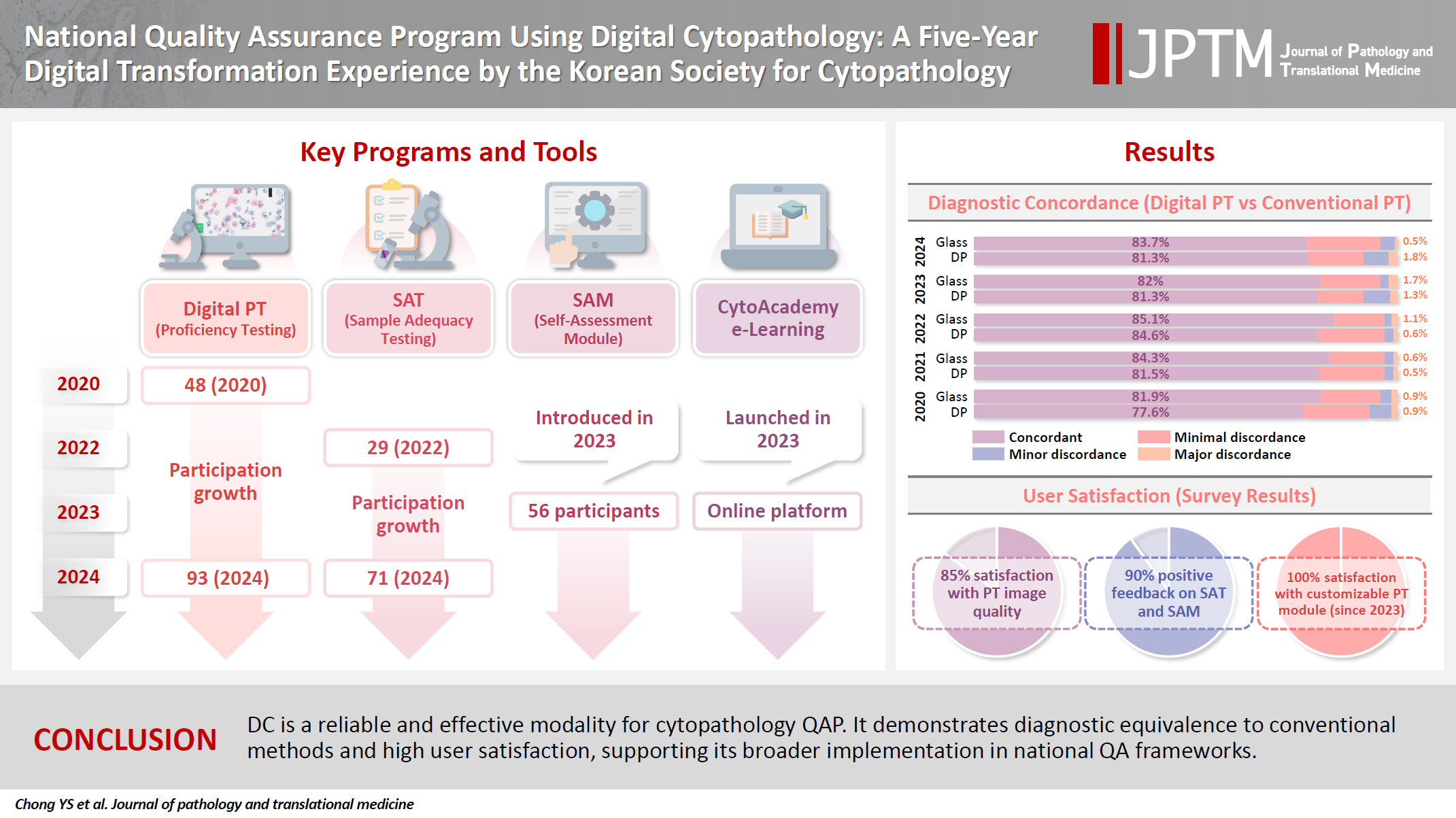
- National quality assurance program using digital cytopathology: a 5-year digital transformation experience by the Korean Society for Cytopathology
- Yosep Chong, Hyeong Ju Kwon, Soon Auck Hong, Sung Soon Kim, Bo-Sung Kim, Younghee Choi, Yoon Jung Choi, Jung-Soo Pyo, Ji Yun Jeong, Soo Jin Jung, Hoon Kyu Oh, Seung-Sook Lee
- J Pathol Transl Med. 2025;59(5):320-333. Published online September 15, 2025
- DOI: https://doi.org/10.4132/jptm.2025.06.27
- 4,937 View
- 114 Download
- 2 Web of Science
- 3 Crossref
-
Abstract
PDF
 Supplementary Material
 - Background
Digital cytopathology (DC) is emerging as a transformative approach in quality assurance programs (QAP), though its comprehensive evaluation remains limited. Since 2020, the Korean Society for Cytopathology has progressively incorporated DC into its national QAP, including digital proficiency testing (PT), sample adequacy testing (SAT), a customizable PT module, and a self-assessment module (SAM), aiming for full digital implementation by 2026. Methods: This 5-year study assessed diagnostic concordance between conventional and digital PT formats and analyzed participant feedback on service quality and digital image usability across PT, SAT, and SAM. Parallel testing was conducted during the transitional phase, and satisfaction was measured through structured surveys. Results: Participation in digital PT increased from 48 institutions in 2020 to 93 in 2024, while digital SAT participation rose from 29 to 71 between 2022 and 2024. In 2023, 56 institutions joined SAM. Diagnostic concordance rates were comparable between digital and conventional PTs (78.6%–84.6% vs. 82.0%–85.1%), including similar category C (major discordance) rates. Satisfaction with digital PT services and image quality exceeded 85%, and over 90% of institutions reported positive feedback on SAT and SAM. Over 80% were satisfied with the customizable PT module. Conclusions: DC is a reliable and effective modality for cytopathology QAP. It demonstrates diagnostic equivalence to conventional methods and high user satisfaction, supporting its broader implementation in national quality assurance frameworks. -
Citations
Citations to this article as recorded by
- Practice of Cytopathology in Korea: A 40‐Year Evolution Through Standardization, Digital Transformation, and Global Partnership
Yosep Chong, Ran Hong, Hyeong Ju Kwon, Haeryoung Kim, Lucia Kim, Soon Jae Kim, Yoon Jung Choi
Diagnostic Cytopathology.2026; 54(2): 146. CrossRef - Validation of Digital Cytology for Primary Diagnosis Across a Range of Specimen Types
Talisa Mistry, Harriet Hunter, Dahmane Oukrif, Sabine Pomplun, Reena Khiroya, Mary Falzon, Tanya Alan, Manuel Rodriguez‐Justo, Adam P. Levine
Cytopathology.2026; 37(3): 222. CrossRef - Review of the Changing Roles of Clinical Laboratory Scientists and Strategies for Curricular Innovation in the Era of Artificial Intelligence
Hee Sung KIM
Korean Journal of Clinical Laboratory Science.2026; 58(1): 1. CrossRef
- Practice of Cytopathology in Korea: A 40‐Year Evolution Through Standardization, Digital Transformation, and Global Partnership
- A standardized pathology report for gastric cancer: 2nd edition
- Young Soo Park, Myeong-Cherl Kook, Baek-hui Kim, Hye Seung Lee, Dong-Wook Kang, Mi-Jin Gu, Ok Ran Shin, Younghee Choi, Wonae Lee, Hyunki Kim, In Hye Song, Kyoung-Mee Kim, Hee Sung Kim, Guhyun Kang, Do Youn Park, So-Young Jin, Joon Mee Kim, Yoon Jung Choi, Hee Kyung Chang, Soomin Ahn, Mee Soo Chang, Song-Hee Han, Yoonjin Kwak, An Na Seo, Sung Hak Lee, Mee-Yon Cho
- J Pathol Transl Med. 2023;57(1):1-27. Published online January 15, 2023
- DOI: https://doi.org/10.4132/jptm.2022.12.23

- 42,247 View
- 1,615 Download
- 26 Web of Science
- 23 Crossref
-
Abstract
PDFSupplementary Material
- The first edition of ‘A Standardized Pathology Report for Gastric Cancer’ was initiated by the Gastrointestinal Pathology Study Group of the Korean Society of Pathologists and published 17 years ago. Since then, significant advances have been made in the pathologic diagnosis, molecular genetics, and management of gastric cancer (GC). To reflect those changes, a committee for publishing a second edition of the report was formed within the Gastrointestinal Pathology Study Group of the Korean Society of Pathologists. This second edition consists of two parts: standard data elements and conditional data elements. The standard data elements contain the basic pathologic findings and items necessary to predict the prognosis of GC patients, and they are adequate for routine surgical pathology service. Other diagnostic and prognostic factors relevant to adjuvant therapy, including molecular biomarkers, are classified as conditional data elements to allow each pathologist to selectively choose items appropriate to the environment in their institution. We trust that the standardized pathology report will be helpful for GC diagnosis and facilitate large-scale multidisciplinary collaborative studies.
-
Citations
Citations to this article as recorded by- GAST-NET: A multi-modal and multi-task deep learning framework for preoperative prediction of perineural invasion and prognostic risk in gastric cancer
Shidi Miao, Hexiang Dong, Jinyang Feng, Yuyang Jiang, Mengzhuo Sun, Zengyao Liu, Qiujun Wang, Xuemei Ding, Ruitao Wang
International Journal of Medical Informatics.2026; 212: 106348. CrossRef - Poorly cohesive carcinoma diffusely involving the whole gastrointestinal tract: a case report
Wei Gao, Yusheng Yang, Xinyi Hu, Yujuan Shi, Kai Liu, Minmin Gu, Jing Wang
Discover Oncology.2026;[Epub] CrossRef - Comprehensive Overview of Gastric Cancer Immunohistochemistry: Key Biomarkers, Advanced Detection Methods, and Perspectives
Bogdan Oprea
Medicina.2026; 62(4): 683. CrossRef - Tumor Budding in Gastric Carcinoma: Beyond Counting Cells at the Invasive Front—A Review of Current Evidence and Biological Perspectives
Catalin-Bogdan Satala, Gabriela Gurau, Alina-Mihaela Gurau, Gabriela Patrichi, Daniela Mihalache
International Journal of Molecular Sciences.2026; 27(9): 3787. CrossRef - Spatial and Temporal Tumor Heterogeneity in Gastric Cancer: Discordance of Predictive Biomarkers
Hye Seung Lee
Journal of Gastric Cancer.2025; 25(1): 192. CrossRef - PD-L1 as a Biomarker in Gastric Cancer Immunotherapy
Yunjoo Cho, Soomin Ahn, Kyoung-Mee Kim
Journal of Gastric Cancer.2025; 25(1): 177. CrossRef - Korean Gastric Cancer Association-Led Nationwide Survey on Surgically Treated Gastric Cancers in 2023
Dong Jin Kim, Jeong Ho Song, Ji-Hyeon Park, Sojung Kim, Sin Hye Park, Cheol Min Shin, Yoonjin Kwak, Kyunghye Bang, Chung-sik Gong, Sung Eun Oh, Yoo Min Kim, Young Suk Park, Jeesun Kim, Ji Eun Jung, Mi Ran Jung, Bang Wool Eom, Ki Bum Park, Jae Hun Chung, S
Journal of Gastric Cancer.2025; 25(1): 115. CrossRef - A Comprehensive and Comparative Review of Global Gastric Cancer Treatment Guidelines: 2024 Update
Sang Soo Eom, Keun Won Ryu, Hye Sook Han, Seong-Ho Kong
Journal of Gastric Cancer.2025; 25(1): 153. CrossRef - Korea, Japan, Europe, and the United States: Why are guidelines for gastric cancer different?
Emily E. Stroobant, Seong-Ho Kong, Maria Bencivenga, Takahiro Kinoshita, Tae-Han Kim, Takeshi Sano, Giovanni de Manzoni, Han-Kwang Yang, Yuko Kitagawa, Vivian E. Strong
Gastric Cancer.2025; 28(4): 559. CrossRef - Can the Japanese guidelines for endoscopic submucosal dissection be safely applied to Korean gastric cancer patients? A multicenter retrospective study based on the Korean Gastric Cancer Association nationwide survey
Hayemin Lee, Mi Ryeong Park, Junhyun Lee
Annals of Surgical Treatment and Research.2025; 109(2): 81. CrossRef - Double optimal transport for differential gene regulatory network inference with unpaired samples
Mengyu Li, Bencong Zhu, Cheng Meng, Xiaodan Fan, Laura Cantini
Bioinformatics.2025;[Epub] CrossRef - A Randomized Controlled Trial to Evaluate the Effect of Fibrin Glue on Bleeding after Gastric Endoscopic Submucosal Dissection
Tae-Se Kim, Tae-Jun Kim, Yang Won Min, Hyuk Lee, Byung-Hoon Min, Jun Haeng Lee, Poong-Lyul Rhee, Jae J. Kim
Gut and Liver.2025; 19(5): 677. CrossRef - Diagnostic accuracy of stereomicroscopy assessment of invasion depth in ex vivo specimens of early gastric cancer
Jing Wang, Lin Chang, Dong-Feng Niu, Yan Yan, Chang-Qi Cao, Shi-Jie Li, Qi Wu
World Journal of Gastroenterology.2025;[Epub] CrossRef - SMMILe enables accurate spatial quantification in digital pathology using multiple-instance learning
Zeyu Gao, Anyu Mao, Yuxing Dong, Hannah Clayton, Jialun Wu, Jiashuai Liu, ChunBao Wang, Kai He, Tieliang Gong, Chen Li, Mireia Crispin-Ortuzar
Nature Cancer.2025; 6(12): 2025. CrossRef - Genomic and Transcriptomic Characterization of Gastric Cancer with Bone Metastasis
Sujin Oh, Soo Kyung Nam, Keun-Wook Lee, Hye Seung Lee, Yujun Park, Yoonjin Kwak, Kyu Sang Lee, Ji-Won Kim, Jin Won Kim, Minsu Kang, Young Suk Park, Sang-Hoon Ahn, Yun-Suhk Suh, Do Joong Park, Hyung Ho Kim
Cancer Research and Treatment.2024; 56(1): 219. CrossRef - Microscopic tumor mapping of post-neoadjuvant therapy pancreatic cancer specimens to predict post-surgical recurrence: A prospective cohort study
Yeshong Park, Yeon Bi Han, Jinju Kim, MeeYoung Kang, Boram Lee, Eun Sung Ahn, Saemi Han, Haeryoung Kim, Hee-Young Na, Ho-Seong Han, Yoo-Seok Yoon
Pancreatology.2024; 24(4): 562. CrossRef - Effect of Neoadjuvant Chemotherapy on Tumor-Infiltrating Lymphocytes in Resectable Gastric Cancer: Analysis from a Western Academic Center
Elliott J. Yee, Danielle Gilbert, Jeffrey Kaplan, Sachin Wani, Sunnie S. Kim, Martin D. McCarter, Camille L. Stewart
Cancers.2024; 16(7): 1428. CrossRef - Interpretation of PD-L1 expression in gastric cancer: summary of a consensus meeting of Korean gastrointestinal pathologists
Soomin Ahn, Yoonjin Kwak, Gui Young Kwon, Kyoung-Mee Kim, Moonsik Kim, Hyunki Kim, Young Soo Park, Hyeon Jeong Oh, Kyoungyul Lee, Sung Hak Lee, Hye Seung Lee
Journal of Pathology and Translational Medicine.2024; 58(3): 103. CrossRef - Expression of claudin 18.2 in poorly cohesive carcinoma and its association with clinicopathologic parameters in East Asian patients
Moonsik Kim, Byung Woog Kang, Jihyun Park, Jin Ho Baek, Jong Gwang Kim
Pathology - Research and Practice.2024; 263: 155628. CrossRef - Clinicopathological analysis of claudin 18.2 focusing on intratumoral heterogeneity and survival in patients with metastatic or unresectable gastric cancer
T.-Y. Kim, Y. Kwak, S.K. Nam, D. Han, D.-Y. Oh, S.-A. Im, H.S. Lee
ESMO Open.2024; 9(12): 104000. CrossRef - Pathological Interpretation of Gastric Tumors in Endoscopic Submucosal Dissection
Jung Yeon Kim
Journal of Digestive Cancer Research.2023; 11(1): 15. CrossRef - Histopathology of Gastric Cancer
Baek-hui Kim, Sung Hak Lee
The Korean Journal of Helicobacter and Upper Gastrointestinal Research.2023; 23(2): 143. CrossRef - Endoscopic submucosal dissection hands-on training with artificial mucosal layer EndoGEL
Tae-Se Kim, Jun Haeng Lee
Journal of Innovative Medical Technology.2023; 1(1): 5. CrossRef
- GAST-NET: A multi-modal and multi-task deep learning framework for preoperative prediction of perineural invasion and prognostic risk in gastric cancer
- Early Colorectal Epithelial Neoplasm in Korea: A Multicenter Survey of Pathologic Diagnosis
- Yun Kyung Kang, So-Young Jin, Mee Soo Chang, Jung Yeon Kim, Gyeong Hoon Kang, Hye Seung Lee, Jin Hee Sohn, Ho Sung Park, Kye Won Kwon, Mi Jin Gu, Young Hee Maeng, Jong Eun Joo, Haeng Ji Kang, Hee Kyung Kim, Kee-Taek Jang, Mi Ja Lee, Hee Kyung Chang, Joon Mee Kim, Hye Seung Han, Won Ae Lee, Yoon Jung Choi, Dong Wook Kang, Sunhoo Park, Jae Hyuk Lee, Mee-Yon Cho
- Korean J Pathol. 2013;47(3):245-251. Published online June 25, 2013
- DOI: https://doi.org/10.4132/KoreanJPathol.2013.47.3.245
- 12,071 View
- 56 Download
- 1 Crossref
-
Abstract
PDF
Background The incidence of early colorectal epithelial neoplasm (ECEN) is increasing, and its pathologic diagnosis is important for patient care. We investigated the incidence of ECEN and the current status of its pathologic diagnosis.
Methods We collected datasheets from 25 institutes in Korea for the incidence of colorectal adenoma with high grade dysplasia (HGD) and low grade dysplasia in years 2005, 2007, and 2009; and early colorectal carcinoma in the year 2009. We also surveyed the diagnostic terminology of ECEN currently used by the participating pathologists.
Results The average percentage of diagnoses of adenoma HGD was 7.0%, 5.0%, and 3.4% in years 2005, 2007, and 2009, respectively. The range of incidence rates of adenoma HGD across the participating institutes has gradually narrowed over the years 2005 to 2009. The incidence rate of early colorectal carcinoma in the year 2009 was 21.2%. The participants did not share a single criterion or terminology for the diagnosis of adenoma HGD. The majority accepted the diagnostic terms that distinguished noninvasive, mucosal confined, and submucosal invasive carcinoma.
Conclusions Further research requirements suggested are a diagnostic consensus for the histopathologic diagnosis of ECEN; and standardization of diagnostic terminology critical for determining the disease code.
-
Citations
Citations to this article as recorded by- Diminutive and Small Colorectal Polyps: The Pathologist's Perspective
Yun Kyung Kang
Clinical Endoscopy.2014; 47(5): 404. CrossRef
- Diminutive and Small Colorectal Polyps: The Pathologist's Perspective
- Cytologic findings of pancreatic islet cell tumor with lymph node metastasis.
- Yee Jeong Kim, Yoon Jung Choi, Kyu Rae Kim, Woo Hee Jung, Kwang Gil Lee
- J Pathol Transl Med. 1992;3(2):60-66.
- 2,513 View
- 12 Download
- Histopathological and Immunohistochemical Features of Wilms' Tumor.
- Yoon Jung Choi, Woo Hee Jung, Dong Whan Shin, Chan Il Park, Chuhl Joo Lyu
- Korean J Pathol. 1993;27(4):339-348.
- 6,261 View
- 283 Download
-
Abstract
PDF
- Wilms' tumor is one of the most common primary malignant tumors of the kidney during infancy and childhood and is known to be originated from the primitive cells of metanephric blastema. It presents difficulties when encountered in deciding the presence of anaplasia or in differentiating it from other renal tumors of childhood with different biologic behavior because of its diverse histologic patterns and varying degrees of differentiation. Evaluation of clinical and histopathologic features in terms of prognostication was done of 32 cases of Wilms' tumor which were surgically resected and diagnosed in the period from January 1979 through June 1992. Immunohistochemical reaction for cytokeratin, vimentin, actin and desmin was also analysed on all cases of Wilms' tumor in conjunction with clear cell sarcoma of the kidney(CCSK), malignant rhabdoid tumor of the kidney(MRTK) and congenital mesoblastic nephroma(CMN) to assess the validity of immunohistochemistry in differentiating Wilms' tumor from these renal tumors. Twenty four(75%) cases were diagnosed before the age of 5 and 40.7% were under 2 years old. Mixed type was most common(62.5%), followed by epithelial, blastemal and stromal predominant type in descending order of frequency. Anaplasia was observed in 3 cases(9.4%), two of which were epithelial predominant type and one blastemal predominant type. Treatment modality and presence of anaplasia were significantly correlated with 5 year survival rate of patients. Immunohistochemical stain revealed that all epithelial component of Wilms' tumor were positive for cytokeratin and 56.3% of Wilms' tumor had blastemal component which were positive for both cytokeratin and vimentin. Twenty cases(62.5%) of Wilms' tumor had blastemal component which were positive for cytokeratin with a proportion of more than 5% of reactive cells. Stromal component of Wilms' tumor generally did not show differentiation into the specialized type of tissue and all revealed positive reactions for vimentin among which some revealed positive reactions for actin. Only 3 out of 6 cases with rhabdomyoblastic differentiation were positive for desmin. CCSK, MRTK and CMN which have different biologic behavior and treatment modality compared to Wilm's tumor showed positivity only for vimentin and/or actin. In summary, treatment modality and presence of anaplasia are significantly correlated with patients' survival and the immunohistochemical stain for cytokeratin is very helpful in confirming the presence of blastemal component and useful in the differential diagnosis of Wilms' tumor from other kinds of pediatric renal tumors.
- Fine needle aspiration cytology of proliferative fasciitis.
- Yoon Jung Choi, Sang Yeop Yi, Woo Ick Yang, Soon Hee Jung, Kwang Gil Lee
- J Pathol Transl Med. 1993;4(1):52-56.
- 2,776 View
- 14 Download
- Papillary Neoplasm of the Endolymphatic Sac: A report of two cases.
- Jai Hyang Go, Yoon Jung Choi, Tae Seung Kim, Chan Il Park
- Korean J Pathol. 1996;30(2):150-154.
- 2,166 View
- 12 Download
-
Abstract
PDF
- Papillary tumor of the temporal bone or middle ear has been recognized as an aggressive neoplasm because of its invasive growth pattern. The site of origin is controversial so that most cases have been reported under various diagnostic terms. Recently, Heffner(1989) suggested that the endolymphatic sac is a possible site of origin, because the tumor resembles the endolymphatic sac in several aspects. We report two such cases. One patient was a 34-year-old female presenting with tinnitus and hearing difficulty for 1 year. Temporal bone CT revealed extensive bone destruction by the tumor which was located in the posterolateral aspect of temporal bone. The other patient was a 56-year-old female who complained of tinnitus, dizziness and otalgia for 2 years. Cranial MR imaging showed an irregularly marginated mass in the left jugular fossa with extension to the petrous bone. Histologically, both cases showed a papillary pattern and locally destructive growth that are typical of papillary tumor of the endolymphatic sac. The papillae were lined by a single layer of bland-looking cuboidal to low columnar cells. Immunohistochemically the lining cells expressed cytokeratin, epithelial membrane antigen, neuron specific antigen and in one case, S-100 protein, supporting the thesis that these neoplasms might be of endolymphatic sac origin.
- Clinicopathologic Features of Granulomatous Mastitis.
- Yee Jeong Kim, Yoon Jung Choi, Ji Young Kim, Hee Jung Kim, Yang Soon Park, Soon Won Hong, Chanil Park, Doyil Kim, Hyde Lee, Woo Hee Jung
- Korean J Pathol. 2005;39(3):181-186.
- 2,516 View
- 26 Download
-
Abstract
PDF
- BACKGROUND
Granulomatous mastitis (GM) is a rare chronic inflammatory condition that clinically mimics a carcinoma. The diagnosis of idiopathic GM depends on the exclusion of other granulomatous inflammations. The purpose of this study is to correlate the clinicopathological features of GM with etiologies.
METHODS
We reviewed the clinical records of 58 cases that were histologically diagnosed as GM. We performed special stains for microorganisms such as Ziehl-Neelsen, periodic acid Schiff and gram stains, and polymerase chain reaction (PCR) for Mycobacterium tuberculosis (TB PCR).
RESULTS
The mean age of patients was 35.3 years. Most patients were parous except three. Seven patients (12.1%) were related with pregnancy or lactation. TB PCR was positive in nine patients (15.5%). Five patients (8.6%) had gram positive bacilli that were recognizable as coryneform bacteria. Culture study demonstrated Staphylococcus aureus in only one case. Infectious GM had a greater tendency to form abscesses. Fat necrosis was more likely to be present in idiopathic GM, but other histological features were similar to each other. Twenty-two cases (37.9%) showed recurrence.
CONCLUSIONS
We suggest that TB PCR and gram stain are essential tests for the differential diagnosis of GM, because the histologic features considerably overlap irrespective of the various etiologies.
- Urinary Cytologic Findings of Urothelial Lesions.
- Yoon Jung Choi, Kwang Gil Lee
- J Pathol Transl Med. 1994;5(2):130-136.
- 2,194 View
- 17 Download
-
Abstract
PDF
- Urinary cytology is increasingly accepted as a diagnostic tool in the detection and follow-up of patients with bladder cancer. However, its value is reduced by several limitations, especially by the tack of cytologic criteria specifically reflecting the morphology of low-grade urothelial neoplasm. We reviewed histologically proven 50 cases of urine cytology with emphasis on cytologic findings of benign atypia and differential findings of urothelial neoplasm according to the grade. The diagnoses included 17 benign lesions (including 5 cases of urine calculi) and 33 malignant lesions(including 28 transitional cell carcinomas. 3 squamous cell carcinomas, 1 adenocarcinoma and 1 prostate adenocarcinoma). Diagnostic accuracy was 92%. Important cytodiagnostic criteria for benign atypia and low grade malignancy were cellularity, number of cell clusters, and morphology and arrangement of urothelial cells. The cytologic findings of urothelial neoplasms according to histologic grade were relatively well correlated with the histologic findings. However, the cytologic criteria were not sufficient to readily distinguish grade I from grade II. In view of this, we think that cytologic nomenclature "low-grade" and "high-grade" is a more reliable criterion. Recognition of subtle cellular morphologic features specific for urothelial lesions(including benign or malignancy) and proper fixation, processing and staining of specimen can expand the role of urinary cytology in detection and follow-up of patients.
- Leiomyoma of the Urinary Bladder.
- Kye Weon Kwon, Hee Jung Ahn, Yoon Jung Choi, Young Kwon Hong, Jae Seop Shin
- Korean J Pathol. 1997;31(12):1320-1323.
- 2,273 View
- 19 Download
-
Abstract
PDF
- Leiomyoma is commonly found in the female genital tract, but occurrence in the urinary bladder is very rare with only 235 cases reported in the literature. These tumors have been classified as intravesical (63%), intramural (7%) and extravesical (30%) depending on the direction of the growth. We report a case of intravesical leiomyoma of the urinary bladder in a 36 year-old woman who exhibited dysuria and urinary retention. The gross and microscopical findings of leiomyoma of the bladder are similar to those of the uterus. Immunohistochemical stains for estrogen receptor (ER) and progesterone receptor (PR) revealed diffuse nuclear staining in smooth muscle cells, supporting the hypothesis of hormonal influence in tumorigenesis.
- Histopathological Analysis of Posterior Fossa Tumor.
- Yoon Jung Choi, Tai Seung Kim
- Korean J Pathol. 1994;28(3):228-234.
- 2,659 View
- 10 Download
-
Abstract
- The posterior fossa, containing roughly 6ne fourth of the intracranial contents, is the site of about 30-35% of the intracranial tumors. The incidence of primary tumors in the posterior fossa is quite different from that of the cerebivm. We analysed 124 cases of posterior fossa tumor, over a 10 year period, to understand the status of posterior fossa tumor and its histologic characteristics. Medulloblastoma was most common(37cases, 29.8%), followed by astrocytoma, hemangiobla-stoma, ependymoma, meningioma, metastatic tumor, arteriovenous malformation and choroid plexus papilloma in descending order of frequency. Tumors were found most frequently between the ages of two and ten years(28.2%) and sixty seven(54.0%) cases were diagnosed before the age of fifteen. The ratio of male to female was 60 : 64. Astrocytoma revealed a characteristic juvenile pilocytic type and a microcystic change. Hemangioblastoma showed higher frequency(17.7%) than previous reports and the origin of tumor cells is still equivocal. Other tumors revealed the same histologic features as other intracranial tumors.
- Optic Nerve sheath Meningioma: A case report.
- Yoon Jung Choi, Yong Hee Lee, Tai Seung Kim
- Korean J Pathol. 1994;28(4):430-432.
- 2,488 View
- 40 Download
-
Abstract
PDF
- Optic nerve sheath meningioma arises from the arachnoid cap cell of optic nerve sheath and comprises most of primary orbital meningioma. We experienced a rare case of optic nerve sheath meningioma originating in the left orbit. A 44-year-old woman had suffered from visual disturbance in the left eye for 3 years and had only light perceptibility for the recent 6 months. The right eye was normal. Brain CT scan and MRI revealed a 2x0.8 cm sized fusiform solid mass in the left retrobulbar area. Under the impression of optic nerve tumor. excision of the mass including a small segment of the optic nerve was performed. The tumor was a yellowish firm, relatively well demarcated mass that encircled the optic nerve without infiltrating it. Microscopically it was a typical meningioma, meningothelial type. The recovery of visual acuity was impossible due to resection of optic nerve but there was no evidence of recurrence for u months.
- Wegener's Granulomatosis Involving Lung and Middle Ear: A case report.
- Kye Weon Kwon, Yoon Jung Choi, Hee Jeong Ahn, Min Soo Han, Dong Hwan Shin
- Korean J Pathol. 1998;32(6):470-473.
- 2,225 View
- 10 Download
-
Abstract
- A case of Wegener's granulomatosis is described, with special attention focused on the typical histologic findings and involvement of both middle ear and lung. The patient is a 37-year-old man presented with four-month history of cough and sputum. He had a past history of surgery of both ears because of otitis media followed by left facial palsy. Chest radiographs showed variable sized ill defined nodules in both lower lobes with internal airspace consolidation. Histologic preparations of the open lung biopsy specimens demonstrated a diffusely scattered palisading micro and macrogranulomas with central focus of neutrophils and necrotic collagen surrounded by histiocytes, histiocytic giant cells. Fibrinoid necrosis involved blood vessels and lung parenchyma. Chronic inflammation, diffuse granulation tissue formation and irregular fibrosis are also found in the lung parenchyma. The histologic findings of middle ear which was previously biopsied showed scattered palisading ill defined microgranulomas mixed with fibrotic tissue.
- The Effect of Preoperative Treatment on Cell Kinetics and Patients Survival in Hepatocellular Carcinoma.
- Yoon Jung Choi, Ho Guen Kim, Chan Il Park, Woo Hee Jung
- Korean J Pathol. 1994;28(6):605-611.
- 2,091 View
- 13 Download
-
Abstract
PDF
- To evaluate the effect of preoperative treatment on proliferative activity and prognosis of the hepatocellular carcinomas(HCCs), fifty-three surgically resected HCCs were studied. Twenty cases were treated preoperatively and thirty-three were not treated before surgery. The proliferation index(PI, % of proliferating cell nuclear antigen positive cells) of the remaining cancer cases(35.41). Although PI was similar among gross types and among histologic grades, tumors of the expanding type and of the histologic grade I revealed distinctly low PI in pretreated cases. Two-year survival rate was not significantly different between pretreated and not-pretreated cases(67.4 vs 52.7). But the differences between gross types(p<0.05) and between histologic grades(p<0.01) were significant. Total necrosis of tumor occurred in five pretreated patients, all of whom were alive during two-year follow-up. Smaller HCCs showed better prognosis(p<0.01). Although PI appeared not correlated well with the two tear survival rate, the pretreated HCCs preoperative modalities induce tumor necrosis, but do not reduce the proliferative activity of tumor cells significantly, and that pretreatment does not affect the long-term prognosis of HCCs except for the accasions of total necrosis of tumor.
- Atypical Polypoid Adenomyomas of the Endometrium: 2 case reports.
- Hee Jeong Ahn, Kyu Rae Kim, Yoon Jung Choi, Bok Soo Kim
- Korean J Pathol. 1996;30(11):1034-1039.
- 2,214 View
- 20 Download
-
Abstract
PDF
- Atypical polypoid adenomyoma(APA) is a rare benign polypoid tumor arising in the uterine endometrium which was first designated by Mazur in 1981. Microscopic examination reveals architecturally and cytologically atypical endometrial glands separated by intersecting fascicles of smooth muscle cells. The tumor can be misdiagnosed as endometrial adenocarcinoma with myometrial invasion, especially on microscopic examination of a curettage specimen. However a granulation tissue-like stromal response is absent and the smooth muscle in APA is more cellular than that of normal myometrium. We are reporting 2 cases of APA of the uterine endometrium which are 1.5 cm and 1.7 cm in size, respectively, in a 30 and a 22 year-old women. This is the first report in Korean literature using the immunohistochemical staining for smooth muscle actin and desmin.
- Expression of Matrix Metalloproteinase and Tissue Inhibitor of Metallproteinase in Breast Carcinoma Related to Angiogenesis and Invasion.
- Yoon Jung Choi, Woo Hee Jung, Hy De Lee, Kwang Gil Lee
- Korean J Pathol. 2000;34(9):652-664.
- 2,375 View
- 22 Download
-
Abstract
PDF
- Among the enzymes which are responsible for basement membrane breakdown, matrix metalloproteinases (MMP) form a family of neutral proteases that are regulated at the levels of gene transcription, proenzyme activation by the cleavage of protein, and the inhibition of the active enzyme by tissue inhibitors of matrix metalloproteinases (TIMP). Recent reports have demonstrated that the expression of these proteolytic enzymes are elevated in several solid tumors and that it can be associated with invasiveness and poor prognosis. We examined the expression of MMP-2, MMP-9, TIMP-1 and TIMP-2 by immunohistochemistry in 160 cases of infiltrating ductal carcinoma. And we compared these data with the established prognostic parameters - tumor size, nodal status, clinical stage, hormonal receptor status, microvessel density, and TGF-beta1 expression in order to evaluate how MMP and TIMP expression are associated with breast cancer progression and prognosis. Microvessel density in invasive breast carcinoma was significantly correlated with tumor size and recurrence (p<0.05). The immunohistochemical expression of TGF-beta1 was significantly associated with tumor size, lymph node metastasis, and clinical stage (p<0.05). The microvessel density was significantly correlated with TGF-beta1 expression in more than 50% of tumor cells. The immunohistochemical expression of MMP-2 and MMP-9 were significantly correlated with nodal metastasis and absence of immunoreactivity for estrogen and progesterone receptors. The immunohistochemical expression of TIMP-1 was inversely correlated with clinical stage and microvessel density while that of TIMP-2 was inversely correlated with clinical stage (p<0.05). Small size of tumor, presence of progesterone receptor, highly differentiated histologic grade, and absence of immunoreactivity for MMP-9 were significantly associated with higher survival rate, but in multivariate analysis only tumor size and MMP-9 expression appeared to affect survival independently.
 First
First Prev
Prev


