Articles
- Page Path
- HOME > J Pathol Transl Med > Volume 49(4); 2015 > Article
-
Review
Pathology Reporting of Thyroid Core Needle Biopsy: A Proposal of the Korean Endocrine Pathology Thyroid Core Needle Biopsy Study Group - Chan Kwon Jung1, Hye Sook Min2,3, Hyo Jin Park2, Dong Eun Song4, Jang Hee Kim5, So Yeon Park2, Hyunju Yoo6, Mi Kyung Shin7, Korean Endocrine Pathology Thyroid Core Needle Biopsy Study Group
-
Journal of Pathology and Translational Medicine 2015;49(4):288-299.
DOI: https://doi.org/10.4132/jptm.2015.06.04
Published online: June 17, 2015
1Department of Hospital Pathology, College of Medicine, The Catholic University of Korea, Seoul, Korea
2Department of Pathology, Seoul National University College of Medicine, Seoul, Korea
3Department of Epidemiology and Preventive Medicine, Graduate School of Public Health, Seoul National University, Seoul, Korea
4Department of Pathology, University of Ulsan College of Medicine, Seoul, Korea
5Department of Pathology, Ajou University School of Medicine, Suwon, Korea
6Department of Pathology, Daerim Saint Mary’s Hospital, Seoul, Korea
7Department of Pathology, Hallym University College of Medicine, Seoul, Korea
-
Corresponding Authors Chan Kwon Jung, MD, PhD Department of Hospital Pathology, Seoul St. Mary’s Hospital, College of Medicine, The Catholic University of Korea, 222 Banpo-daero, Seocho-gu, Seoul 137-701, Korea Tel: +82-2-2258-1622 Fax: +82-2-2258-1627 E-mail: ckjung@catholic.ac.kr
Corresponding Authors Mi Kyung Shin, MD, PhD Department of Pathology, Hallym University Kangnam Sacred Heart Hospital, Hallym University College of Medicine, 1 Singil-ro, Yeongdeungpo-gu, Seoul 150-950, Korea Tel: +82-2-829-5443 Fax: +82-2-829-5268 E-mail: smk0103@yahoo.co.kr
© 2015 The Korean Society of Pathologists/The Korean Society for Cytopathology
This is an Open Access article distributed under the terms of the Creative Commons Attribution Non-Commercial License (http://creativecommons.org/licenses/by-nc/3.0/) which permits unrestricted noncommercial use, distribution, and reproduction in any medium, provided the original work is properly cited.
Abstract
- In recent years throughout Korea, the use of ultrasound-guided core needle biopsy (CNB) has become common for the preoperative diagnosis of thyroid nodules. However, there is no consensus on the pathology reporting system for thyroid CNB. The Korean Endocrine Pathology Thyroid Core Needle Biopsy Study Group held a conference on thyroid CNB pathology and developed guidelines through contributions from the participants. This article discusses the outcome of the discussions that led to a consensus on the pathology reporting of thyroid CNB.
- Since May 2013, the Korean Endocrine Pathology Thyroid Core Needle Biopsy Study Group has held three consensus conferences on thyroid CNB pathology reporting in Seoul, Korea. The purpose of the conferences was to bring together endocrine pathologists who were thyroid specialists (1) to review existing reporting formats and published literature related to thyroid CNB, (2) to review 25 thyroid CNB specimens using virtual microscopy on each slide, (3) to reach a consensus regarding the histological diagnostic criteria and reporting format of thyroid CNB pathology, and (4) to develop a standard pathology reporting system for thyroid CNB that would be useful for clinical management and pathologic diagnosis. Twenty-one Korean endocrine pathologists participated in the conferences. In Korea, pathologists and clinicians are familiar with TBSRTC terminology and the management guidelines of each diagnostic category [3]. Therefore, all participants in the conferences agreed to use the standardized diagnostic categories for thyroid CNB.
CONSENSUS CONFERENCE
- Most participants decided to use the same six-level system as TBSRTC for categorizing the results of thyroid CNB. However, the following concerns exist with regard to the use of the same terminology: (1) some clinicians and pathologists may understand the meaning of terms in a CNB pathology report differently from the terms in an FNAC report; (2) the phrase “atypia of undetermined significance” in TBSRTC is inappropriate terminology in the field of histopathology; (3) there is no consensus regarding specimen adequacy; and (4) the sample representativity of the CNB may be different from that of the FNAC. Therefore, we recommend that the limitations of CNB should be explained in the pathology report.
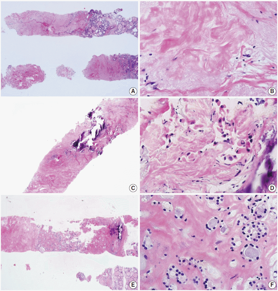
- Most participants in the conferences agreed that a sample composed primarily of an acellular/paucicellular fibrotic nodule should be considered nondiagnostic if it did not contain any atypical cells (Fig. 1). However, a categorical diagnosis should be rendered, irrespective of the number of follicular cells, if a sample contains atypical cells (Fig. 1).
- Another dilemma is the differential diagnosis of follicular proliferative lesions without nuclear atypia in a CNB sample. Histologically, hyperplastic nodules are usually found in multiples and are partially encapsulated by a fibrous capsule. Follicular adenoma is a completely encapsulated follicular nodule that is mostly solitary and has microscopic features that are different from those of the surrounding normal thyroid tissue [14]. Fibrous capsules of follicular neoplasms vary in thickness. Tumor capsules are usually thin on follicular adenomas and tend to be thicker on follicular carcinomas [14]. It is not possible to differentiate between follicular adenoma and carcinoma using FNAC or CNB samples because the diagnosis of follicular carcinoma is based on the presence of tumor capsular invasion or vascular invasion. Nonetheless, in CNB samples, it may be possible to determine whether a nodule is a follicular neoplasm or a non-neoplastic lesion [15]. Most participants agreed that a diagnosis of “follicular neoplasm or suspicious for a follicular neoplasm” is appropriate when a follicular proliferative lesion is separated from the surrounding thyroid parenchyma by a definite fibrous capsule, has features that are architecturally and cytologically different from the adjacent thyroid glands, and lacks the nuclear features of a papillary carcinoma. However, the CNB specimen of a widely invasive follicular carcinoma may not show a fibrous capsule because the tumor is not encapsulated although it has invaded into the thyroid parenchyma and extrathyroidal tissue. Some hyperplastic nodules have a cellular microfollicular or solid growth pattern and a fibrous capsule. Therefore, misdiagnosing a widely invasive follicular carcinoma as a benign follicular lesion or a hyperplastic nodule as a follicular neoplasm on the CNB specimens remains a concern.
- No study has yet been conducted to define the number of tissue cores that are required for an accurate diagnosis of a thyroid nodule. However, most conference participants agreed that two cores are adequate to diagnose a lesion.
CONTROVERSIAL SUBJECTS
- In the histologic examination of CNB samples, a diagnostic category is selected from the following six standardized options. This proposal was approved by the Korean Endocrine Pathology Thyroid Core Needle Biopsy Study Group (Table 1).
- I. Nondiagnostic or unsatisfactory
- This category includes specimens with an insufficient number of follicular cells to provide an appropriate diagnosis or when the specimen is not representative of the ultrasound image of the lesion. This diagnosis is subjective; and therefore, the report should explain why the sample is nondiagnostic or unsatisfactory. For example, the sample may be normal thyroid tissue only, extrathyroid tissue only (e.g., skeletal muscle and mature adipose tissue), a virtually acellular specimen, an acellular/paucicellular fibrotic nodule, a blood clot only, or it may show other nondiagnostic or unsatisfactory findings. However, any CNB specimen containing atypical cells should not be considered nondiagnostic or unsatisfactory.
- Normal thyroid tissue or extrathyroid tissue only implies that the thyroid lesion has not been sampled. For some benign follicular lesions with normofollicular structures, it may be difficult to assess specimen adequacy. It is important to find the transition between the follicular lesion and the surrounding normal parenchyma and compare between the pathologic finding and its ultrasound images.
- II. Benign
- This category includes all benign thyroidal and nonthyroidal diseases. A CNB specimen can be classified, based on the specific diagnosis for the lesion, as benign. For example, the sample may be a benign follicular nodule or consistent with a benign follicular nodule, Hashimoto’s thyroiditis, subacute granulomatous thyroiditis, a nonthyroidal lesion (e.g., a parathyroid lesion, benign neurogenic tumors, and benign lymph node), or another benign lesion.
- A benign follicular nodule encompasses nodular hyperplasias (adenomatoid nodule), colloid nodules, a nodule in Graves’ disease, nodular Hashimoto’s thyroiditis, and a subset of follicular adenomas. CNB specimens of these lesions show a benign-appearing normofollicular or macrofollicular structure and do not have a well-defined fibrous capsule.
- III. Indeterminate lesion
- The cytologic atypia and histologic growth patterns in this category are of uncertain significance and insufficient to be classified under other diagnostic categories. The diagnostic category III “indeterminate lesion” corresponds to “atypia of undetermined significance” or “follicular lesion of undetermined significance” in TBSRTC. However, it was considered inappropriate to use the term “atypia of undetermined significance” in the histopathologic diagnosis of CNB specimens.
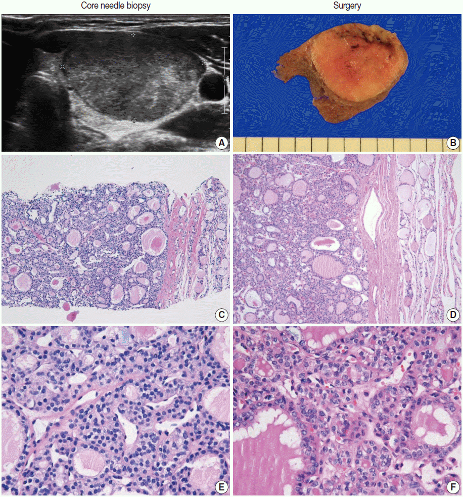
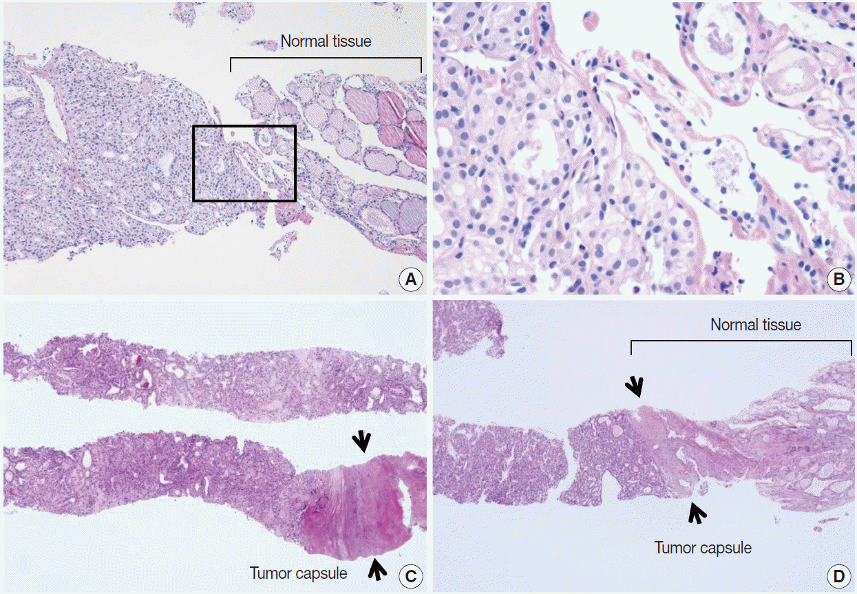
- The diagnosis of category III is appropriate when a follicular proliferative lesion shows focal nuclear atypia such as nuclear enlargement with pale chromatin, irregular nuclear membranes, and nuclear grooves in a background of predominantly benign-appearing follicles. If a microfollicular proliferative lesion is separated by a fibrous capsule from the surrounding normal parenchyma it is diagnosed as a follicular neoplasm (see category IV). If a fibrous capsule or adjacent nonlesional tissue is not identified in a CNB specimen that shows a predominantly microfollicular or trabecular growth pattern, it is reasonable to classify the lesion as diagnostic category III because it cannot be determined whether the nodule has a fibrous capsule. However, if sonographic features suggest a follicular neoplasm in such lesions, the sample can be considered a category IV, “follicular neoplasm” (Fig. 2) [15]. A multidisciplinary approach to a thyroid nodule can improve the preoperative diagnostic accuracy of FNAC and CNB specimens [16].
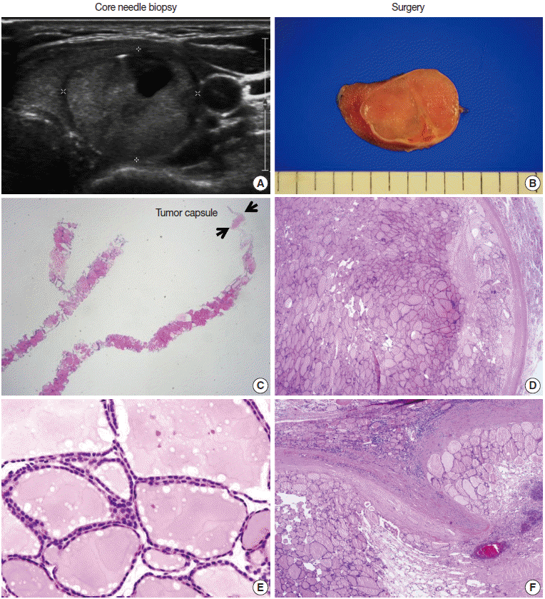
- FNAC specimens of macrofollicular lesions are usually diagnosed as benign. However, when ultrasound images show the typical features of a follicular neoplasm and the CNB specimen microscopically shows a fibrous capsule, then the CNB specimen can be diagnosed as a follicular neoplasm, even in a macrofollicular lesion (Fig. 3). However, in surgical pathologies, most participants agreed that category III is appropriate in a macrofollicular proliferative lesion with a definite fibrous capsule (Fig. 3).
- The following are common scenarios that may be encountered in case of follicular proliferative lesions:
- IIIA. Indeterminate follicular lesion with nuclear atypia
- Examples in this category are follicular proliferative lesions with focal nuclear atypia, follicular proliferative lesions with equivocal or questionable nuclear atypia, and atypical follicular cells embedded in fibrotic stroma.
- IIIB. Indeterminate follicular lesion with architectural atypia
- Examples in this category are microfollicular proliferative lesions lacking a fibrous capsule or the adjacent nonlesional tissue in the specimen; solid or trabecular follicular lesions lacking a fibrous capsule or the adjacent nonlesional tissue in the specimen; macrofollicular proliferative lesions with a fibrous capsule; Hürthle cell proliferative lesions lacking a fibrous capsule or the adjacent nonlesional tissue in the specimen.
- IIIC. Other indeterminate lesions
- IV. Follicular neoplasm or suspicious for a follicular neoplasm
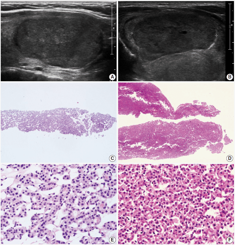
- In CNB and FNAC, the term “follicular neoplasm or suspicious for a follicular neoplasm” is used to encompass neoplastic lesions with follicular proliferative patterns (e.g., follicular adenoma, follicular carcinoma, follicular variant of papillary carcinoma, follicular variant of medullary carcinoma) [2,3] The histologic diagnosis of “follicular neoplasm or suspicious for a follicular neoplasm” in a CNB specimen is based on the presence of a fibrous capsule and microscopic features that differ from the adjacent thyroid parenchyma (Fig. 4). Follicular cells do not show the typical nuclear features of papillary carcinomas. It is important to identify a well-formed fibrous capsule in the CNB specimen. The FNAC diagnosis for follicular neoplasm is primarily based on the presence of microfollicular or trabecular architecture and the lack of colloid. In a CNB specimen, the growth patterns of a follicular neoplasm can be microfollicular, normofollicular, solid, or trabecular when a fibrous tumor capsule is identified in the sample. Examples in this category include microfollicular proliferative lesions with fibrous capsules, mixed microfollicular and normofollicular proliferative lesions with fibrous capsules, solid or trabecular follicular lesions with fibrous capsules, Hürthle cell proliferative lesions with fibrous capsules, and follicular neoplasms or samples suspicious for follicular neoplasms with focal nuclear atypia.
- A recent study reported that the neoplasm or malignancy rate was not different among these architectural patterns of follicular lesions [15]. A CNB cannot discriminate between a follicular carcinoma and a follicular adenoma because the diagnosis of these neoplasms requires examination of the entire thick fibrous capsule. However, in a subset of this category, focal nuclear atypia raises the possibility of a follicular variant of papillary carcinoma (Fig. 5) [15,17,18]. A follicular variant of papillary carcinoma is reportedly the most common malignancy found after surgery of thyroid nodules with a preoperative diagnosis of follicular neoplasm [17,18].
- Histological findings of a follicular neoplasm on the CNB sample should be correlated with ultrasound imaging features. Typical ultrasound images of a follicular neoplasm show a well-circumscribed, solid, ovoid or round mass with a surrounding halo or a hypoechoic rim around the thyroid nodule (Figs. 3, 5) [19,20]. The neoplasm can be hypoechoic, isoechoic, hyperechoic, or mixed [20]. The sonographic halo corresponds with the fibrous capsule surrounding the follicular lesion (Figs. 3, 5) [19,20]. Focal cystic change can be present, but calcification is rare [20].
- V. Suspicious for malignancy
- The “suspicious for malignancy” diagnosis is given when histologic features are strongly suspicious for malignancy, but they are insufficient for a definitive diagnosis of malignancy. In this category, a lesion may be suspicious for papillary thyroid carcinoma, suspicious for medullary thyroid carcinoma, suspicious for poorly differentiated thyroid carcinoma, suspicious for metastatic carcinoma, suspicious for lymphoma, or show other suspicious findings.
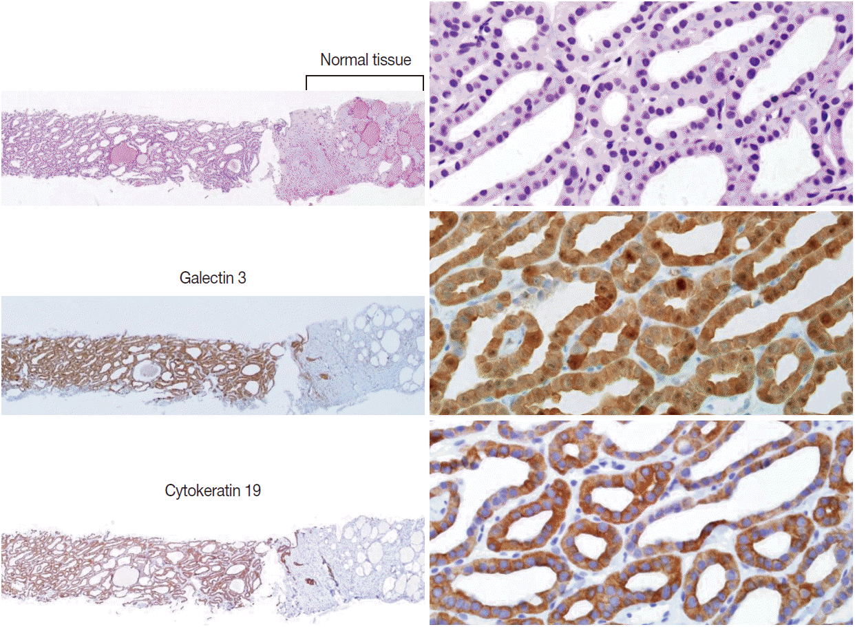
- Ancillary immunohistochemical or molecular studies can be helpful for the diagnosis of CNB specimens with findings that are suspicious for malignancy. An immunostaining panel consisting of galectin-3, HBME1, cytokeratin 19, or CD56 is helpful in the diagnosis of lesions suspicious for papillary thyroid carcinoma (Fig. 6) [21-23]. A combination of at least two immunostaining markers is recommended to improve the diagnostic accuracy [22]. Presence of the BRAF V600E mutation, detected by molecular test or immunostaining, strongly suggests a diagnosis of the papillary carcinoma [21].
- A diagnosis of medullary thyroid carcinoma can be confirmed with positive immunostaining for calcitonin on a CNB specimen. A diagnosis of lymphoma can be rendered using immunophenotyping studies on a CNB specimen that is suspicious for lymphoma [5].
- VI. Malignant
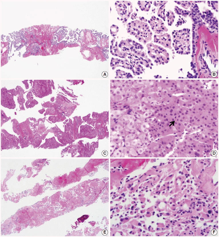
- Most thyroid malignancies, except for follicular carcinoma, have typical histologic features and are easily diagnosed as a malignancy on a CNB specimen. The following diagnoses are included in this category: papillary thyroid carcinoma (Fig. 7A, B), poorly differentiated carcinoma (Fig. 7C, D), undifferentiated (anaplastic) carcinoma, medullary thyroid carcinoma (Fig. 7E, F), lymphoma, and metastatic carcinoma.
CONSENSUS DIAGNOSTIC CATEGORIES
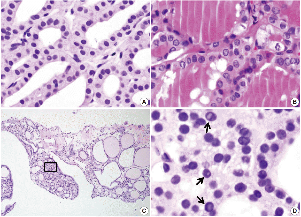
- The follicular cells in a CNB specimen appear smaller and show darker chromatin than in typical surgical specimens (Fig. 8A, B). Nuclear artifacts that mimic intranuclear cytoplasmic pseudoinclusions in papillary carcinoma may also be present in benign follicular cells (Fig. 8C, D). The artifactual vacuoles are irregularly shaped and appear pale on staining, whereas pseudoinclusions in papillary carcinoma are round and sharply delineated by the rim of the nuclear membrane.
COMMON PITFALLS OF A DIAGNOSIS OF CORE NEEDLE BIOPSY
Acknowledgments


- 1. American Thyroid Association (ATA) Guidelines Taskforce on Thyroid Nodules and Differentiated Thyroid Cancer, Cooper DS, Doherty GM, et al. Revised American Thyroid Association management guidelines for patients with thyroid nodules and differentiated thyroid cancer. Thyroid 2009; 19: 1167-214. ArticlePubMed
- 2. Ali SZ. Thyroid cytopathology: Bethesda and beyond. Acta Cytol 2011; 55: 4-12. ArticlePubMedPDF
- 3. Cibas ES, Ali SZ; NCI Thyroid FNA State of the Science Conference. The Bethesda System for Reporting Thyroid Cytopathology. Am J Clin Pathol 2009; 132: 658-65. ArticlePubMed
- 4. Lee SH, Kim MH, Bae JS, Lim DJ, Jung SL, Jung CK. Clinical outcomes in patients with non-diagnostic thyroid fine needle aspiration cytology: usefulness of the thyroid core needle biopsy. Ann Surg Oncol 2014; 21: 1870-7. ArticlePubMedPDF
- 5. Hahn SY, Shin JH, Han BK, Ko EY, Ko ES. Ultrasonography-guided core needle biopsy for the thyroid nodule: does the procedure hold any benefit for the diagnosis when fine-needle aspiration cytology analysis shows inconclusive results? Br J Radiol 2013; 86: 20130007.ArticlePubMedPMC
- 6. Trimboli P, Crescenzi A. Thyroid core needle biopsy: taking stock of the situation. Endocrine 2015; 48: 779-85. ArticlePubMedPDF
- 7. Sung JY, Na DG, Kim KS, et al. Diagnostic accuracy of fine-needle aspiration versus core-needle biopsy for the diagnosis of thyroid malignancy in a clinical cohort. Eur Radiol 2012; 22: 1564-72. ArticlePubMedPDF
- 8. Na DG, Kim JH, Sung JY, et al. Core-needle biopsy is more useful than repeat fine-needle aspiration in thyroid nodules read as nondiagnostic or atypia of undetermined significance by the Bethesda system for reporting thyroid cytopathology. Thyroid 2012; 22: 468-75. ArticlePubMed
- 9. Baek JH, Na DG, Lee JH, et al. Core needle biopsy of thyroid nodules: consensus statement and recommendations. J Korean Soc Ultrasound Med 2013; 32: 95-102.
- 10. Ha EJ, Baek JH, Lee JH, et al. Core needle biopsy can minimise the non-diagnostic results and need for diagnostic surgery in patients with calcified thyroid nodules. Eur Radiol 2014; 24: 1403-9. ArticlePubMedPDF
- 11. Yeon JS, Baek JH, Lim HK, et al. Thyroid nodules with initially nondiagnostic cytologic results: the role of core-needle biopsy. Radiology 2013; 268: 274-80. ArticlePubMed
- 12. Trimboli P, Nasrollah N, Guidobaldi L, et al. The use of core needle biopsy as first-line in diagnosis of thyroid nodules reduces false negative and inconclusive data reported by fine-needle aspiration. World J Surg Oncol 2014; 12: 61.ArticlePubMedPMCPDF
- 13. Nasrollah N, Trimboli P, Rossi F, et al. Patient’s comfort with and tolerability of thyroid core needle biopsy. Endocrine 2014; 45: 79-83. ArticlePubMedPDF
- 14. Baloch ZW, LiVolsi VA. Our approach to follicular-patterned lesions of the thyroid. J Clin Pathol 2007; 60: 244-50. ArticlePubMedPMC
- 15. Min HS, Kim JH, Ryoo I, Jung SL, Jung CK. The role of core needle biopsy in the preoperative diagnosis of follicular neoplasm of the thyroid. APMIS 2014; 122: 993-1000. ArticlePubMed
- 16. Yassa L, Cibas ES, Benson CB, et al. Long-term assessment of a multidisciplinary approach to thyroid nodule diagnostic evaluation. Cancer 2007; 111: 508-16. ArticlePubMed
- 17. Bae JS, Choi SK, Jeon S, et al. Impact of NRAS mutations on the diagnosis of follicular neoplasm of the thyroid. Int J Endocrinol 2014; 2014: 289834.ArticlePubMedPMCPDF
- 18. Ustun B, Chhieng D, Van Dyke A, et al. Risk stratification in follicular neoplasm: a cytological assessment using the modified Bethesda classification. Cancer Cytopathol 2014; 122: 536-45. ArticlePubMed
- 19. Sillery JC, Reading CC, Charboneau JW, Henrichsen TL, Hay ID, Mandrekar JN. Thyroid follicular carcinoma: sonographic features of 50 cases. AJR Am J Roentgenol 2010; 194: 44-54. ArticlePubMed
- 20. Reading CC, Charboneau JW, Hay ID, Sebo TJ. Sonography of thyroid nodules: a “classic pattern” diagnostic approach. Ultrasound Q 2005; 21: 157-65. PubMed
- 21. Crescenzi A, Guidobaldi L, Nasrollah N, et al. Immunohistochemistry for BRAF(V600E) antibody VE1 performed in core needle biopsy samples identifies mutated papillary thyroid cancers. Horm Metab Res 2014; 46: 370-4. ArticlePubMed
- 22. Alshenawy HA. Utility of immunohistochemical markers in diagnosis of follicular cell derived thyroid lesions. Pathol Oncol Res 2014; 20: 819-28. ArticlePubMedPDF
- 23. El Demellawy D, Nasr AL, Babay S, Alowami S. Diagnostic utility of CD56 immunohistochemistry in papillary carcinoma of the thyroid. Pathol Res Pract 2009; 205: 303-9. ArticlePubMed
REFERENCES
Figure & Data
References
Citations

- Predictive US findings for differentiating between malignant and benign thyroid nodules: Doppler and microvascular imaging according to the TI-RADS and pathologic subtypes of malignancy
Younghee Yim, Hye Shin Ahn, Min Ji Hong, Hyun Jeong Park, Sung Bin Park
Ultrasonography.2026; 45(1): 38. CrossRef - Risk Stratification of Thyroid Nodules Diagnosed as Follicular Neoplasm on Core Needle Biopsy
Byeong-Joo Noh, Won Jun Kim, Jin Yub Kim, Ha Young Kim, Jong Cheol Lee, Myoung Sook Shim, Yong Jin Song, Kwang Hyun Yoon, In-Hye Jung, Hyo Sang Lee, Wooyul Paik, Dong Gyu Na
Endocrinology and Metabolism.2025; 40(4): 610. CrossRef - Diagnostic performances of five US risk stratification systems for malignancy in focal [18F]FDG-PET/CT thyroid incidentalomas
Chae Young Shin, Hye Shin Ahn, Dong Gyu Na, Hyo Sang Lee, Eon-Woo Shin
European Radiology.2025; 35(12): 7701. CrossRef - Malignancy risk in AUS thyroid lesions: comparison between FNA and CNB with implications for NIFTP diagnosis
Yeseul Kim, Jae Ho Shin, You-Na Sung, Dawon Park, Harim Oh, Hyo Seon Ryu, Kyeong Jin Kim, Hyun Joo Kim, Sin Gon Kim, Hoon Yub Kim, Kwang Yoon Jung, Seung-Kuk Baek, Sangjeong Ahn
Frontiers in Endocrinology.2025;[Epub] CrossRef - The Diagnostic Role of Repeated Biopsy of Thyroid Nodules with Atypia of Undetermined Significance with Architectural Atypia on Core-Needle Biopsy
Hye Hyeon Moon, Sae Rom Chung, Young Jun Choi, Tae-Yon Sung, Dong Eun Song, Tae Yong Kim, Jeong Hyun Lee, Jung Hwan Baek
Endocrinology and Metabolism.2024; 39(2): 300. CrossRef - Core needle biopsy for thyroid nodules assessment-a new horizon?
David D Dolidze, Serghei Covantsev, Grigorii M Chechenin, Natalia V Pichugina, Anastasia V Bedina, Anna Bumbu
World Journal of Clinical Oncology.2024; 15(5): 580. CrossRef - Ultrasonographic features and diagnostic accuracy of FNA and CNB in secondary thyroid malignancies: A retrospective study
Zhen Xia, Xiaochen Huang, Ting Zhang, Zhigang Gao, Xiuliang Tang, Wei Zhang, Qing Miao
Heliyon.2024; 10(16): e36305. CrossRef - Current role of interventional radiology in thyroid nodules
Onur Taydas, Erbil Arik, Omer Faruk Sevinc, Ahmet Burak Kara, Mustafa Ozdemir, Hasret Cengiz, Zulfu Bayhan, Mehmet Halil Ozturk
Frontiers in Endocrinology.2024;[Epub] CrossRef - A simplified four-tier classification for thyroid core needle biopsy
M. Paja, J. L. Del Cura, R. Zabala, I. Korta, Mª T. Gutiérrez, A. Expósito, A. Ugalde
Journal of Endocrinological Investigation.2024; 48(4): 895. CrossRef - Follow-up of benign thyroid nodules confirmed by ultrasound-guided core needle biopsy after inconclusive cytology on fine-needle aspiration biopsy
Yoon Ji Hwang, Hye Ryoung Koo, Jeong Seon Park
Ultrasonography.2023; 42(1): 121. CrossRef - Subcategorization of intermediate suspicion thyroid nodules based on suspicious ultrasonographic findings
Haejung Kim, Jung Hee Shin, Ka Eun Kim, Myoung Kyoung Kim, Jiyun Oh, Soo Yeon Hahn
Ultrasonography.2023; 42(2): 307. CrossRef - Preoperative Risk Stratification of Follicular-patterned Thyroid Lesions on Core Needle Biopsy by Histologic Subtyping and RAS Variant-specific Immunohistochemistry
Meejeong Kim, Sora Jeon, Chan Kwon Jung
Endocrine Pathology.2023; 34(2): 247. CrossRef - Cytological and Ultrasound Features of Thyroid Nodules Correlate With Histotypes and Variants of Thyroid Carcinoma
Daniele Sgrò, Alessandro Brancatella, Giuseppe Greco, Liborio Torregrossa, Paolo Piaggi, Nicola Viola, Teresa Rago, Fulvio Basolo, Riccardo Giannini, Gabriele Materazzi, Rossella Elisei, Ferruccio Santini, Francesco Latrofa
The Journal of Clinical Endocrinology & Metabolism.2023; 108(11): e1186. CrossRef - Reevaluating diagnostic categories and associated malignancy risks in thyroid core needle biopsy
Chan Kwon Jung
Journal of Pathology and Translational Medicine.2023; 57(4): 208. CrossRef - Contrast Enhancement Ultrasound Improves Diagnostic Accuracy for Thyroid Nodules: A Prospective Multicenter Study
Jianming Li, Jianping Dou, Huarong Li, Fan Xiao, Jie Yu, Mingxing Xie, Ping Zhou, Lei Liang, Guiming Zhou, Ying Che, Cun Liu, Zhibin Cong, Fangyi Liu, Zhiyu Han, Ping Liang
Journal of the Endocrine Society.2023;[Epub] CrossRef - Core needle biopsy and ultrasonography are superior to fine needle aspiration in the management of follicular variant papillary thyroid carcinomas
Ji-Ye Kim, Sunhee Chang, Ah-Young Kwon, Eun Young Park, Tae Hyuk Kim, Sangjoon Choi, Minju Lee, Young Lyun Oh
Endocrine.2022; 75(2): 437. CrossRef - Fine Needle Aspiration Cytology vs. Core Needle Biopsy for Thyroid Nodules: A Prospective, Experimental Study Using Surgical Specimen
Hyuk Kwon, Jandee Lee, Soon Won Hong, Hyeong Ju Kwon, Jin Young Kwak, Jung Hyun Yoon
Journal of the Korean Society of Radiology.2022; 83(3): 645. CrossRef - Diagnostic efficacy, performance and safety of side-cut core needle biopsy for thyroid nodules: comparison of automated and semi-automated biopsy needles
Ji Yeon Park, Seong Yoon Yi, Soo Heui Baek, Yu Hyun Lee, Heon-Ju Kwon, Hee Jin Park
Endocrine.2022; 76(2): 341. CrossRef - Approach to Bethesda system category III thyroid nodules according to US-risk stratification
Jieun Kim, Jung Hee Shin, Young Lyun Oh, Soo Yeon Hahn, Ko Woon Park
Endocrine Journal.2022; 69(1): 67. CrossRef - Quantitative analysis of vascularity for thyroid nodules on ultrasound using superb microvascular imaging
Min Ji Hong, Hye Shin Ahn, Su Min Ha, Hyun Jeong Park, Jiyun Oh
Medicine.2022; 101(5): e28725. CrossRef - Diagnostic Performance of Thyroid Core Needle Biopsy Using the Revised Reporting System: Comparison with Fine Needle Aspiration Cytology
Kwangsoon Kim, Ja Seong Bae, Jeong Soo Kim, So Lyung Jung, Chan Kwon Jung
Endocrinology and Metabolism.2022; 37(1): 159. CrossRef - A Matched-Pair Analysis of Nuclear Morphologic Features Between Core Needle Biopsy and Surgical Specimen in Thyroid Tumors Using a Deep Learning Model
Faridul Haq, Andrey Bychkov, Chan Kwon Jung
Endocrine Pathology.2022; 33(4): 472. CrossRef - Role of echogenic foci in ultrasonographic risk stratification of thyroid nodules: Echogenic focus scoring in the American College of Radiology Thyroid Imaging Reporting and Data System
Renxu Li, Zhenwei Liang, Xiangyu Wang, Luzeng Chen
Frontiers in Oncology.2022;[Epub] CrossRef - Ultrasonographic characteristics of medullary thyroid carcinoma according to nodule size: application of the Korean Thyroid Imaging Reporting and Data System and American Thyroid Association guidelines
Soo Yeon Hahn, Jung Hee Shin, Young Lyun Oh, Ko Woon Park
Acta Radiologica.2021; 62(4): 474. CrossRef - Malignancy risk of thyroid nodules with nonshadowing echogenic foci
Yu-Mee Sohn, Dong Gyu Na, Wooyul Paik, Hye Yun Gwon, Byeong-Joo Noh
Ultrasonography.2021; 40(1): 115. CrossRef - Assessing the diagnostic performance of thyroid biopsy with recommendations for appropriate interpretation
Su Min Ha, Jung Hwan Baek, Dong Gyu Na, Chan-Kwon Jung, Chong Hyun Suh, Young Kee Shong, Tae Yon Sung, Dong Eun Song, Jeong Hyun Lee
Ultrasonography.2021; 40(2): 228. CrossRef - Usage and Diagnostic Yield of Fine-Needle Aspiration Cytology and Core Needle Biopsy in Thyroid Nodules: A Systematic Review and Meta-Analysis of Literature Published by Korean Authors
Soon-Hyun Ahn
Clinical and Experimental Otorhinolaryngology.2021; 14(1): 116. CrossRef - Comparison of the Efficacy and Safety of the American Thyroid Association Guidelines and American College of Radiology TI-RADS
Jinghua Liu, Yajun Guo, Jiangxi Xiao, Luzeng Chen, Zhenwei Liang
Endocrine Practice.2021; 27(7): 661. CrossRef - Malignancy Rate of Bethesda Class III Thyroid Nodules Based on the Presence of Chronic Lymphocytic Thyroiditis in Surgical Patients
Yoon Young Cho, Yun Jae Chung, Hee Sung Kim
Frontiers in Endocrinology.2021;[Epub] CrossRef - Efficacy of Differential Diagnosis of Thyroid Nodules by Shear Wave Elastography—the Stiffness Map
Myung Hi Yoo, Hye Jeong Kim, In Ho Choi, Suyeon Park, Sumi Yun, Hyeong Kyu Park, Dong Won Byun, Kyoil Suh
Journal of the Endocrine Society.2021;[Epub] CrossRef - The relationship of thyroid nodule size on malignancy risk according to histological type of thyroid cancer
Sae Rom Chung, Jung Hwan Baek, Young Jun Choi, Tae-Yon Sung, Dong Eun Song, Tae Yong Kim, Jeong Hyun Lee
Acta Radiologica.2020; 61(5): 620. CrossRef - Concordance of Three International Guidelines for Thyroid Nodules Classified by Ultrasonography and Diagnostic Performance of Biopsy Criteria
Younghee Yim, Dong Gyu Na, Eun Ju Ha, Jung Hwan Baek, Jin Yong Sung, Ji-hoon Kim, Won-Jin Moon
Korean Journal of Radiology.2020; 21(1): 108. CrossRef - 2019 Practice guidelines for thyroid core needle biopsy: a report of the Clinical Practice Guidelines Development Committee of the Korean Thyroid Association
Chan Kwon Jung, Jung Hwan Baek, Dong Gyu Na, Young Lyun Oh, Ka Hee Yi, Ho-Cheol Kang
Journal of Pathology and Translational Medicine.2020; 54(1): 64. CrossRef - Core-Needle Biopsy Does Not Show Superior Diagnostic Performance to Fine-Needle Aspiration for Diagnosing Thyroid Nodules
Ilah Shin, Eun-Kyung Kim, Hee Jung Moon, Jung Hyun Yoon, Vivian Youngjean Park, Si Eun Lee, Hye Sun Lee, Jin Young Kwak
Yonsei Medical Journal.2020; 61(2): 161. CrossRef - Ultrasound‐guided fine‐needle aspiration or core needle biopsy for diagnosing follicular thyroid carcinoma?
Ko Woon Park, Jung Hee Shin, Soo Yeon Hahn, Young Lyun Oh, Sun Wook Kim, Tae Hyuk Kim, Jae Hoon Chung
Clinical Endocrinology.2020; 92(5): 468. CrossRef - Distribution and malignancy risk of six categories of the pathology reporting system for thyroid core-needle biopsy in 1,216 consecutive thyroid nodules
Hye Min Son, Ji-hoon Kim, Soo Chin Kim, Roh-Eul Yoo, Jeong Mo Bae, Hyobin Seo, Dong Gyu Na
Ultrasonography.2020; 39(2): 159. CrossRef - CT features of thyroid nodules with isolated macrocalcifications detected by ultrasonography
Wooyul Paik, Dong Gyu Na, Hye Yun Gwon, Jinna Kim
Ultrasonography.2020; 39(2): 130. CrossRef - Contribution of cytologic examination to diagnosis of poorly differentiated thyroid carcinoma
Na Rae Kim, Jae Yeon Seok, Yoo Seung Chung, Joon Hyop Lee, Dong Hae Chung
Journal of Pathology and Translational Medicine.2020; 54(2): 171. CrossRef - Comparison Between Fine Needle Aspiration and Core Needle Biopsy for the Diagnosis of Thyroid Nodules: Effective Indications According to US Findings
Soo Yeon Hahn, Jung Hee Shin, Young Lyun Oh, Ko Woon Park, Yaeji Lim
Scientific Reports.2020;[Epub] CrossRef - Thyroid Nodules with Isolated Macrocalcifications: Malignancy Risk of Isolated Macrocalcifications and Postoperative Risk Stratification of Malignant Tumors Manifesting as Isolated Macrocalcifications
Hye Yun Gwon, Dong Gyu Na, Byeong-Joo Noh, Wooyul Paik, So Jin Yoon, Soo-Jung Choi, Dong Rock Shin
Korean Journal of Radiology.2020; 21(5): 605. CrossRef - Diagnostic Efficacy and Safety of Core Needle Biopsy as a First-Line Diagnostic Method for Thyroid Nodules: A Prospective Cohort Study
Min Ji Hong, Dong Gyu Na, Hunkyung Lee
Thyroid.2020; 30(8): 1141. CrossRef - Re: The 2019 core-needle biopsy practice guidelines
Ji-hoon Kim
Ultrasonography.2020; 39(3): 313. CrossRef - Cytopathologic criteria and size should be considered in comparison of fine-needle aspiration vs. core-needle biopsy for thyroid nodules: results based on large surgical series
Jung Hyun Yoon, Hye Sun Lee, Eun-Kyung Kim, Hee Jung Moon, Vivian Youngjean Park, Jin Young Kwak
Endocrine.2020; 70(3): 558. CrossRef - Preoperative diagnostic categories of fine needle aspiration cytology for histologically proven thyroid follicular adenoma and carcinoma, and Hurthle cell adenoma and carcinoma: Analysis of cause of under- or misdiagnoses
Hee Young Na, Jae Hoon Moon, June Young Choi, Hyeong Won Yu, Woo-Jin Jeong, Yeo Koon Kim, Ji-Young Choe, So Yeon Park, Paula Soares
PLOS ONE.2020; 15(11): e0241597. CrossRef - The Role of Core Needle Biopsy for the Evaluation of Thyroid Nodules with Suspicious Ultrasound Features
Sae Rom Chung, Jung Hwan Baek, Young Jun Choi, Tae-Yon Sung, Dong Eun Song, Tae Yong Kim, Jeong Hyun Lee
Korean Journal of Radiology.2019; 20(1): 158. CrossRef - Pathological diagnosis of thyroid nodules based on core needle biopsies: comparative study between core needle biopsies and resected specimens in 578 cases
Yan Xiong, Limin Yan, Lin Nong, Yalin Zheng, Ting Li
Diagnostic Pathology.2019;[Epub] CrossRef - The Current Histologic Classification of Thyroid Cancer
Sylvia L. Asa
Endocrinology and Metabolism Clinics of North America.2019; 48(1): 1. CrossRef - Core-needle biopsy in thyroid nodules: performance, accuracy, and complications
Miguel Paja, Jose Luis del Cura, Rosa Zabala, Igone Korta, Aitziber Ugalde, José I. López
European Radiology.2019; 29(9): 4889. CrossRef - Ultrasound‐guided needle biopsy of large thyroid nodules: Core needle biopsy yields more reliable results than fine needle aspiration
Hyeon Jin Lee, Young Joong Kim, Hye Yeon Han, Jae Young Seo, Cheol Mog Hwang, KeumWon Kim
Journal of Clinical Ultrasound.2019; 47(5): 255. CrossRef - Tumor Volume Doubling Time in Active Surveillance of Papillary Thyroid Carcinoma
Hye-Seon Oh, Hyemi Kwon, Eyun Song, Min Ji Jeon, Tae Yong Kim, Jeong Hyun Lee, Won Bae Kim, Young Kee Shong, Ki-Wook Chung, Jung Hwan Baek, Won Gu Kim
Thyroid.2019; 29(5): 642. CrossRef - Role of Immunohistochemistry in Fine Needle Aspiration and Core Needle Biopsy of Thyroid Nodules
Seulki Song, Hyojin Kim, Soon-Hyun Ahn
Clinical and Experimental Otorhinolaryngology.2019; 12(2): 224. CrossRef - Echogenic foci in thyroid nodules: diagnostic performance with combination of TIRADS and echogenic foci
Su Min Ha, Yun Jae Chung, Hye Shin Ahn, Jung Hwan Baek, Sung Bin Park
BMC Medical Imaging.2019;[Epub] CrossRef - Risk of Malignancy According to the Sub-classification of Atypia of Undetermined Significance and Suspicious Follicular Neoplasm Categories in Thyroid Core Needle Biopsies
Sae Rom Chung, Jung Hwan Baek, Jeong Hyun Lee, Yu-Mi Lee, Tae-Yon Sung, Ki-Wook Chung, Suck Joon Hong, Min Ji Jeon, Tae Yong Kim, Young Kee Shong, Won Bae Kim, Won Gu Kim, Dong Eun Song
Endocrine Pathology.2019; 30(2): 146. CrossRef - Thyroid core needle biopsy: patients’ pain and satisfaction compared to fine needle aspiration
Hyo Jin Kim, Yeo Koon Kim, Jae Hoon Moon, June Young Choi, Sang Il Choi
Endocrine.2019; 65(2): 365. CrossRef - Does Radiofrequency Ablation Induce Neoplastic Changes in Benign Thyroid Nodules: A Preliminary Study
Su Min Ha, Jun Young Shin, Jung Hwan Baek, Dong Eun Song, Sae Rom Chung, Young Jun Choi, Jeong Hyun Lee
Endocrinology and Metabolism.2019; 34(2): 169. CrossRef - Differential Diagnosis of Thyroid Follicular Neoplasm from Nodular Hyperplasia by Shear Wave Elastography
Myung Hi Yoo, Hye Jeong Kim, In Ho Choi, Ji-Oh Mok, Hyeong Kyu Park, Dong Won Byun, Kyoil Suh
Soonchunhyang Medical Science.2019; 25(1): 10. CrossRef - Ultrasound-Guided Core Needle Biopsy Techniques for Intermediate or Low Suspicion Thyroid Nodules: Which Method is Effective for Diagnosis?
Soo Yeon Hahn, Jung Hee Shin, Young Lyun Oh, Ko Woon Park
Korean Journal of Radiology.2019; 20(10): 1454. CrossRef - Preoperative Diagnostic Categories of Noninvasive Follicular Thyroid Neoplasm with Papillary-Like Nuclear Features in Thyroid Core Needle Biopsy and Its Impact on Risk of Malignancy
Hee Young Na, Ji Won Woo, Jae Hoon Moon, June Young Choi, Woo-Jin Jeong, Yeo Koon Kim, Ji-Young Choe, So Yeon Park
Endocrine Pathology.2019; 30(4): 329. CrossRef - Utility of a formatted pathologic reporting system in thyroid core needle biopsy: A validation study of 1998 consecutive cases
Ji‐Young Choe, Yoonjin Kwak, Mimi Kim, Yul Ri Chung, Hyun Jeong Kim, Yeo Koon Kim, So Yeon Park
Clinical Endocrinology.2018; 88(1): 96. CrossRef - Nuclear features of papillary thyroid carcinoma: Comparison of Core needle biopsy and thyroidectomy specimens
Jae Yeon Seok, Jungsuk An, Hyun Yee Cho, Younghye Kim, Seung Yeon Ha
Annals of Diagnostic Pathology.2018; 32: 35. CrossRef - Prevention of total thyroidectomy in noninvasive follicular thyroid neoplasm with papillary‐like nuclear features (NIFTP) based on combined interpretation of ultrasonographic and cytopathologic results
Sung‐Hye You, Kyu Eun Lee, Roh‐Eul Yoo, Hye Jeong Choi, Kyeong Cheon Jung, Jae‐Kyung Won, Koung Mi Kang, Tae Jin Yoon, Seung Hong Choi, Chul‐Ho Sohn, Ji‐hoon Kim
Clinical Endocrinology.2018; 88(1): 114. CrossRef - Statement and Recommendations on Interventional Ultrasound as a Thyroid Diagnostic and Treatment Procedure
Christoph F. Dietrich, Thomas Müller, Jörg Bojunga, Yi Dong, Giovanni Mauri, Maija Radzina, Manjiri Dighe, Xin-Wu Cui, Frank Grünwald, Andreas Schuler, Andre Ignee, Huedayi Korkusuz
Ultrasound in Medicine & Biology.2018; 44(1): 14. CrossRef - Core needle biopsy of thyroid nodules: outcomes and safety from a large single-center single-operator study
Jooae Choe, Jung Hwan Baek, Hye Sun Park, Young Jun Choi, Jeong Hyun Lee
Acta Radiologica.2018; 59(8): 924. CrossRef - Improvement of diagnostic performance of pathologists by reducing the number of pathologists responsible for thyroid fine needle aspiration cytology: An institutional experience
Jae Yeon Seok, Jungsuk An, Hyun Yee Cho
Diagnostic Cytopathology.2018; 46(7): 561. CrossRef - Comparison of Immunohistochemistry and Direct Sanger Sequencing for Detection of theBRAFV600EMutation in Thyroid Neoplasm
Hye-Seon Oh, Hyemi Kwon, Suyeon Park, Mijin Kim, Min Ji Jeon, Tae Yong Kim, Young Kee Shong, Won Bae Kim, Jene Choi, Won Gu Kim, Dong Eun Song
Endocrinology and Metabolism.2018; 33(1): 62. CrossRef - Comparison of the Diagnostic Efficacy of Ultrasound‐Guided Core Needle Biopsy With 18‐ Versus 20‐Gauge Needles for Thyroid Nodules
Hye Shin Ahn, Mirinae Seo, Su Min Ha, Hee Sung Kim
Journal of Ultrasound in Medicine.2018; 37(11): 2565. CrossRef - Impact of Nodule Size on Malignancy Risk Differs according to the Ultrasonography Pattern of Thyroid Nodules
Min Ji Hong, Dong Gyu Na, Jung Hwan Baek, Jin Yong Sung, Ji-Hoon Kim
Korean Journal of Radiology.2018; 19(3): 534. CrossRef - Web‐based thyroid imaging reporting and data system: Malignancy risk of atypia of undetermined significance or follicular lesion of undetermined significance thyroid nodules calculated by a combination of ultrasonography features and biopsy results
Young Jun Choi, Jung Hwan Baek, Jung Hee Shin, Woo Hyun Shim, Seon‐Ok Kim, Won‐Hong Lee, Dong Eun Song, Tae Yong Kim, Ki‐Wook Chung, Jeong Hyun Lee
Head & Neck.2018; 40(9): 1917. CrossRef - Thyroid Incidentalomas Detected on18F-Fluorodeoxyglucose Positron Emission Tomography with Computed Tomography: Malignant Risk Stratification and Management Plan
Sae Rom Chung, Young Jun Choi, Chong Hyun Suh, Hwa Jung Kim, Jong Jin Lee, Won Gu Kim, Tae Yon Sung, Yu-mi Lee, Dong Eun Song, Jeong Hyun Lee, Jung Hwan Baek
Thyroid.2018; 28(6): 762. CrossRef - The role of core needle biopsy in the diagnosis of initially detected thyroid nodules: a systematic review and meta-analysis
Sae Rom Chung, Chong Hyun Suh, Jung Hwan Baek, Young Jun Choi, Jeong Hyun Lee
European Radiology.2018; 28(11): 4909. CrossRef - The History of Korean Thyroid Pathology
Soon Won Hong, Chan Kwon Jung
International Journal of Thyroidology.2018; 11(1): 15. CrossRef - Ultrasonographic Echogenicity and Histopathologic Correlation of Thyroid Nodules in Core Needle Biopsy Specimens
Ji-hoon Kim, Dong Gyu Na, Hunkyung Lee
Korean Journal of Radiology.2018; 19(4): 673. CrossRef - Evaluation of Modified Core-Needle Biopsy in the Diagnosis of Thyroid Nodules
Soomin Ahn, Sejin Jung, Ji-Ye Kim, Jung Hee Shin, Soo Yeon Hahn, Young Lyun Oh
Korean Journal of Radiology.2018; 19(4): 656. CrossRef - Role of core needle biopsy as a first-line diagnostic tool for thyroid nodules: a retrospective cohort study
Min Ji Hong, Dong Gyu Na, Soo Jin Kim, Dae Sik Kim
Ultrasonography.2018; 37(3): 244. CrossRef - Active Surveillance of Low-Risk Papillary Thyroid Microcarcinoma: A Multi-Center Cohort Study in Korea
Hye-Seon Oh, Jeonghoon Ha, Hye In Kim, Tae Hyuk Kim, Won Gu Kim, Dong-Jun Lim, Tae Yong Kim, Sun Wook Kim, Won Bae Kim, Young Kee Shong, Jae Hoon Chung, Jung Hwan Baek
Thyroid.2018; 28(12): 1587. CrossRef - Risk of malignancy according to sub‐classification of the atypia of undetermined significance or follicular lesion of undetermined significance (AUS/FLUS) category in the Bethesda system for reporting thyroid cytopathology
S. J. Kim, J. Roh, J. H. Baek, S. J. Hong, Y. K. Shong, W. B. Kim, D. E. Song
Cytopathology.2017; 28(1): 65. CrossRef - Fine-needle aspiration versus core needle biopsy for diagnosis of thyroid malignancy and neoplasm: a matched cohort study
Soo-Yeon Kim, Hye Sun Lee, Jieun Moon, Eun-Kyung Kim, Hee Jung Moon, Jung Hyun Yoon, Jin Young Kwak
European Radiology.2017; 27(2): 801. CrossRef - Follicular variant of papillary thyroid carcinoma: comparison of ultrasound-guided core needle biopsy and ultrasound-guided fine needle aspiration in a multicentre study
Soo Yeon Hahn, Jung Hee Shin, Hyun Kyung Lim, So Lyung Jung
Clinical Endocrinology.2017; 86(1): 113. CrossRef - Core‐needle biopsy versus repeat fine‐needle aspiration for thyroid nodules initially read as atypia/follicular lesion of undetermined significance
Young Jun Choi, Jung Hwan Baek, Chong Hyun Suh, Woo Hyun Shim, Boseul Jeong, Jae Kyun Kim, Dong Eun Song, Tae Yong Kim, Ki‐Wook Chung, Jeong Hyun Lee
Head & Neck.2017; 39(2): 361. CrossRef - Preoperative differentiation between noninvasive follicular thyroid neoplasm with papillary-like nuclear features (NIFTP) and non-NIFTP
Soo Yeon Hahn, Jung Hee Shin, Hyun Kyung Lim, So Lyung Jung, Young Lyun Oh, In Ho Choi, Chan Kwon Jung
Clinical Endocrinology.2017; 86(3): 444. CrossRef - Core Needle Biopsy of the Thyroid: 2016 Consensus Statement and Recommendations from Korean Society of Thyroid Radiology
Dong Gyu Na, Jung Hwan Baek, So Lyung Jung, Ji-hoon Kim, Jin Yong Sung, Kyu Sun Kim, Jeong Hyun Lee, Jung Hee Shin, Yoon Jung Choi, Eun Ju Ha, Hyun Kyung Lim, Soo Jin Kim, Soo Yeon Hahn, Kwang Hwi Lee, Young Jun Choi, Inyoung Youn, Young Joong Kim, Hye Sh
Korean Journal of Radiology.2017; 18(1): 217. CrossRef - First-Line Use of Core Needle Biopsy for High-Yield Preliminary Diagnosis of Thyroid Nodules
H.C. Kim, Y.J. Kim, H.Y. Han, J.M. Yi, J.H. Baek, S.Y. Park, J.Y. Seo, K.W. Kim
American Journal of Neuroradiology.2017; 38(2): 357. CrossRef - Ultrasound-Pathology Discordant Nodules on Core-Needle Biopsy: Malignancy Risk and Management Strategy
Sae Rom Chung, Jung Hwan Baek, Hye Sun Park, Young Jun Choi, Tae-Yon Sung, Dong Eun Song, Tae Yong Kim, Jeong Hyun Lee
Thyroid.2017; 27(5): 707. CrossRef - Current status of core needle biopsy of the thyroid
Jung Hwan Baek
Ultrasonography.2017; 36(2): 83. CrossRef - Cytology-Ultrasonography Risk-Stratification Scoring System Based on Fine-Needle Aspiration Cytology and the Korean-Thyroid Imaging Reporting and Data System
Min Ji Hong, Dong Gyu Na, Jung Hwan Baek, Jin Yong Sung, Ji-Hoon Kim
Thyroid.2017; 27(7): 953. CrossRef - Preoperative clinicopathological characteristics of patients with solitary encapsulated follicular variants of papillary thyroid carcinomas
Hyemi Kwon, Min Ji Jeon, Jong Ho Yoon, Suck Joon Hong, Jeong Hyun Lee, Tae Yong Kim, Young Kee Shong, Won Bae Kim, Won Gu Kim, Dong Eun Song
Journal of Surgical Oncology.2017; 116(6): 746. CrossRef - Active Surveillance for Patients With Papillary Thyroid Microcarcinoma: A Single Center’s Experience in Korea
Hyemi Kwon, Hye-Seon Oh, Mijin Kim, Suyeon Park, Min Ji Jeon, Won Gu Kim, Won Bae Kim, Young Kee Shong, Dong Eun Song, Jung Hwan Baek, Ki-Wook Chung, Tae Yong Kim
The Journal of Clinical Endocrinology & Metabolism.2017; 102(6): 1917. CrossRef - Comparison of Consecutive Results from Fine Needle Aspiration and Core Needle Biopsy in Thyroid Nodules
Soon-Hyun Ahn, So-Yeon Park, Sang Il Choi
Endocrine Pathology.2017; 28(4): 332. CrossRef - Efficacy and safety of core-needle biopsy in initially detected thyroid nodules via propensity score analysis
Chong Hyun Suh, Jung Hwan Baek, Young Jun Choi, Tae Yong Kim, Tae Yon Sung, Dong Eun Song, Jeong Hyun Lee
Scientific Reports.2017;[Epub] CrossRef - Comparison of Core-Needle Biopsy and Fine-Needle Aspiration for Evaluating Thyroid Incidentalomas Detected by 18F-Fluorodeoxyglucose Positron Emission Tomography/Computed Tomography: A Propensity Score Analysis
Chong Hyun Suh, Young Jun Choi, Jong Jin Lee, Woo Hyun Shim, Jung Hwan Baek, Han Cheol Chung, Young Kee Shong, Dong Eun Song, Tae Yon Sung, Jeong Hyun Lee
Thyroid.2017; 27(10): 1258. CrossRef - Thyroid Fine-Needle Aspiration Cytology Practice in Korea
Yoon Jin Cha, Ju Yeon Pyo, SoonWon Hong, Jae Yeon Seok, Kyung-Ju Kim, Jee-Young Han, Jeong Mo Bae, Hyeong Ju Kwon, Yeejeong Kim, Kyueng-Whan Min, Soonae Oak, Sunhee Chang
Journal of Pathology and Translational Medicine.2017; 51(6): 521. CrossRef - Recent Advances in Core Needle Biopsy for Thyroid Nodules
Chan Kwon Jung, Jung Hwan Baek
Endocrinology and Metabolism.2017; 32(4): 407. CrossRef - Core-needle biopsy for the preoperative diagnosis of follicular neoplasm in thyroid nodule screening: A validation study
Sung Hak Lee, Gyeong Sin Park, So Lyung Jung, Min-Hee Kim, Ja Seong Bae, Dong Jun Lim, Chan Kwon Jung
Pathology - Research and Practice.2016; 212(1): 44. CrossRef - The Role of Core-Needle Biopsy as a First-Line Diagnostic Tool for Initially Detected Thyroid Nodules
Chong Hyun Suh, Jung Hwan Baek, Jeong Hyun Lee, Young Jun Choi, Jae Kyun Kim, Tae-Yon Sung, Jong Ho Yoon, Young Kee Shong
Thyroid.2016; 26(3): 395. CrossRef - Thyroid nodules with minimal cystic changes have a low risk of malignancy
Dong Gyu Na, Ji-hoon Kim, Dea Sik Kim, Soo Jin Kim
Ultrasonography.2016; 35(2): 153. CrossRef - Thyroid nodules with isolated macrocalcification: malignancy risk and diagnostic efficacy of fine-needle aspiration and core needle biopsy
Dong Gyu Na, Dae Sik Kim, Soo Jin Kim, Jae Wook Ryoo, So Lyung Jung
Ultrasonography.2016; 35(3): 212. CrossRef - Thyroid Imaging Reporting and Data System Risk Stratification of Thyroid Nodules: Categorization Based on Solidity and Echogenicity
Dong Gyu Na, Jung Hwan Baek, Jin Yong Sung, Ji-Hoon Kim, Jae Kyun Kim, Young Jun Choi, Hyobin Seo
Thyroid.2016; 26(4): 562. CrossRef - Fine‐needle aspiration and core needle biopsy: An update on 2 common minimally invasive tissue sampling modalities
Paul A. VanderLaan
Cancer Cytopathology.2016; 124(12): 862. CrossRef - The role of core-needle biopsy in the diagnosis of thyroid malignancy in 4580 patients with 4746 thyroid nodules: a systematic review and meta-analysis
Chong Hyun Suh, Jung Hwan Baek, Jeong Hyun Lee, Young Jun Choi, Kyung Won Kim, Jayoun Lee, Ki-Wook Chung, Young Kee Shong
Endocrine.2016; 54(2): 315. CrossRef - The Role of Core-Needle Biopsy for Thyroid Nodules with Initially Nondiagnostic Fine-Needle Aspiration Results: A Systematic Review and Meta-Analysis
Chong Hyun Suh, Jung Hwan Baek, Kyung Won Kim, Tae Yon Sung, Tae Yong Kim, Dong Eun Song, Young Jun Choi, Jeong Hyun Lee
Endocrine Practice.2016; 22(6): 679. CrossRef - Should Core Needle Biopsy be Used in the Evaluation of Thyroid Nodules?
Beril Guler, Tugce Kiran, Dilek Sema Arici, Erhan Aysan, Fatma Cavide Sonmez
Endocrine Pathology.2016; 27(4): 352. CrossRef - A Multicenter Prospective Validation Study for the Korean Thyroid Imaging Reporting and Data System in Patients with Thyroid Nodules
Eun Ju Ha, Won-Jin Moon, Dong Gyu Na, Young Hen Lee, Nami Choi, Soo Jin Kim, Jae Kyun Kim
Korean Journal of Radiology.2016; 17(5): 811. CrossRef - Impact of Reclassification on Thyroid Nodules with Architectural Atypia: From Non-Invasive Encapsulated Follicular Variant Papillary Thyroid Carcinomas to Non-Invasive Follicular Thyroid Neoplasm with Papillary-Like Nuclear Features
Min Ji Jeon, Dong Eun Song, Chan Kwon Jung, Won Gu Kim, Hyemi Kwon, Yu-Mi Lee, Tae-Yon Sung, Jong Ho Yoon, Ki-Wook Chung, Suck Joon Hong, Jung Hwan Baek, Jeong Hyun Lee, Tae Yong Kim, Young Kee Shong, Won Bae Kim, Rafael Rosell
PLOS ONE.2016; 11(12): e0167756. CrossRef
 PubReader
 ePub Link
-
 Cite this Article
- Cite this Article
-
- Close
- Download Citation
- Close
- Figure


Fig. 1.
Fig. 2.
Fig. 3.
Fig. 4.
Fig. 5.
Fig. 6.
Fig. 7.
Fig. 8.
| I. Nondiagnostic or unsatisfactory |
| • Normal thyroid tissue only |
| • Extrathyroid tissue only (e.g., skeletal muscle, mature adipose tissue) |
| • A virtually acellular specimen |
| • Acellular/paucicellular fibrotic nodule |
| • Blood clot only |
| • Other |
| II. Benign lesion |
| • Benign follicular nodule or consistent with a benign follicular nodule |
| • Hashimoto's thyroiditis |
| • Granulomatous (subacute) thyroiditis |
| • Nonthyroidal lesion (e.g., parathyroid lesions, benign neurogenic tumors, benign lymph node) |
| • Other |
| III. Indeterminate lesion |
| IIIA. Indeterminate follicular lesion with nuclear atypia |
| • Follicular proliferative lesions with focal nuclear atypia |
| • Follicular proliferative lesions with equivocal or questionable nuclear atypia |
| • Atypical follicular cells embedded in a fibrotic stroma |
| IIIB. Indeterminate follicular lesion with architectural atypia |
| • Microfollicular proliferative lesion lacking a fibrous capsule or the adjacent nonlesional tissue in the specimen |
| • Solid or trabecular follicular lesion lacking a fibrous capsule or the adjacent nonlesional tissue in the specimen |
| • Macrofollicular proliferative lesion with a fibrous capsule |
| • Hürthle cell proliferative lesion lacking a fibrous capsule or the adjacent nonlesional tissue in the specimen |
| IIIC. Other indeterminate lesions |
| IV. Follicular neoplasm or suspicious for a follicular neoplasm |
| • Microfollicular proliferative lesion with a fibrous capsule |
| • Mixed microfollicular and normofollicular proliferative lesion with a fibrous capsule |
| • Solid/trabecular follicular proliferative lesion with a fibrous capsule |
| • Hürthle cell proliferative lesion with a fibrous capsule |
| • Follicular neoplasm with focal nuclear atypia |
| V. Suspicious for malignancy |
| • Suspicious for papillary carcinoma, medullary carcinoma, poorly differentiated carcinoma, metastatic carcinoma, lymphoma, etc. |
| VI. Malignant |
| • Papillary thyroid carcinoma, poorly differentiated carcinoma, undifferentiated (anaplastic carcinoma), medullary thyroid carcinoma, lymphoma, metastatic carcinoma, etc. |
| Comments |
| 1. The core needle biopsy provides an accurate diagnosis in most cases; however, it may miss some cancers or sometimes may be inconclusive. |
| 2. Definitive therapeutic surgery (i.e., a total thyroidectomy) should not be undertaken as a result of a category III, IV, or V core needle biopsy diagnosis. |
| 3. The management of a thyroid lesion must be based on a multidisciplinary approach. |

 E-submission


