Articles
- Page Path
- HOME > J Pathol Transl Med > Volume 49(6); 2015 > Article
-
Review
Dysembryoplastic Neuroepithelial Tumors - Yeon-Lim Suh
-
Journal of Pathology and Translational Medicine 2015;49(6):438-449.
DOI: https://doi.org/10.4132/jptm.2015.10.05
Published online: October 23, 2015
Department of Pathology, Samsung Medical Center, Sungkyunkwan University School of Medicine, Seoul, Korea
- Corresponding Author: Yeon-Lim Suh, MD, PhD Department of Pathology, Samsung Medical Center, Sungkyunkwan University School of Medicine, 81 Irwon-ro, Gangnam-gu, Seoul 06351, Korea Tel: +82-2-3410-2800 Fax: +82-2-3410-0025 E-mail: ylsuh76@skku.edu
• Received: September 16, 2015 • Accepted: October 5, 2015
© 2015 The Korean Society of Pathologists/The Korean Society for Cytopathology
This is an Open Access article distributed under the terms of the Creative Commons Attribution Non-Commercial License (http://creativecommons.org/licenses/by-nc/3.0/) which permits unrestricted non-commercial use, distribution, and reproduction in any medium, provided the original work is properly cited.
Abstract
- Dysembryoplastic neuroepithelial tumor (DNT) is a benign glioneuronal neoplasm that most commonly occurs in children and young adults and may present with medically intractable, chronic seizures. Radiologically, this tumor is characterized by a cortical topography and lack of mass effect or perilesional edema. Partial complex seizures are the most common presentation. Three histologic subtypes of DNTs have been described. Histologically, the recognition of a unique, specific glioneuronal element in brain tumor samples from patients with medically intractable, chronic epilepsy serves as a diagnostic feature for complex or simple DNT types. However, nonspecific DNT has diagnostic difficulty because its histology is indistinguishable from conventional gliomas and because a specific glioneuronal element and/or multinodularity are absent. This review will focus on the clinical, radiographic, histopathological, and immunohistochemical features as well as the molecular genetics of all three variants of DNTs. The histological and cytological differential diagnoses for this lesion, especially the nonspecific variant, will be discussed.
- DNTs are found in approximately 17.8%–20% of patients who undergo surgical resection for chronic epilepsy [11,12]. DNTs are the second most common tumors in surgically resected cases for intractable epileptic seizures [13,14]. Gangliogliomas and DNTs account for 65% of 1,551 tumors collected at the European Epilepsy Brain Bank [13]. DNTs comprised 87% of 31 cases with tumor-associated, temporal lobe epilepsy [15]. In children (below 18 years of age), the frequency of DNTs was reported to be 0.6% among 340 primary CNS tumors [16]. In another report, the estimated prevalence of DNTs was 0.8% in 233 children with hemato-oncologic problems [17].
- Partial complex seizures are the most common clinical manifestation, followed by generalized tonic-clonic, simple partial, and partial seizures with secondary generalization [18]. Secondary generalized seizures are more common in simple DNT cases [6]. An additional neurologic symptom is headache only. Over 90% of patients with DNTs have epilepsy before the age of 20 years [11]. In one of the largest series, the mean age at seizure onset and surgery was 14.6 years (range, 3 months to 54 years), and 30.5 years (range, 6 to 65 years), respectively, with no significant difference among the DNT types [6]. In pediatric DNTs, the mean age at seizure onset and surgery was 8.1 years (2 months–14 years) and 12.4 years (3.25–18.5 years), respectively [19]. Patients generally do not have neurological deficits or evidence of elevated intracranial pressure. Males are more frequently affected, and tumors have a predilection for the temporal lobe, followed by the frontal and parietal or occipital lobes. Multifocal DNTs affecting the different sites in the CNS have been reported [20] and unusual locations for DNTs include the septum pellucidum, caudate nucleus, thalamus, pons, cerebellum, brainstem, and ventricles [21-23]. Familial occurrence of DNTs has been described [24].
- Lesionectomy alone controls seizures and tumors in most cases [25]. Seizure recurrence is associated with the presence of residual tumors or FCD in the peritumoral cortex. Therefore, complete removal of tumors and FCD in the adjacent cortex achieves seizure-free outcomes [18]. Chassoux et al. [26,27] reported that favorable prognostic factors for seizure-free outcomes are complete removal of tumor and epileptogenic zones, shorter epilepsy duration, and absence of cortico-subcortical damage at the resection site. The surgical outcome is not different in the three histological types of DNTs. Rarely, tumor recurrence has been reported in cases with gross total or subtotal resection. The histology of recurrent tumors is found to be similar to those of primary tumors. Malignant transformation into high-grade astrocytoma is also reported and may be induced by radiation therapy [6,28]. Aggressive histopathological findings such as increased mitosis, necrosis, microvascular proliferation or high Ki-67 labeling index, and incomplete removal of tumors have not correlated with poor outcomes.
CLINICAL FEATURES
- Cortical topography and the lack of mass effect and perilesional edema are common characteristics of neuroimaging [29]. In a large series of DNT, cystic change (23.9%), expansion of the overlying calvarium (19.5%), focal enhancement (19.5%), and calcification (15.2%) are common radiologic features [6]. On noncontrast computed tomography scans, tumors are hypodense with a cystic appearance in half of the cases and can appear as a calcific hyperdensity and frequently show focal contrast enhancement. Deformity can also be seen in the overlying calvarium that is indicative of chronic focal pressure. Magnetic resonance imaging (MRI) shows a well-delineated tumor with pseudocystic or multicystic appearance, and low signal intensity on T1-weighted images but high signal intensity on T2-weighted images. Focal ring enhancement can rarely be seen on contrast-enhanced T1-weighted images.
- Recently, MRI features in histologic variants of DNTs are classified into three types as follows: type 1 (cystic/polycystic-like, well-delineated, strongly hypointense on T1), type 2 (nodularlike, heterogeneous signal), or type 3 (dysplastic-like, isosignal/hyposignal T1, poor delineation, gray-white matter blurring) [29] (Fig. 1A–F). Simple or complex DNTs are always seen as type 1 on MRI, whereas nonspecific DNTs are seen as either type 2 or type 3 on MRI. Epileptogenic zones are found to be significantly different among MRI subtypes. The epileptogenic zone co-localizes to the tumor in type 1 MRI and involves the peritumoral cortex in type 2 MRI, while there are more extensive areas involved in type 3 MRI.
NEUROIMAGING
- Lesions vary in size from 10 to 25 mm, although occasionally larger tumors of up to 70 mm have been reported [11]. Grossly, tumors appear as well-defined, solitary nodular masses or poorly demarcated lesions (Fig. 2A). On the cut section, most tumors are cortically located and may extend into the underlying subcortical white matter in larger tumors. Multi-nodular appearance or cystic changes are commonly found (Fig. 2B, C).
- Among three histologic variants of DNTs (Fig. 3), both simple and complex subtypes are characterized by the presence of specific GN elements that consist of small, round monotonous cells, so-called oligodendroglia-like cells (OLCs) and floating neurons in an abundant mucinous matrix. The specific GN elements may form typical nodules (Fig. 3A), but they may also show a diffuse pattern. The OLCs are arranged in a columnar pattern perpendicular to the cortical surface and separated by a mucinous matrix. Depending on the amount of mucinous matrix in the specific GN elements, OLCs may show their arrangement in various patterns including microcystic, alveolar, compact, or targetoid structure (Fig. 3B–D). Floating neurons that are observed within the mucin pool typically lack perineuronal satellitosis or nuclear atypia (Fig. 3E). The simple type of DNT consists only of the specific GN elements, and the complex type additionally has glial nodules and/or FCD of the adjacent cortex. The glial nodules show morphological similarity to low-grade gliomas, including oligodendroglioma, astrocytoma, mixed oligoastrocytoma, pleomorphic xanthoastrocytoma, and pilocytic astrocytoma (Fig. 3F, G). Frank nuclear atypia and multinucleated cells are commonly observed in glial components (Fig. 3H). The internodular areas are frequently abnormal, and may contain oligodendroglial and/or astrocytic components, although in some tumors, they are normal. Nonspecific DNT consists of poorly demarcated, diffuse cortical lesions with blurred normal anatomical landmarks (Fig. 3I). The various glial components seen in this type of DNT are similar to those seen in the glial nodules of the complex forms of DNTs or resemble diffuse components observed in the internodular areas of classical multi-nodular DNTs [4,6,7] (Fig. 3J). Nuclear pleomorphism of glial cells is commonly found in all three types of tumors and predominantly affects the astrocytic component. In one of the largest series studied, rarefaction of the underlying white matter (34%), leptomeningeal involvement (51%), and hemosiderin pigmentation (53%) are frequently found but are less common in simple DNTs. Calcification (25%) is not uncommon. Necrosis, mitosis, and microvascular proliferation are rarely observed [6]. Foci of FCD in the adjacent neocortex are found in roughly two-thirds of DNT cases [11].
HISTOPATHOLOGY
- In specific GN elements, floating neurons have expressed neuronal markers including synaptophysin neurofilament, NeuN, neuron specific enolase, MAP2, and class-III beta-tubulin (Fig. 4A, B). The majority of OLCs are strongly positive for S-100 protein and Oligo-2 but generally negative for glial fibrillary acidic protein (GFAP) (Fig. 4C, D). Rarely, OLCs may show immunoexpression of NeuN. Glial nodules contain variable numbers of GFAP-positive astrocytes. In the nonspecific type of DNTs, synaptophysin granular staining is slightly decreased in the lesion compared with that seen in the adjacent normal cortex (Fig. 4E). Other neuronal markers except MAP2 are negative in nonspecific DNTs, but MAP2 is frequently expressed [8]. The expression of CD34 has been reported in 25% to 61% of cases [6-8]. In our study, CD34 expression was more frequently observed in nonspecific types (83.3%) than in simple (10%) and complex types (30.8%). CD34 was positive in the neuronal cell membrane, pericellular stroma, and cytoplasm of OLCs and stellate cells with astroglial morphology (Fig. 4F). CD34 positive cells were focally identified in specific GN elements, whereas most nonspecific tumors and glial nodules of some complex DNTs showed a focal, multifocal, or diffuse pattern of CD34 immunoreactivity (Fig. 4G, H). CD34 was expressed in the peritumoral cortex (Fig. 5A–C), which is more frequent in nonspecific (94.4%) than in classic (26.1%) DNTs. Different expression patterns of nestin and MAP2 in three subtypes of DNTs have also been demonstrated [8,30]. Combined analysis of CD34 and MAP2 is useful in differential diagnosis between nonspecific DNTs and diagnostically challenging mimickers, which will be discussed in the section on differential diagnosis. The Ki-67 labeling index is generally low, below 1% or 2%. The immunohistochemical detection of BRAFV600E has been described in 30% of DNTs, including specific and nonspecific tumors [31]. BRAFV600E immunostaining is diffusely and strongly positive in glial nodules and usually negative in the floating neurons. However, dysplastic neurons show strong BRAFV600E immunoreactivity in complex DNTs associated with FCD.
IMMUNOHISOCHEMISTRY
- Ultrastucturally, OLCs exhibit round to oval invaginated nuclei with evenly dispersed chromatin, marginally condensed heterochromatin, and frequently a single small nucleolus (Fig. 6A). The cytoplasm of OLCs has scanty numbers of organelles, such as rough endoplasmic reticulum, mitochondria, microtubules, and clear vesicles. Unlike oligodendroglial cells, OLCs do not show abundant microtubules, although oligodendroglial differentiation such as pericellular lamination of cell processes has been demonstrated in one case [32]. OLCs may show features of neuronal differentiation [33]. The neuronal features include scant dense core granules (45–60 µm), clear vesicles, synaptic junctions, or neuropil-like cellular processes (Fig. 6B, C). OLCs may show astrocytic features with small numbers of intermediate filaments. Ganglion cells are also observed but never exhibit the dysplastic features or abundant dense core granules of gangliogliomas. Ribosome-lamellae complexes can be found in a few OLCs (Fig. 6D). They are cylindrical and resemble “laboratory tubes with cone-like endings [33].” The presence of these inclusions in OLCs suggests an astrocytic differentiation, because they have been demonstrated in tumors of astrocytic lineage, including glioblastomas.
ULTRASTRUCTURAL FINDINGS
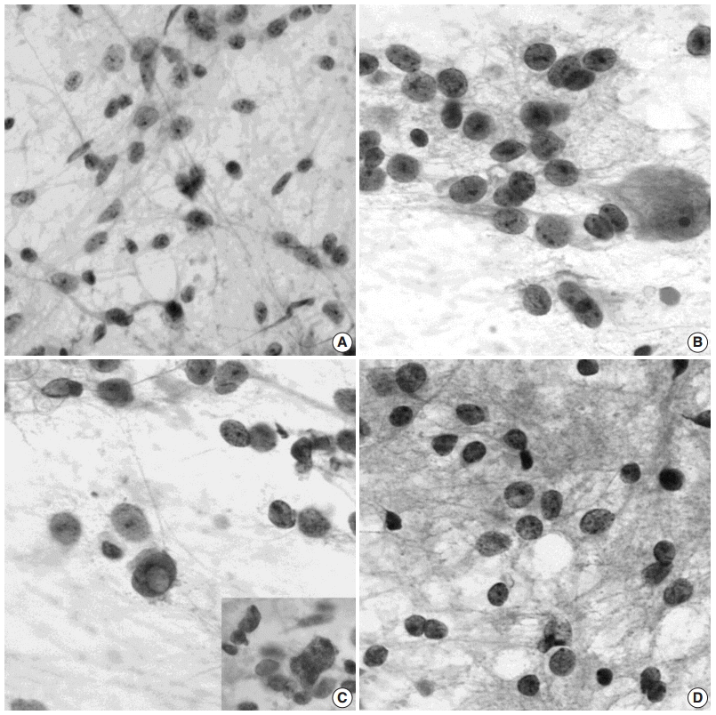
- The cytological features of squash preparations of DNT during intraoperative consultation are fairly characteristic and reliable for correct intraoperative diagnosis, which helps to determine the appropriate neurosurgical procedure. The floating neurons and extracellular mucin that are typical of DNT are more easily demonstrated in cytological preparations than in frozen sections [34] (Fig. 7A, B). OLCs appear as aggregates or are dispersed around abundant arborizing capillaries or in the mucinous or focal fibrillary background. Round to oval naked nuclei of OLCs are larger than non-neopalstic and neoplastic oligodendrocytes. Cytologically, distinguishing features of OLCs from oligodendrogliomas include frequent indentation of the nuclear membrane and multiple, small nucleoli (Fig. 7C). The nuclei of oligodendrogliomas are round with a smooth outline and contain one or two occasional nucleoli (Fig. 7D). The presence of eosinophilic granular bodies in the background favors DNTs rather than oligodendrogliomas.
SQUASH CYTOLOGICAL FEATURES
- Histologically, the recognition of a unique, specific GN element in brain tumor samples from patients with medically intractable, chronic epilepsy serves as a diagnostic feature for complex or simple forms of DNT. The appreciation of unique histological features may be difficult in fragmented specimens. The nonspecific forms, which lack specific GN elements and multi-nodular architecture, are often not distinguished from epilepsy-associated gliomas. Prominent perineuronal satellitosis and subpial aggregates of tumor cells favor gliomas, although a limited degree of secondary structure formation can be seen in DNTs. Distinguishing DNTs, particularly simple and nonspecific types, from gangliogliomas is often difficult because of overlap in their clinicopathological features, including an association with FCD. Gangliogliomas can be distinguished by the presence of neuronal atypia, perivascular lymphocytic infiltration, and reticulin networks surrounding glial cells. DNTs may rarely show perivascular lymphocytic infiltration and mild neuronal atypia, but they do not show reticulin deposition within tumors.
- It is important to differentiate DNTs from gliomas or gangliogliomas that may undergo malignant transformation. Combined immunostains for CD34 and MAP2 can facilitate the differential diagnosis between DNTs and epilepsy-associated tumors that mimic DNTs [8]. Nonspecific DNTs and gangliogliomas share similar immunoreactivity patterns for CD34, but show different immunoreactivity for MAP2 in their glial components. MAP2 immunostaining is faintly positive or negative in the glial component of gangliogliomas, whereas it is frequently seen in DNTs. Most ordinary glial tumors, such as astrocytomas and oligodendrogliomas, are diffusely positive for MAP2 but negative for CD34. The “isomorphic subtype of long-term epilepsy-associated astrocytomas” described by Blumcke et al. [35] shows the absence of both MAP2 and CD34 immunoreactivity. Since BRAFV600E mutations have not been detected in oligodendrogliomas, identification of BRAFV600E mutation by immunohistochemistry or molecular study has diagnostic utility for distinguishing DNT from oligodendroglioma [9].
DIFFERENTIAL DIAGNOSIS
- In a study of 13 DNTs, none of the tumors showed 1p19q deletion, IDH1-2, or p53 mutation [36], but a large series of DNTs found IDH1 mutation in three cases, loss of heterozygosity (LOH) 1p/19q in 10 cases, isolated LOH 19q in two cases, LOH 10q (PTEN locus) in three cases, and combined LOH 1p/19q and 10q in one case [6]. Other studies, however, including this one, did not show IDH1 mutation [9,26]. IDH2 mutation or epidermal growth factor receptor amplification has not been identified. Recently, BRAFV600E mutations were identified in 30%–51% of DNTs [9,31]. In our study, the frequency of BRAFV600E in DNTs was related to the location of tumors and was more frequent in extratemporal location (68.2% vs 37.9%). BRAFV600E mutation was more common in classical DNTs (59.4%) than nonspecific DNTs (36.8%). Recently, copy number aberrations (CNAs) have been identified in DNTs, and the most frequent are gains at chromosomes 5 and 7, often concurrent, and at chromosome 6. CNAs in DNTs are not correlated with their histological subtypes, CD34 expression or clinical features [37].
GENETICS
- DNTs were initially believed to be a hamartoma in nature [2-4], but recent identification of genetic alterations in DNTs, including classical and nonspecific types, indicate that this tumor is a neoplastic condition. The histogenesis of DNTs remains unknown. The developmental origin from the secondary germinal layer has been proposed based on the mixed cellularity of DNTs, their preponderance in the temporal lobe, and their association with FCD [2]. The origin of DNTs from pluripotent precursor cells is also suggested based on ultrastructural findings of OLCs showing neuronal and glial differentiation. This hypothesis is supported by the immunoexpression of both nestin and MAP2 in OLCs, which has been identified in neural and glial precursor cells during human development. Higher expression levels of nestin, MAP2 and CD34, stem cell markers, in nonspecific rather than simple or complex DNTs suggests an earlier developmental origin of the nonspecific DNTs [8,30].
- Because DNTs share CD34 expression, BRAFV600E mutations, and chromosomal copy number profiles with gangliogliomas, and occurrence of composite tumors with ganglioglioma and DNT, both tumors may represent different morphological variants of the same tumor entity [38].
HISTOGENESIS
Acknowledgments
Fig. 1.Three different types of magnetic resonance imaging in dysembryoplastic neuroepithelial tumors. (A, B) Type 1 shows a well-delineated, polycystic-like tumor with strongly hypointense on T1- and hyperintense on T2-weighted images. (C, D) Type 2 shows a nodular-like, heterogeneous lesion. (E, F) Type 3 shows a poorly delineated, dysplastic-like, iso/hyposignal T1 with gray-white matter blurring. Type 1 is mainly found in simple or complex forms, and type 2 and 3 are observed in nonspecific forms. (A, C, E) T1-weighted images. (B, D, F) T2 FLARE images.
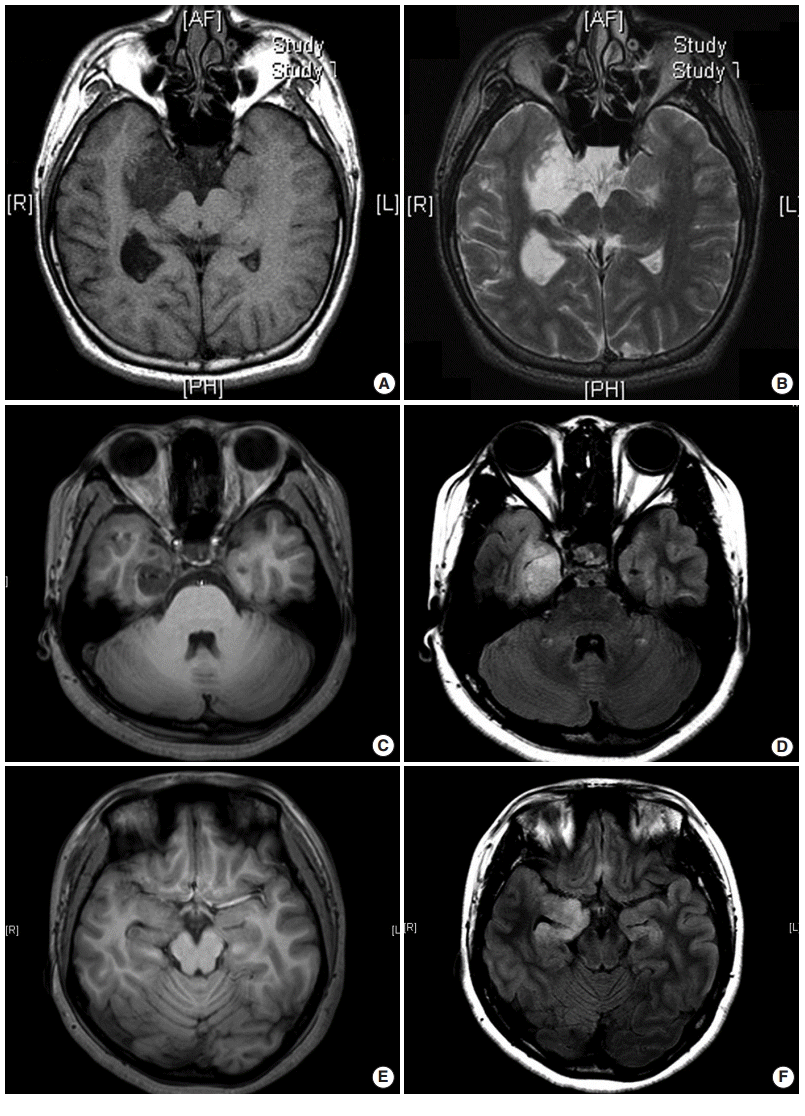

Fig. 2. Gross findings of dysembryoplastic neuroepithelial tumors (DNTs). (A) A hippocampectomy specimen shows a well circumscribed, gray white mass with two small satellite nodules. (B) On the cut section, a complex type of DNT shows multiple gray-white or gelatinous nodules affecting the cortex and white matter. (C) Nonspecific DNT shows a poorly demarcated, cortical thickening with underlying area of white matter rarefaction and cyst formation.
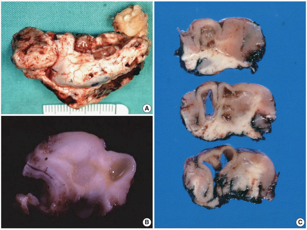

Fig. 3.Histopathological findings of dysembryoplastic neuroepithelial tumors (DNTs). (A) Multi-nodular appearance typical of complex DNTs. (B) Column arrangement of oligodendroglioma-like tumor cells (OLCs) in the specific glioneuronal element. (C) The characteristic appearance of DNTs with OLCs and mature neurons. (D) Targetoid structure of the specific glioneuronal element. (E) Floating neurons in the mucinous matrix. (F) The histology of glial nodules resembling oligodendroglioma. (G) This glial nodule with predominantly astrocytic differentiation. (H)Nuclear pleomorphism in glial component of DNTs. Histopathological findings of dysembryoplastic neuroepithelial tumors (DNTs). (I) A poorly demarcated cortical lesion of nonspecific DNT. (J) The similar histology of nonspecific DNTs to that observed within the glial nodules of complex DNTs.
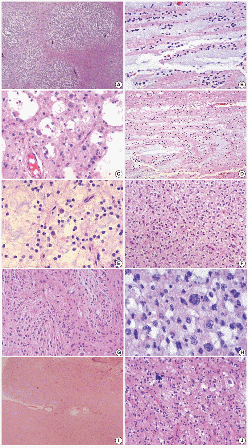

Fig. 4. Immunohistochemical findings of dysembryoplastic neuroepithelial tumors (DNTs). (A, B) Floating neurons are positive for synaptophysin (A) and phosphorylated neurofilament (B). (C, D) The oligodendroglioma-like cells (OLCs) are diffusely positive for S-100 (C) but negative for glial fibrillary acidic protein (D). (E) Nonspecific DNTs show slightly decreased synaptophysin granular staining compared with that seen in the adjacent normal cortex. (F) In the specific glioneuronal element, CD34 is expressed along the perikarya and in membrane of the floating neurons, pericellular stroma, and cytoplasm of OLCs. (G, H) The specific glioneuronal element shows cluster staining pattern of CD34 (G), while diffuse CD34 immunoreactivity in glial nodules of complex DNTs (H).
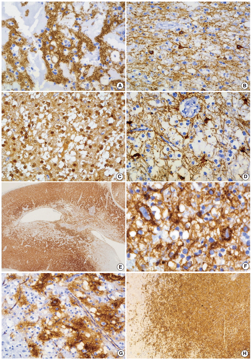

Fig. 5.The histology and CD34 immunoreactivity of focal cortical dysplasia (FCD) in the peritumoral cortex. (A) Dysplastic neurons and abnormalities in cortical lamination are observed. (B, C) CD34 immunoreactivity patterns of FCD are similar to that seen in dysembryoplastic neuroepithelial tumors.
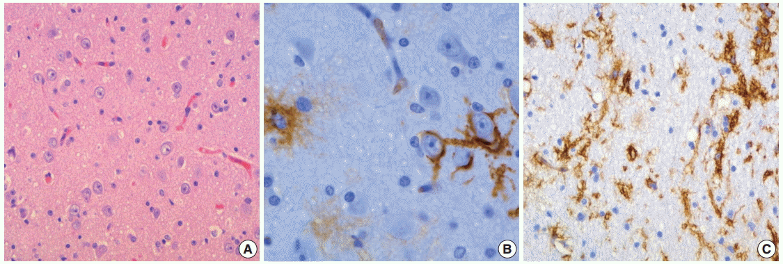

Fig. 6. Ultrastructural findings of dysembryoplastic neuroepithelial tumors. (A) Oligodendroglioma-like cells (OLCs) show oval nuclei with small indentation, marginal aggregates of heterochromatin, and their cytoplasm with scanty organelles (×7,000). (B, C) Neuropil-like network of cellular process with a synaptic contact (arrow) (B, ×17,000) and scant dense core granules (arrow) (C, ×30,000) indicates neuronal differentiation of OLCs. (D) A few OLCs contain ribosome-lamellae complex inclusions (arrow) (×7,000).
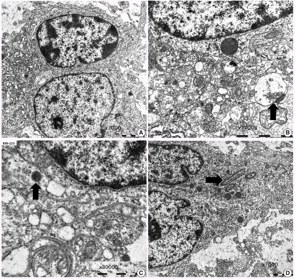

Fig. 7.Squash cytological findings of dysembryoplastic neuroepithelial tumors. (A) Squash preparation shows round, oval to elongated naked nuclei in the mucinous background. (B) The nuclei of oligodendroglioma-like cells (OLCs) are irregular with small indentations or deep grooves, and fine, granular chromatin and 1–4 small nucleoli. There is a large, normal-looking neuron in the mucinous background. (C) OLCs shows multinuclear giant cell formation (inset) and an intranuclear pseudoinclusion. (D) Squash preparation of oligodendroglioma shows smaller, dark nuclei without nucleoli and larger nuclei with granular chromatin and micronucleoli.


- 1. Burneo JG, Tellez-Zenteno J, Steven DA, et al. Adult-onset epilepsy associated with dysembryoplastic neuroepithelial tumors. Seizure 2008; 17: 498-504. ArticlePubMed
- 2. Daumas-Duport C, Scheithauer BW, Chodkiewicz JP, Laws ER Jr, Vedrenne C. Dysembryoplastic neuroepithelial tumor: a surgically curable tumor of young patients with intractable partial seizures. Report of thirty-nine cases. Neurosurgery 1988; 23: 545-56. PubMed
- 3. Daumas-Duport C. Dysembryoplastic neuroepithelial tumours. Brain Pathol 1993; 3: 283-95. ArticlePubMed
- 4. Daumas-Duport C, Varlet P, Bacha S, Beuvon F, Cervera-Pierot P, Chodkiewicz JP. Dysembryoplastic neuroepithelial tumors: nonspecific histological forms: a study of 40 cases. J Neurooncol 1999; 41: 267-80. ArticlePubMedPDF
- 5. Louis DN, Ohgaki H, Wiestler OD, et al. The 2007 WHO classification of tumours of the central nervous system. Acta Neuropathol 2007; 114: 97-109. ArticlePubMedPMCPDF
- 6. Thom M, Toma A, An S, et al. One hundred and one dysembryoplastic neuroepithelial tumors: an adult epilepsy series with immunohistochemical, molecular genetic, and clinical correlations and a review of the literature. J Neuropathol Exp Neurol 2011; 70: 859-78. ArticlePubMed
- 7. Honavar M, Janota I, Polkey CE. Histological heterogeneity of dysembryoplastic neuroepithelial tumour: identification and differential diagnosis in a series of 74 cases. Histopathology 1999; 34: 342-56. ArticlePubMedPDF
- 8. Bodi I, Selway R, Bannister P, et al. Diffuse form of dysembryoplastic neuroepithelial tumour: the histological and immunohistochemical features of a distinct entity showing transition to dysembryoplastic neuroepithelial tumour and ganglioglioma. Neuropathol Appl Neurobiol 2012; 38: 411-25. ArticlePubMed
- 9. Sung CO, Suh YL, Hong SC. CD34 and microtubule-associated protein 2 expression in dysembryoplastic neuroepithelial tumours with an emphasis on dual expression in non-specific types. Histopathology 2011; 59: 308-17. ArticlePubMed
- 10. Lee D, Cho YH, Kang SY, Yoon N, Sung CO, Suh YL. BRAF V600E mutations are frequent in dysembryoplastic neuroepithelial tumors and subependymal giant cell astrocytomas. J Surg Oncol 2015; 111: 359-64. ArticlePubMedPDF
- 11. Chassoux F, Landré E, Mellerio C, Laschet J, Devaux B, DaumasDuport C. Dysembryoplastic neuroepithelial tumors: epileptogenicity related to histologic subtypes. Clin Neurophysiol 2013; 124: 1068-78. ArticlePubMed
- 12. Devaux B, Chassoux F, Guenot M, et al. Epilepsy surgery in France. Neurochirurgie 2008; 54: 453-65. ArticlePubMed
- 13. Blumcke I, Aronica E, Urbach H, Alexopoulos A, Gonzalez-Martinez JA. A neuropathology-based approach to epilepsy surgery in brain tumors and proposal for a new terminology use for long-term epilepsy-associated brain tumors. Acta Neuropathol 2014; 128: 39-54. ArticlePubMedPMCPDF
- 14. Piao YS, Lu DH, Chen L, et al. Neuropathological findings in intractable epilepsy: 435 Chinese cases. Brain Pathol 2010; 20: 902-8. ArticlePubMedPMC
- 15. Kirkpatrick PJ, Honavar M, Janota I, Polkey CE. Control of temporal lobe epilepsy following en bloc resection of low-grade tumors. J Neurosurg 1993; 78: 19-25. ArticlePubMed
- 16. Rickert CH, Paulus W. Epidemiology of central nervous system tumors in childhood and adolescence based on the new WHO classification. Childs Nerv Syst 2001; 17: 503-11. ArticlePubMedPDF
- 17. Spalice A, Ruggieri M, Grosso S, et al. Dysembryoplastic neuroepithelial tumors: a prospective clinicopathologic and outcome study of 13 children. Pediatr Neurol 2010; 43: 395-402. ArticlePubMed
- 18. Chang EF, Christie C, Sullivan JE, et al. Seizure control outcomes after resection of dysembryoplastic neuroepithelial tumor in 50 patients. J Neurosurg Pediatr 2010; 5: 123-130. ArticlePubMed
- 19. Lee J, Lee BL, Joo EY, et al. Dysembryoplastic neuroepithelial tumors in pediatric patients. Brain Dev 2009; 31: 671-81. ArticlePubMed
- 20. Leung SY, Gwi E, Ng HK, Fung CF, Yam KY. Dysembryoplastic neuroepithelial tumor: a tumor with small neuronal cells resembling oligodendroglioma. Am J Surg Pathol 1994; 18: 604-14. PubMed
- 21. Cervera-Pierot P, Varlet P, Chodkiewicz JP, Daumas-Duport C. Dysembryoplastic neuroepithelial tumors located in the caudate nucleus area: report of four cases. Neurosurgery 1997; 40: 1065-9. ArticlePubMed
- 22. Fujimoto K, Ohnishi H, Tsujimoto M, Hoshida T, Nakazato Y. Dysembryoplastic neuroepithelial tumor of the cerebellum and brainstem: case report. J Neurosurg 2000; 93: 487-9. PubMed
- 23. Ongürü O, Deveci S, Sirin S, Timurkaynak E, Günhan O. Dysembryoplastic neuroepithelial tumor in the left lateral ventricle. Minim Invasive Neurosurg 2003; 46: 306-9. ArticlePubMed
- 24. Hasselblatt M, Kurlemann G, Rickert CH, et al. Familial occurrence of dysembryoplastic neuroepithelial tumor. Neurology 2004; 62: 1020-1. ArticlePubMed
- 25. Ranger A, Diosy D. Seizures in children with dysembryoplastic neuroepithelial tumors of the brain: a review of surgical outcomes across several studies. Childs Nerv Syst 2015; 31: 847-55. ArticlePubMedPMCPDF
- 26. Chassoux F, Daumas-Duport C. Dysembryoplastic neuroepithelial tumors: where are we now? Epilepsia 2013; 54 Suppl 9: 129-34. ArticlePubMed
- 27. Chassoux F, Rodrigo S, Mellerio C, et al. Dysembryoplastic neuroepithelial tumors: an MRI-based scheme for epilepsy surgery. Neurology 2012; 79: 1699-707. ArticlePubMed
- 28. Moazzam AA, Wagle N, Shiroishi MS. Malignant transformation of DNETs: a case report and literature review. Neuroreport 2014; 25: 894-9. PubMed
- 29. Stanescu Cosson R, Varlet P, Beuvon F, et al. Dysembryoplastic neuroepithelial tumors: CT, MR findings and imaging follow-up: a study of 53 cases. J Neuroradiol 2001; 28: 230-40. PubMed
- 30. Sung CO, Suh YL. Different pattern of expression of nestin in the non-specific form of dysembryoplastic neuroepithelial tumors compared to the simple and complex forms. J Neurooncol 2009; 92: 7-13. ArticlePubMedPDF
- 31. Chappé C, Padovani L, Scavarda D, et al. Dysembryoplastic neuroepithelial tumors share with pleomorphic xanthoastrocytomas and gangliogliomas BRAF(V600E) mutation and expression. Brain Pathol 2013; 23: 574-83. PubMedPMC
- 32. Hirose T, Scheithauer BW, Lopes MB, VandenBerg SR. Dysembryoplastic neuroeptihelial tumor (DNT): an immunohistochemical and ultrastructural study. J Neuropathol Exp Neurol 1994; 53: 184-95. PubMed
- 33. Biernat W, Liberski PP, Kordek R, Zakrzewski K, Polis L, Budka H. Dysembryoplastic neuroectodermal tumor: an ultrastructural study of six cases. Ultrastruct Pathol 2001; 25: 455-67. ArticlePubMed
- 34. Park JY, Suh YL, Han J. Dysembryoplastic neuroepithelial tumor: features distinguishing it from oligodendroglioma on cytologic squash preparations. Acta Cytol 2003; 47: 624-9. PubMed
- 35. Blümcke I, Luyken C, Urbach H, Schramm J, Wiestler OD. An isomorphic subtype of long-term epilepsy-associated astrocytomas associated with benign prognosis. Acta Neuropathol 2004; 107: 381-8. ArticlePubMedPDF
- 36. Padovani L, Colin C, Fernandez C, et al. Search for distinctive markers in DNT and cortical grade II glioma in children: same clinicopathological and molecular entities? Curr Top Med Chem 2012; 12: 1683-92. ArticlePubMed
- 37. Prabowo AS, van Thuijl HF, Scheinin I, et al. Landscape of chromosomal copy number aberrations in gangliogliomas and dysembryoplastic neuroepithelial tumours. Neuropathol Appl Neurobiol 2015; 41: 743-55. ArticlePubMed
- 38. Prayson RA, Napekoski KM. Composite ganglioglioma/dysembryoplastic neuroepithelial tumor: a clinicopathologic study of 8 cases. Hum Pathol 2012; 43: 1113-8. ArticlePubMed
REFERENCES
Figure & Data
References
Citations
Citations to this article as recorded by 

- A Case of Dysembryoplastic Neuroepithelial Tumor in an HIV-Positive Adult: Diagnostic Lessons for Clinicians
Mariana Lobo, Susana Viana, Andreia Sá Lima, Isabel Monteiro, Carolina M Cerqueira, Frederico Duarte, Luís M Ribeiro, Sara Camões
Cureus.2026;[Epub] CrossRef - An Imaging Review of Common Pediatric Brain Tumors
Joseph Yang, Brandon Collins, Matthew Beniuk, Alexandra Hodder, Dani Bahnam, Angela Pickles
Roentgen Ray Review.2025;[Epub] CrossRef - Ruptured intratumoral arteriovenous malformation in a patient with dysembryoplastic neuroepithelial tumor: A case report
Takashi Aoka, Masaaki Nishimoto, Hideki Ogiwara
Surgical Neurology International.2025; 16: 375. CrossRef - Pediatric Neuroglial Tumors: A Review of Ependymoma and Dysembryoplastic Neuroepithelial Tumor
Melissa Arfuso, Sandeepkumar Kuril, Harshal Shah, Derek Hanson
Pediatric Neurology.2024; 156: 139. CrossRef - From bedside to bench: New insights in epilepsy‐associated tumors based on recent classification updates and animal models on brain tumor networks
Silvia Cases‐Cunillera, Lea L. Friker, Philipp Müller, Albert J. Becker, Gerrit H. Gielen
Molecular Oncology.2024; 18(12): 2951. CrossRef - Imaging of pediatric glioneuronal and neuronal tumors
Vivek Pai, Suzanne Laughlin, Birgit Ertl-Wagner
Child's Nervous System.2024; 40(10): 3007. CrossRef - Dysembryoplastic Neuroepithelial Tumor: A Case Report of A Benign Intracranial Lesion Masquerading as Seizure Disorder
Garima S Agarwal, Anil K Agrawal, Daksh Singhal, Jayashree Bhawani
Cureus.2024;[Epub] CrossRef - Super T2-FLAIR mismatch sign: a prognostic imaging biomarker for non-enhancing astrocytoma, IDH-mutant
Iori Ozono, Shumpei Onishi, Ushio Yonezawa, Akira Taguchi, Novita Ikbar Khairunnisa, Vishwa Jeet Amatya, Fumiyuki Yamasaki, Yukio Takeshima, Nobutaka Horie
Journal of Neuro-Oncology.2024; 169(3): 571. CrossRef - Genotype-relevant neuroimaging features in low-grade epilepsy-associated tumors
Keiya Iijima, Hiroyuki Fujii, Fumio Suzuki, Kumiko Murayama, Yu-ichi Goto, Yuko Saito, Terunori Sano, Hiroyoshi Suzuki, Hajime Miyata, Yukio Kimura, Takuma Nakashima, Hiromichi Suzuki, Masaki Iwasaki, Noriko Sato
Frontiers in Neurology.2024;[Epub] CrossRef - Extra-temporal pediatric low-grade gliomas and epilepsy
José Hinojosa, Victoria Becerra, Santiago Candela-Cantó, Mariana Alamar, Diego Culebras, Carlos Valencia, Carlos Valera, Jordi Rumiá, Jordi Muchart, Javier Aparicio
Child's Nervous System.2024; 40(10): 3309. CrossRef - Atypical Presentation of Dysembryoplastic Neuroepithelial Tumor
Varis S. Khalilov, Aleksey N. Kislyakov, Natalia A. Medvedeva, Natalia S. Serova
Annals of Clinical and Experimental Neurology.2024; 18(3): 109. CrossRef - Unusual low-grade neuroepithelial tumour in a child
Leia Salongo, Ali Nael, Pournima Navalkele, John Ross Crawford
BMJ Case Reports.2024; 17(10): e262692. CrossRef - Glioneuronal and Neuronal Tumors: Who? When? Where? An Update Based on the 2021 World Health Organization Classification
A.S. Ayres, G.A. Bandeira, S.F. Ferraciolli, J.T. Takahashi, R.A. Moreno, L.F. de Souza Godoy, Y.R. Casal, L.G.C.A. de Lima, F.P. Frasseto, L.T. Lucato
Neurographics.2023; 13(1): 1. CrossRef - Biological functions of the Olig gene family in brain cancer and therapeutic targeting
Jenny I. Szu, Igor F. Tsigelny, Alexander Wojcinski, Santosh Kesari
Frontiers in Neuroscience.2023;[Epub] CrossRef - Aspekte der Bildgebung des Hippokampus
Isabela S. Alves, Artur M. N. Coutinho, Ana Vieira, Bruno P. Rocha, Ula L. Passos, Vinicius T. Gonçalves, Paulo D. S. Silva, Malia X. Zhan, Paula C. Pinho, Daniel S. Delgado, Marcos F. L. Docema, Hae W. Lee, Bruno A. Policeni, Claudia C. Leite, Maria G. M
Neuroradiologie Scan.2023; 13(03): 197. CrossRef - T2-FLAIR Mismatch Sign in Pediatric Low-Grade Glioma
M.W. Wagner, L. Nobre, K. Namdar, F. Khalvati, U. Tabori, C. Hawkins, B.B. Ertl-Wagner
American Journal of Neuroradiology.2023; 44(7): 841. CrossRef - Clinicopathological features of dysembryoplastic neuroepithelial tumor: a case series
Shabina Rahim, Nasir Ud Din, Jamshid Abdul-Ghafar, Qurratulain Chundriger, Poonum Khan, Zubair Ahmad
Journal of Medical Case Reports.2023;[Epub] CrossRef - Imaging Aspects of the Hippocampus
Isabela S. Alves, Artur M. N. Coutinho, Ana P. F. Vieira, Bruno P. Rocha, Ula L. Passos, Vinicius T. Gonçalves, Paulo D. S. Silva, Malia X. Zhan, Paula C. Pinho, Daniel S. Delgado, Marcos F. L. Docema, Hae W. Lee, Bruno A. Policeni, Claudia C. Leite, Mari
RadioGraphics.2022; 42(3): 822. CrossRef - Dysembryoplastic neuroepithelial tumors: A single-institutional series with special reference to glutamine synthetase expression
Chiara Caporalini, Mirko Scagnet, Selene Moscardi, Gioia Di Stefano, Gianna Baroni, Flavio Giordano, Federico Mussa, Carmen Barba, Iacopo Sardi, Lorenzo Genitori, Anna Maria Buccoliero
Annals of Diagnostic Pathology.2021; 54: 151774. CrossRef - Unusual case of occipital lobe dysembryoplastic neuroepithelial tumour with GNAi1-BRAF fusion
Jennifer H Yang, Denise M Malicki, Michael L Levy, John Ross Crawford
BMJ Case Reports.2021; 14(1): e241440. CrossRef - Malformations of Cortical Development, Cognitive Involvementand Epilepsy: A Single Institution Experience in 19 Young Patients
Valeria Venti, Maria Chiara Consentino, Pierluigi Smilari, Filippo Greco, Claudia Francesca Oliva, Agata Fiumara, Raffaele Falsaperla, Martino Ruggieri, Piero Pavone
Children.2021; 8(8): 637. CrossRef - A Case of Dysembryoplastic Neuroepithelial Tumor in an Adolescent Male
Marcel Yibirin, Diana De Oliveira, Isabella Suarez, Gabriela Lombardo, Carlos Perez
Cureus.2021;[Epub] CrossRef - Clinical and histopathological profile of dysembryoplastic neuroepithelial tumor
Pooja Gupta, Fouzia Siraj, Akanksha Malik, K. B. Shankar
Journal of Cancer Research and Therapeutics.2021; 17(4): 912. CrossRef - Neuroradiological and pathomorphological features of epilepsy associated brain tumors
V. S. Khalilov, A. A. Kholin, A. N. Kisyakov, N. A. Medvedeva, B. R. Bakaeva
Diagnostic radiology and radiotherapy.2021; 12(2): 7. CrossRef - Evaluación prequirúrgica mediante resonancia magnética funcional en pacientes con tumores neuroepiteliales disembrioplásicos: una serie de casos
Natalia García-Casares, Francisco Alfaro-Rubio, José Ramón Ramos-Rodríguez, Álvaro Ocaña-Ledesma, Bernarda Márquez-Márquez, Victoria E. Fernández-Sánchez, Guillermo Ibáñez-Botella, Miguel Ángel Arráez-Sánchez, Pedro J. Serrano-Castro
Neurocirugía.2020; 31(4): 158. CrossRef - Features of the neuroradiological picture of ganglioglioma on the example of 20 clinical cases
V.S. Khalilov, A.A. Kholin, Kh.Sh. Gazdieva, A.N. Kislyakov, N.N. Zavadenko
Zhurnal nevrologii i psikhiatrii im. S.S. Korsakova.2020; 120(11): 90. CrossRef - Malignant Glial Neuronal Tumors After West Nile Virus Neuroinvasive Disease: A Coincidence or a Clue?
Akanksha Sharma, Marie F. Grill, Scott Spritzer, A. Arturo Leis, Mark Anderson, Parminder Vig, Alyx B. Porter
The Neurohospitalist.2019; 9(3): 160. CrossRef - Particularities in differential diagnostics of epileptogenic brain malformations on the low-field MRI-device
V. S. Khalilov, A. A. Kholin, B. R. Bakaeva, M. Yu. Bobylova, Kh. Sh. Gazdieva
Russian Journal of Child Neurology.2019; 13(4): 23. CrossRef - Dysembryoplastic Neuroepithelial Tumors: What You Need to Know
Sabino Luzzi, Angela Elia, Mattia Del Maestro, Samer K. Elbabaa, Sergio Carnevale, Francesco Guerrini, Massimo Caulo, Patrizia Morbini, Renato Galzio
World Neurosurgery.2019; 127: 255. CrossRef - The miR‐139‐5p regulates proliferation of supratentorial paediatric low‐grade gliomas by targeting the PI3K/AKT/mTORC1 signalling
G. Catanzaro, Z. M. Besharat, E. Miele, M. Chiacchiarini, A. Po, A. Carai, C. E. Marras, M. Antonelli, M. Badiali, A. Raso, S. Mascelli, D. Schrimpf, D. Stichel, M. Tartaglia, D. Capper, A. von Deimling, F. Giangaspero, A. Mastronuzzi, F. Locatelli, E. Fe
Neuropathology and Applied Neurobiology.2018; 44(7): 687. CrossRef - Dysembryoplastic Neuroectodermal Tumor: An Analysis from the Surveillance, Epidemiology, and End Results Program, 2004–2013
Ha Son Nguyen, Ninh Doan, Michael Gelsomino, Saman Shabani
World Neurosurgery.2017; 103: 380. CrossRef - Common Histologically Benign Tumors of the Brain
Roy E. Strowd, Jaishri O. Blakeley
Continuum.2017; 23(6): 1680. CrossRef
 PubReader
 ePub Link
-
 Cite this Article
- Cite this Article
-
- Close
- Download Citation
- Close
- Figure
Dysembryoplastic Neuroepithelial Tumors


Fig. 1. Three different types of magnetic resonance imaging in dysembryoplastic neuroepithelial tumors. (A, B) Type 1 shows a well-delineated, polycystic-like tumor with strongly hypointense on T1- and hyperintense on T2-weighted images. (C, D) Type 2 shows a nodular-like, heterogeneous lesion. (E, F) Type 3 shows a poorly delineated, dysplastic-like, iso/hyposignal T1 with gray-white matter blurring. Type 1 is mainly found in simple or complex forms, and type 2 and 3 are observed in nonspecific forms. (A, C, E) T1-weighted images. (B, D, F) T2 FLARE images.
Fig. 2. Gross findings of dysembryoplastic neuroepithelial tumors (DNTs). (A) A hippocampectomy specimen shows a well circumscribed, gray white mass with two small satellite nodules. (B) On the cut section, a complex type of DNT shows multiple gray-white or gelatinous nodules affecting the cortex and white matter. (C) Nonspecific DNT shows a poorly demarcated, cortical thickening with underlying area of white matter rarefaction and cyst formation.
Fig. 3. Histopathological findings of dysembryoplastic neuroepithelial tumors (DNTs). (A) Multi-nodular appearance typical of complex DNTs. (B) Column arrangement of oligodendroglioma-like tumor cells (OLCs) in the specific glioneuronal element. (C) The characteristic appearance of DNTs with OLCs and mature neurons. (D) Targetoid structure of the specific glioneuronal element. (E) Floating neurons in the mucinous matrix. (F) The histology of glial nodules resembling oligodendroglioma. (G) This glial nodule with predominantly astrocytic differentiation. (H)Nuclear pleomorphism in glial component of DNTs. Histopathological findings of dysembryoplastic neuroepithelial tumors (DNTs). (I) A poorly demarcated cortical lesion of nonspecific DNT. (J) The similar histology of nonspecific DNTs to that observed within the glial nodules of complex DNTs.
Fig. 4. Immunohistochemical findings of dysembryoplastic neuroepithelial tumors (DNTs). (A, B) Floating neurons are positive for synaptophysin (A) and phosphorylated neurofilament (B). (C, D) The oligodendroglioma-like cells (OLCs) are diffusely positive for S-100 (C) but negative for glial fibrillary acidic protein (D). (E) Nonspecific DNTs show slightly decreased synaptophysin granular staining compared with that seen in the adjacent normal cortex. (F) In the specific glioneuronal element, CD34 is expressed along the perikarya and in membrane of the floating neurons, pericellular stroma, and cytoplasm of OLCs. (G, H) The specific glioneuronal element shows cluster staining pattern of CD34 (G), while diffuse CD34 immunoreactivity in glial nodules of complex DNTs (H).
Fig. 5. The histology and CD34 immunoreactivity of focal cortical dysplasia (FCD) in the peritumoral cortex. (A) Dysplastic neurons and abnormalities in cortical lamination are observed. (B, C) CD34 immunoreactivity patterns of FCD are similar to that seen in dysembryoplastic neuroepithelial tumors.
Fig. 6. Ultrastructural findings of dysembryoplastic neuroepithelial tumors. (A) Oligodendroglioma-like cells (OLCs) show oval nuclei with small indentation, marginal aggregates of heterochromatin, and their cytoplasm with scanty organelles (×7,000). (B, C) Neuropil-like network of cellular process with a synaptic contact (arrow) (B, ×17,000) and scant dense core granules (arrow) (C, ×30,000) indicates neuronal differentiation of OLCs. (D) A few OLCs contain ribosome-lamellae complex inclusions (arrow) (×7,000).
Fig. 7. Squash cytological findings of dysembryoplastic neuroepithelial tumors. (A) Squash preparation shows round, oval to elongated naked nuclei in the mucinous background. (B) The nuclei of oligodendroglioma-like cells (OLCs) are irregular with small indentations or deep grooves, and fine, granular chromatin and 1–4 small nucleoli. There is a large, normal-looking neuron in the mucinous background. (C) OLCs shows multinuclear giant cell formation (inset) and an intranuclear pseudoinclusion. (D) Squash preparation of oligodendroglioma shows smaller, dark nuclei without nucleoli and larger nuclei with granular chromatin and micronucleoli.
Fig. 1.
Fig. 2.
Fig. 3.
Fig. 4.
Fig. 5.
Fig. 6.
Fig. 7.
Dysembryoplastic Neuroepithelial Tumors

 E-submission


