 E-submission
Articles
- Page Path
- HOME > J Pathol Transl Med > Volume 51(4); 2017 > Article
-
Original Article
Loss of Progesterone Receptor Expression Is an Early Tumorigenesis Event Associated with Tumor Progression and Shorter Survival in Pancreatic Neuroendocrine Tumor Patients - Sung Joo Kim, Soyeon An, Jae Hoon Lee1, Joo Young Kim2, Ki-Byung Song1, Dae Wook Hwang1, Song Cheol Kim1, Eunsil Yu, Seung-Mo Hong
-
Journal of Pathology and Translational Medicine 2017;51(4):388-395.
DOI: https://doi.org/10.4132/jptm.2017.03.19
Published online: June 8, 2017
Department of Pathology, Asan Medical Center, University of Ulsan College of Medicine, Seoul, Korea
1Division of Hepatobiliary and Pancreatic Surgery, Asan Medical Center, University of Ulsan College of Medicine, Seoul, Korea
2Department of Pathology, Korea University Anam Hospital, Korea University College of Medicine, Seoul, Korea
- Corresponding Author Seung-Mo Hong, MD, PhD Department of Pathology, Asan Medical Center, University of Ulsan College of Medicine, 88 Olympic-ro 43-gil, Songpa-gu, Seoul 05505, Korea Tel: +82-2-3010-4558 Fax: +82-2-472-7898 E-mail: smhong28@gmail.com
© 2017 The Korean Society of Pathologists/The Korean Society for Cytopathology
This is an Open Access article distributed under the terms of the Creative Commons Attribution Non-Commercial License (http://creativecommons.org/licenses/by-nc/4.0) which permits unrestricted non-commercial use, distribution, and reproduction in any medium, provided the original work is properly cited.
Abstract
-
Background
- Pancreatic neuroendocrine tumors (PanNETs) are the second most common pancreatic neoplasms and there is no well-elucidated biomarker to stratify their detection and prognosis. Previous studies have reported that progesterone receptor (PR) expression status was associated with poorer survival in PanNET patients.
-
Methods
- To validate previous studies, PR protein expression was assessed in 21 neuroendocrine microadenomas and 277 PanNETs and compared with clinicopathologic factors including patient survival.
-
Results
- PR expression was gradually decreased from normal islets (49/49 cases, 100%) to neuroendocrine microadenoma (14/21, 66.6%) to PanNETs (60/277, 21.3%; p < .001). PanNETs with loss of PR expression were associated with increased tumor size (p < .001), World Health Organization grade (p = .001), pT classification (p < .001), perineural invasion (p = .028), lymph node metastasis (p = .004), activation of alternative lengthening of telomeres (p = .005), other peptide hormonal expression (p < .001) and ATRX/DAXX expression (p = .015). PanNET patients with loss of PR expression (5-year survival rate, 64.1%) had significantly poorer recurrence-free survival outcomes than those with intact PR expression (90%) by univariate (p = .012) but not multivariate analyses. Similarly, PanNET patients with PR expression loss (5-year survival rate, 76%) had significantly poorer overall survival by univariate (p = .015) but not multivariate analyses.
-
Conclusions
- Loss of PR expression was noted in neuroendocrine microadenomas and was observed in the majority of PanNETs. This was associated with increased grade, tumor size, and advanced pT and pN classification; and was correlated with decreased patient survival time by univariate but not multivariate analyses. Loss of PR expression can provide additional information on shorter disease-free survival in PanNET patients.
- Case selection
- After approval (2015-0387) from the Institutional Review Board of Asan Medical Center, the records of 277 surgically resected primary PanNETs and 21 sporadic neuroendocrine microadenomas resected between January 1995 and December 2015 were retrieved from the Department of Pathology at Asan Medical Center. Primary PanNETs were defined as well-differentiated, nonfunctional neuroendocrine neoplasms with diameters ≥0.5 cm, while neuroendocrine microadenomas were defined as well-differentiated, nonfunctional NETs with diameters <0.5 cm [1,18]. All PanNET cases were classified using the 2010 World Health Organization (WHO) classification scheme with mitotic activity and the Ki- 67 labeling index [1]. The Ki-67 labeling index was measured by manually assessing the tumor’s hottest spot in high-power fields after printing the captured image, as previously described [19]. A minimum of 500 tumor cells were included in the manual count. Poorly differentiated neuroendocrine carcinomas, such as small-cell carcinomas and large-cell carcinomas, were excluded. Pathological data, such as tumor size, extension, lymph node and distant metastases, and perineural and lymphovascular tumor invasion, were extracted from the pathology reports. The clinical data reviewed included patient age, gender, symptoms, and survival outcomes. The expression profiles of specific peptide hormones, including insulin, glucagon, gastrin, serotonin, somatostatin, and glucagon-like peptide 1; other proteins, such as ATRX and DAXX; and the ALT status were used as previously reported [12,14].
- Tissue microarray construction
- Tissue microarrays were constructed from archived, formalin-fixed, paraffin-embedded tissue blocks with a manual tissue microarrayer (Uni TMA Co. Ltd., Seoul, Korea), as previously described [12,14,20]. Briefly, three cores from the tumors and one core from the normal pancreatic parenchyma with a diameter of 2 mm were punched from donor blocks and placed in recipient blocks.
- Immunohistochemical staining
- Immunohistochemical labeling was performed at the immunohistochemical laboratory of the Department of Pathology, Asan Medical Center. In brief, 4-mm-thick tissue sections were deparaffinized and hydrated in xylene and serially diluted in ethanol. Endogenous peroxidase was blocked by incubation in 3% H2O2 for 10 minutes, and then heat-induced antigen retrieval was performed. Primary antibodies were used with a Benchmark autostainer (Ventana Medical Systems, Tucson, AZ, USA) in accordance with the manufacturer’s protocol. Sections were incubated at room temperature for 32 minutes in primary antibodies for PR (1:200, NCL-L-PGR-312, Novocastra, Newcastle upon Tyne, UK), synaptophysin (1:200, DiNona, Seoul, Korea), chromogranin (1:200, DAK-A3, DakoCytomation, Glostrup, Denmark), and Ki-67 (1:100, 7B11, Zymed, San Francisco, CA, USA). The sections were then labeled with an automated immunostaining system and processed with an iView DAB detection kit (Benchmark XT, Ventana Medical Systems). Immunostained sections were lightly counterstained with hematoxylin, dehydrated in ethanol, and cleared in xylene. Immunoreactivity was interpreted by light microscopic examination and independently evaluated by two pathologists, coauthors of this study (S.J.K. and S.M.H.), who were blind to the clinicopathologic information.
- Evaluation of PR labeling
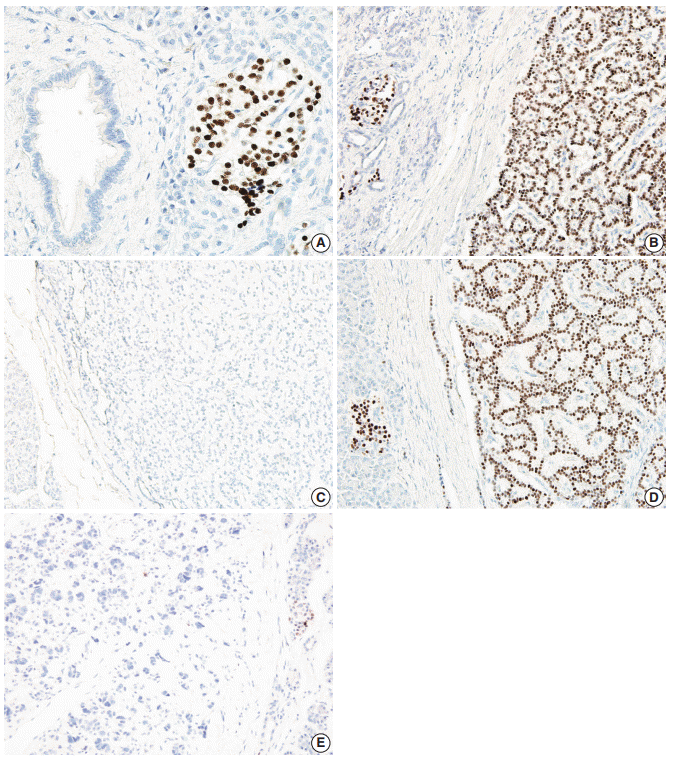
- Immunohistochemical labeling of the PR protein was scored using a previously described histological scoring system, which takes into account the size of the stained area and the intensity of the labeling. To be included in the analyses, a tumor had to have sufficient numbers of PR-labeled cells to permit quantification (>100 PR-positive tumor cells). The labeled area was scored from 0 to 4 for having <5%, 5%–25%, 26%–50%, 51%–75%, or >75% PR-positive cells, respectively. The intensity scale ranged from 0 to 2 as follows: 0, no labeling of tumor cells; 1, weak labeling; and 2, intense labeling, as previously described [21]. The total histological score (H-score) was calculated by multiplying the area score by the intensity score. The resulting H-score ranged from 0 to 8. We considered cases with an H-score below 2 to have loss of PR expression and cases with an H-score greater than or equal to 2 to have intact PR expression. PR labeling in normal islets was used as an internal positive control. Representative images of PR expression in normal islet and in PanNETs are depicted in Fig. 1. Immunohistochemical staining was evaluated by two independent pathologists (S.J.K. and S.M.H.).
- Statistical analyses
- SPSS software ver. 20.0 (IBM Corp., Chicago, IL, USA) was used for statistical analyses. The overall and recurrence-free survival times were calculated from the date of diagnosis of PanNET to that of death from any cause and from the date of diagnosis of PanNET to that of recurrence, respectively. Both overall and recurrence-free survival rates were calculated using the Kaplan-Meier method, and the association between overall survival rate and clinicopathologic factors was compared using the log-rank test. The correlations between PR expression and other prognostic factors were analyzed using the chi-square and Fisher exact tests. Possible prognostic factors associated with survival probability were calculated using the Cox’s proportional hazard regression model; p<.05 was considered statistically significant.
MATERIALS AND METHODS
- Patient characteristics
- Patient characteristics are summarized in Table 1. In the total cohort, 134 patients (49%) were male and 143 (51%) were female. The mean age of the patients was 52.3±12.7 years. According to the WHO classification, there were 85 G1, 95 G2, and 8 G3, respectively. The mean tumor size of the PanNETs and neuroendocrine microadenomas was 3.0±2.2 cm and 0.3±0.1 cm, respectively. In all, 126 cases were classified as pT1, while others had higher pT classifications (95 pT2, 52 pT3, and 4 pT4); 79 cases (28.5%) had lymphovascular invasion and 42 cases (15.2%) had perineural invasion. Metastasis to regional lymph nodes occurred in 39 cases (14.1%) and metastasis to distant organs at the surgical resection of PanNET was observed in 10 cases (3.6%). The median follow-up period was 38±35 months (range, 1 to 188 months).
- PR expression
- All of the normal pancreatic islets in the patient samples expressed the PR protein in various proportions. PR protein expression was observed in 63.5±9.8% of the endocrine cells in the islets. A representative image of PR expression in an islet is depicted in Fig. 1. This expression gradually decreased from normal islets (49/49, 100%) to neuroendocrine microadenoma (14/21, 66.6%) to PanNETs (60/277, 21.3%). The H-scores for PR in normal islets, neuroendocrine microadenomas, and PanNETs were 5.1±2.3, 3.0±3.3, and 1.3±2.5, respectively (p<.001) (Figs. 1, 2). The mean H-score of the PR expression loss group was 0.1±0.3, while that of the PR expression intact group was 5.8±2.3. The majority of PanNETs showed a loss of PR expression (218/277, 78.7%) (Fig. 1).
- Correlations between PR expression and clinicopathologic factors
- The associations between PR expression and clinicopathologic factors are summarized in Table 1. Loss of PR expression was more commonly observed in PanNETs with larger tumors (p<.001), a higher WHO grade (p=.001), higher Ki-67 labeling index (p=.004), higher pT classification (p<.001), frequent perineural invasion (p=.028), and regional lymph node metastasis (p=.004). Loss of PR expression was also strongly associated with other peptide hormonal expression (p<.001), loss of ATRX/DAXX expression (p=.015), and activation of ALT (p=.005). In addition, the loss of PR expression was marginally associated with lymphovascular invasion (p=.077) but not with age, gender, or distant metastasis.
- Survival analyses of PR expression
- The overall 5-year survival rate of 100% among the PanNET patients with intact PR expression was significantly better than for patients with PR expression loss (76%, p=.015) (Fig. 3A). Similarly, the recurrence-free 5-year survival rate in the PanNET patients with intact PR expression (90%) was significantly better than that of those with PR expression loss (64.1%, p=.012) (Fig. 3B).
- Univariate analyses of other clinicopathologic factors
- The relationships found between survival and other clinicopathologic factors are summarized in Table 2. The clinicopathologic factors associated with poorer survival, according to univariate survival analyses, were older age (p=.015), larger tumor size (p=.008), higher WHO grade (p<.001), higher pT classification (p=.015), and frequent lymphovascular (p=.002), perineural (p<.001), and regional lymph node metastasis (p<.001).
- Multivariate analyses of clinicopathologic factors
- The Cox proportional hazard model was employed with other significant clinicopathologic factors to determine the prognostic significance of PR expression as well as other clinicopathologic factors in PanNET patients. Only WHO grade (p=.001) and lymphovascular invasion (p=.020) were independently prognostic, but loss of PR expression was not a prognostic factor in our model (p=.117) (Table 2).
RESULTS
- PR immunohistochemistry has been used to identify ovariantype stroma for the diagnosis of mucinous cystic neoplasms of the pancreas in the field of pancreas pathology [18]. A recent study demonstrated that loss of or decreases in PR expression in ovarian type stroma of invasive carcinoma with mucinous cystic neoplasm and mucinous cystic neoplasm with high-grade dysplasia were associated with decreased volumes of ovarian-type stroma in mucinous cystic neoplasms [22]. These results suggest the possible utility of PR immunolabeling as a surrogate marker for invasion in the diagnosis of mucinous cystic neoplasms.
- In addition to the use of PR expression for diagnosing mucinous cystic neoplasms, this expression has also been reported in all normal islets of Langerhans, and previous studies have detected PR in 40%–75% of islets [16,17]. In our current study, PR expression was noted in 64% of normal islets, a similar proportion to that found in previous studies.
- Neuroendocrine microadenomas are precursors and initiating lesions for PanNETs [23,24]. Only a few previous studies have conducted biomarker evaluations for neuroendocrine microadenomas [12,25,26]. Decreased menin expression and increased cytokeratin 19 expression have been reported in neuroendocrine microadenomas [25,26]. However, controversy exists over the loss of ATRX or DAXX expression in these tumors [12,26,27]. Confirming previous findings, we observed PR expression in 67% of neuroendocrine microadenomas, and our observations suggest that PR expression loss can also be included as an early event in neuroendocrine tumorigenesis.
- PR expression was present in only 21% of the PanNETs examined in our current study, and the majority of PanNETs showed loss of PR expression. Previous studies have found wide ranges of PR expression, from 46% to 76% [16,28,29]. Plausible explanations for the lower PR expression in our study include different ethnic backgrounds (Western vs Korean), antibody clones, and the cut-off point used for positive expression. While previous studies examined European or American populations, the present study was performed on a Korean population. This difference in ethnic background could have affected PR expression. In addition, previous studies considered PR expression loss when the proportion of nuclear PR labeling was <1% or <5% [16,28,29], whereas we used the H-score, multiplying the intensity and proportion of PR expression. Different cutoff points for the evaluation of PR expression could explain the lower levels detected in our study.
- Loss of PR expression was associated with larger tumor size, higher WHO grade, higher pT classification, and frequent lymphovascular and perineural invasion, and regional lymph node metastasis. Our current observations are thus concordant with the results of previous studies [16,28]. Arnason et al. [15] previously examined 40 PanNET cases and observed that PanNETs with strong PR expression were associated with fewer nodal or distant metastases of PanNETs. Viale et al. [16] studied 96 PanNETs and reported that less PR immunoreactivity was more commonly associated with malignant behaviors, including metastasis, invasion of surrounding tissues, or larger vessel involvement. Estrella et al. [28] reviewed 160 PanNET cases and found that loss of PR expression was associated with larger tumor size and advanced American Joint Committee on Cancer tumor staging but was not correlated with age, gender, or WHO grade.
- In our present study series, we observed that PanNET patients with PR expression loss had significantly poorer overall and disease-free survival outcomes by univariate but not multivariate analyses. Thus, PR status can provide additional survival information for PanNET patients but cannot be used as a prognostic indicator. Previous studies have also evaluated PanNETs with PR expression and patient survival [15,28]. Arnason et al. [15] reported that pancreas and small intestinal NET patients with intact PR expression had significantly better disease-free survival (median, 155 months) than those with decreased PR expression (median, 38 months). However, they further found that this was only marginally significant when they restricted their examination to only PanNET patients [15]. Estrella et al. [28] observed no significant differences in overall survival based on PR expression status only. However, when they compared the survival of PanNET patients after combining PTEN and PR expression status, dual PR- and PTEN-negative PanNET patients showed shorter metastasis-free survival than either single PR- or PTEN-positive patients or dual PR- and PTEN-positive patients [28].
- The biological roles of PR in normal islets and PanNETs have not been completely elucidated. One previous study demonstrated that the administration of progesterone to PR knock-out mice with intact gonads induced β-cell proliferation, which suggests anti-proliferation activity for PR [2]. PRs exists as two protein isoforms, PRA and PRB [30]. Recently, Yazdani et al. [31] demonstrated that PanNET tumorigenesis occurred via activation of PRB after its binding to progesterone, which was induced by the activation of transcription factors FOS and Jun and followed by overexpression of CCND1. They also demonstrated that PRA in the progesterone signaling pathway inhibited PanNET tumorigenesis by suppressing the PRB promoter [31].
- In summary, we performed an immunohistochemical study of PR in 21 surgically resected neuroendocrine microadenomas and 277 PanNETs. Our key findings were loss of PR expression in neuroendocrine microadenomas and in the majority of PanNETs and associated with increased WHO grade, tumor size, and advanced pT and pN classification. The loss of PR expression also correlated with decreased patient survival time according to univariate, but not multivariate, analysis. In conclusion, the loss of PR expression can provide additional information on shorter diseasefree survival outcomes in PanNET patients.
DISCUSSION
Acknowledgments


- 1. Bosman FT, Carneiro F, Hruban RH, Theise ND. WHO classification of tumours of the digestive system. 4th ed. Lyon: IARC Press, 2010.
- 2. Picard F, Wanatabe M, Schoonjans K, O'Malley BW, Auwerx J. Progesterone receptor knockout mice have an improved glucose homeostasis secondary to beta -cell proliferation. Proc NatlAcad Sci USA 2002; 99: 15644-8.
- 3. Gastrointestinal Pathology Study Group of Korean Society of Pathologists, Cho MY, Kim JM, et al. Current trends of the incidence and pathological diagnosis of gastroenteropancreatic neuroendocrine tumors (GEP-NETs) in Korea 2000-2009: multicenter study. Cancer Res Treat 2012; 44: 157-65. ArticlePubMedPMCPDF
- 4. Bilimoria KY, Talamonti MS, Tomlinson JS, et al. Prognostic score predicting survival after resection of pancreatic neuroendocrine tumors: analysis of 3851 patients. Ann Surg 2008; 247: 490-500. PubMed
- 5. Fesinmeyer MD, Austin MA, Li CI, De Roos AJ, Bowen DJ. Differences in survival by histologic type of pancreatic cancer. Cancer Epidemiol Biomarkers Prev 2005; 14: 1766-73. ArticlePubMedPDF
- 6. Halfdanarson TR, Rabe KG, Rubin J, Petersen GM. Pancreatic neuroendocrine tumors (PNETs): incidence, prognosis and recent trend toward improved survival. Ann Oncol 2008; 19: 1727-33. ArticlePubMedPMC
- 7. Kulke MH, Shah MH, BensonAB 3rd, et al. Neuroendocrine tumors, version 1.2015. J Natl Compr Canc Netw 2015; 13: 78-108. PubMed
- 8. Amin S, Kim MK. Islet cell tumors of the pancreas. Gastroenterol Clin North Am 2016; 45: 83-100. ArticlePubMed
- 9. Jiao Y, Shi C, Edil BH, et al. DAXX/ATRX, MEN1, and mTOR pathway genes are frequently altered in pancreatic neuroendocrine tumors. Science 2011; 331: 1199-203. PubMedPMC
- 10. Heaphy CM, de Wilde RF, Jiao Y, et al. Altered telomeres in tumors with ATRX and DAXX mutations. Science 2011; 333: 425.PubMedPMC
- 11. Marinoni I, Kurrer AS, Vassella E, et al. Loss of DAXX and ATRX are associated with chromosome instability and reduced survival of patients with pancreatic neuroendocrine tumors. Gastroenterology 2014; 146: 453-60. ArticlePubMed
- 12. Kim JY, Brosnan-Cashman JA, An S, et al. Alternative lengthening of telomeres in primary pancreatic neuroendocrine tumors is associated with aggressive clinical behavior and poor survival. Clin Cancer Res 2017; 23: 1598-606. ArticlePubMedPDF
- 13. Singhi AD, Liu TC, Roncaioli JL, et al. Alternative lengthening of telomeres and loss of DAXX/ATRX expression predicts metastatic disease and poor survival in patients with pancreatic neuroendocrine Tumors. Clin Cancer Res 2017; 23: 600-9. ArticlePubMedPDF
- 14. Kim JY, Kim MS, Kim KS, et al. Clinicopathologic and prognostic significance of multiple hormone expression in pancreatic neuroendocrine tumors. Am J Surg Pathol 2015; 39: 592-601. ArticlePubMed
- 15. Arnason T, Sapp HL, Barnes PJ, Drewniak M, Abdolell M, Rayson D. Immunohistochemical expression and prognostic value of ER, PR and HER2/neu in pancreatic and small intestinal neuroendocrine tumors. Neuroendocrinology 2011; 93: 249-58. ArticlePubMedPDF
- 16. Viale G, Doglioni C, Gambacorta M, Zamboni G, Coggi G, Bordi C. Progesterone receptor immunoreactivity in pancreatic endocrine tumors: an immunocytochemical study of 156 neuroendocrine tumors of the pancreas, gastrointestinal and respiratory tracts, and skin. Cancer 1992; 70: 2268-77. ArticlePubMed
- 17. Doglioni C, Gambacorta M, Zamboni G, Coggi G, Viale G. Immunocytochemical localization of progesterone receptors in endocrine cells of the human pancreas. Am J Pathol 1990; 137: 999-1005. PubMedPMC
- 18. Hruban RH, Pitman MB, Klimstra DS. Tumors of the pancreas. Washington, DC: American Registry of Pathology, 2007.
- 19. Tang LH, Gonen M, Hedvat C, Modlin IM, Klimstra DS. Objective quantification of the Ki67 proliferative index in neuroendocrine tumors of the gastroenteropancreatic system: a comparison of digital image analysis with manual methods. Am J Surg Pathol 2012; 36: 1761-70. PubMed
- 20. Son EM, Kim JY, An S, et al. Clinical and prognostic significances of cytokeratin 19 and KIT expression in surgically resectable pancreatic neuroendocrine tumors. J Pathol Transl Med 2015; 49: 30-6. ArticlePubMedPMCPDF
- 21. Roh J, Knight S, Chung JY, et al. S100A4 expression is a prognostic indicator in small intestine adenocarcinoma. J Clin Pathol 2014; 67: 216-21. ArticlePubMed
- 22. Jang KT, Park SM, Basturk O, et al. Clinicopathologic characteristics of 29 invasive carcinomas arising in 178 pancreatic mucinous cystic neoplasms with ovarian-type stroma: implications for management and prognosis. Am J Surg Pathol 2015; 39: 179-87. PubMedPMC
- 23. Anlauf M, Perren A, Klöppel G. Endocrine precursor lesions and microadenomas of the duodenum and pancreas with and without MEN1: criteria, molecular concepts and clinical significance. Pathobiology 2007; 74: 279-84. ArticlePubMedPDF
- 24. Anlauf M, Schlenger R, Perren A, et al. Microadenomatosis of the endocrine pancreas in patients with and without the multiple endocrine neoplasia type 1 syndrome. Am J Surg Patho 2006; 30: 560-74. Article
- 25. Hackeng WM, Brosens LA, Poruk KE, et al. Aberrant Menin expression is an early event in pancreatic neuroendocrine tumorigenesis. Hum Pathol 2016; 56: 93-100. ArticlePubMed
- 26. Hirabayashi K, Yamada M, et al. Molecular alterations in sporadic pancreatic neuroendocrine microadenomas. Pancreatology 2016; 16: 411-5. ArticlePubMed
- 27. de Wilde RF, Heaphy CM, Maitra A, et al. Loss of ATRX or DAXX expression and concomitant acquisition of the alternative lengthening of telomeres phenotype are late events in a small subset of MEN-1 syndrome pancreatic neuroendocrine tumors. Mod Pathol 2012; 25: 1033-9. ArticlePubMedPMCPDF
- 28. Estrella JS, Broaddus RR, Mathews A, et al. Progesterone receptor and PTEN expression predict survival in patients with low- and intermediate-grade pancreatic neuroendocrine tumors. Arch Pathol Lab Med 2014; 138: 1027-36. ArticlePubMedPDF
- 29. Pelosi G, Bresaola E, Bogina G, et al. Endocrine tumors ofthe pancreas: Ki-67 immunoreactivity on paraffin sections is an independent predictor for malignancy: a comparative study with proliferating-cell nuclear antigen and progesterone receptor protein immunostaining, mitotic index, and other clinicopathologic variables. Hum Pathol 1996; 27: 1124-34. ArticlePubMed
- 30. Li X, O'Malley BW. Unfolding the action of progesterone receptors. J Biol Chem 2003; 278: 39261-4. ArticlePubMed
- 31. Yazdani S, Ogata H, et al. Progesterone receptorisoforms A and B in pancreatic neuroendocrine tumor. Neuroendocrinology 2015; 101: 309-20. ArticlePubMedPDF
REFERENCES
Figure & Data
References
Citations
- Sexual dimorphism and estrogens in neuroendocrine neoplasms
Magdalena Strachowska, Damian Jacenik
Biochimica et Biophysica Acta (BBA) - Reviews on Cancer.2026; 1881(4): 189613. CrossRef - Low Expression of Cell Adhesion Molecule 1 in Resected Gastroenteropancreatic Neuroendocrine Tumors: Relationship With Lymph Node and Distant Metastasis
Ken Miyabe, Michinobu Umakoshi, Yukitsugu Kudo‐Asabe, Kei Koyama, Takahiro Ishinari, Ayana Takahashi, Hikaru Tsukita, Yukinobu Ito, Makoto Yoshida, Masato Takahashi, Tatsuo Sugiyama, Masato Sageshima, Hiroshi Nanjo, Yoshinori Murakami, Akiteru Goto
Pathology International.2026;[Epub] CrossRef - Female sex is associated with improved outcomes after surgery for pancreatic neuroendocrine neoplasms
Fabiola A. Bechtiger, Jörg Kaiser, Ingmar F. Rompen, Ulf Hinz, Magdalena Lewosinska, Carl-Stephan Leonhardt, Mohammed Al-Saeedi, Martin Loos, Markus W. Büchler, Thomas Hank
European Journal of Surgical Oncology.2026; 52(8): 111954. CrossRef - Incidence and Prognostic Implications of Lymphovascular Invasion in Node‐Negative Pancreatic Neuroendocrine Tumors: Results From the US Neuroendocrine Study Group
Kota Sahara, Diamantis I. Tsilimigras, Yuki Homma, Jun Kawashima, Shishir K. Maithel, Flavio Rocha, Sharon Weber, Ryan Fields, Kamran Idrees, George A. Poultsides, Cliff Cho, Itaru Endo, Timothy M. Pawlik
Journal of Surgical Oncology.2025; 131(3): 465. CrossRef - Impact of sex hormones on development and progression in NEN: a new therapeutic target?
Roberta Modica, Alessia Liccardi, Elena Zago, Nevena Mikovic, Franz Sesti, Sofia Ballarini, Renata Simona Auriemma, Annamaria Colao
Endocrine-Related Cancer.2025;[Epub] CrossRef - Sex Differences in the Survival of Patients with Neuroendocrine Neoplasms: A Comparative Study of Two National Databases
Mohamed Mortagy, Marie Line El Asmar, Kandiah Chandrakumaran, John Ramage
Cancers.2024; 16(13): 2376. CrossRef - Association Between Female Sex and Better Survival in Gastroenteropancreatic Neuroendocrine Tumors
Jeremy Chang, Mohammed O. Suraju, Catherine G. Tran, Carlos H.F. Chan, Po Hien Ear, James R. Howe, Scott K. Sherman
Journal of Surgical Research.2024; 302: 53. CrossRef - Venous invasion and lymphatic invasion are correlated with the postoperative prognosis of pancreatic neuroendocrine neoplasm
Sho Kiritani, Junichi Arita, Yuichiro Mihara, Rihito Nagata, Akihiko Ichida, Yoshikuni Kawaguchi, Takeaki Ishizawa, Nobuhisa Akamatsu, Junichi Kaneko, Kiyoshi Hasegawa
Surgery.2023; 173(2): 365. CrossRef - Combined Infiltrative Macroscopic Growth Pattern and Infiltrative Microscopic Tumor Border Status Is a Novel Surrogate Marker of Poor Prognosis in Patients With Pancreatic Neuroendocrine Tumor
Bokyung Ahn, Joo Young Kim, Seung-Mo Hong
Archives of Pathology & Laboratory Medicine.2023; 147(1): 100. CrossRef - HORMONET: a phase II trial of tamoxifen for estrogen/progesterone receptor-positive neuroendocrine tumors
Milton J. Barros, Jonathan Strosberg, Taymeyah Al-Toubah, Victor Hugo F. de Jesus, Lais Durant, Celso A. Mello, Tiago C. Felismino, Louise De Brot, Rodrigo G. Taboada, Mauro D. Donadio, Rachel P. Riechelmann
Therapeutic Advances in Medical Oncology.2023;[Epub] CrossRef - Diagnostic and Prognostic Impact of Progesterone Receptor Immunohistochemistry: A Study Evaluating More Than 16,000 Tumors
Florian Viehweger, Lisa-Marie Tinger, David Dum, Natalia Gorbokon, Anne Menz, Ria Uhlig, Franziska Büscheck, Andreas M. Luebke, Claudia Hube-Magg, Andrea Hinsch, Doris Höflmayer, Christoph Fraune, Patrick Lebok, Sören Weidemann, Maximilian Lennartz, Frank
Analytical Cellular Pathology.2022; 2022: 1. CrossRef - Prognostic Nomograms to Predict Overall Survival and Cancer-Specific Survival of Patients With Pancreatic Neuroendocrine Tumors
Zuoli Song, Sumei Wang, Yujing Wu, Jinjuan Zhang, Shuye Liu
Pancreas.2021; 50(3): 414. CrossRef - Pancreatic High-Grade Neuroendocrine Neoplasms in the Korean Population: A Multicenter Study
Haeryoung Kim, Soyeon An, Kyoungbun Lee, Sangjeong Ahn, Do Youn Park, Jo-Heon Kim, Dong-Wook Kang, Min-Ju Kim, Mee Soo Chang, Eun Sun Jung, Joon Mee Kim, Yoon Jung Choi, So-Young Jin, Hee Kyung Chang, Mee-Yon Cho, Yun Kyung Kang, Myunghee Kang, Soomin Ahn
Cancer Research and Treatment.2020; 52(1): 263. CrossRef - Systemic distribution of progesterone receptor subtypes in human tissues
Teeranut Asavasupreechar, Ryoko Saito, Yasuhiro Miki, Dean P. Edwards, Viroj Boonyaratanakornkit, Hironobu Sasano
The Journal of Steroid Biochemistry and Molecular Biology.2020; 199: 105599. CrossRef - Progesteron receptor expression in insulin producing cells of neuroendocrine neoplasms
Tomoyoshi Tachibana, Atsuko Kasajima, Takeshi Aoki, Tomoaki Tabata, Keely McNamara, Samaneh Yazdani, Sato Satoko, Fumiyoshi Fujishima, Fuyuhiko Motoi, Michiaki Unno, Hironobu Sasano
The Journal of Steroid Biochemistry and Molecular Biology.2020; 201: 105694. CrossRef - Prognostic and predictive factors on overall survival and surgical outcomes in pancreatic neuroendocrine tumors: recent advances and controversies
Lingaku Lee, Tetsuhide Ito, Robert T Jensen
Expert Review of Anticancer Therapy.2019; 19(12): 1029. CrossRef - Immunohistochemistry, carcinomas of unknown primary, and incidence rates
Edward B. Stelow, Hadi Yaziji
Seminars in Diagnostic Pathology.2018; 35(2): 143. CrossRef - Carbonic anhydrase 9 expression in well-differentiated pancreatic neuroendocrine neoplasms might be associated with aggressive behavior and poor survival
Joo Young Kim, Sang Hwa Lee, Soyeon An, Sung Joo Kim, You-Na Sung, Ki-Byung Song, Dae Wook Hwang, Song Cheol Kim, Seung-Mo Hong
Virchows Archiv.2018; 472(5): 739. CrossRef - Prognostic value of progesterone receptor in solid pseudopapillary neoplasm of the pancreas: evaluation of a pooled case series
Feiyang Wang, Zibo Meng, Shoukang Li, Yushun Zhang, Heshui Wu
BMC Gastroenterology.2018;[Epub] CrossRef - Estrogens modulate progesterone receptor expression and may contribute to progesterone-mediated apoptotic β-cell death
Viviane Abreu Nunes
Endocrinology&Metabolism International Journal.2018;[Epub] CrossRef - Diagnostic and prognostic significance of immunohistochemistry in pancreatic tumors
Mohebat H. Gouda, Rasha M. El-Sawy, Gehan M. Elosaily
Egyptian Journal of Pathology.2018; 38(2): 311. CrossRef
 PubReader
 ePub Link
-
 Cite this Article
- Cite this Article
-
- Close
- Download Citation
- Close
- Figure
-

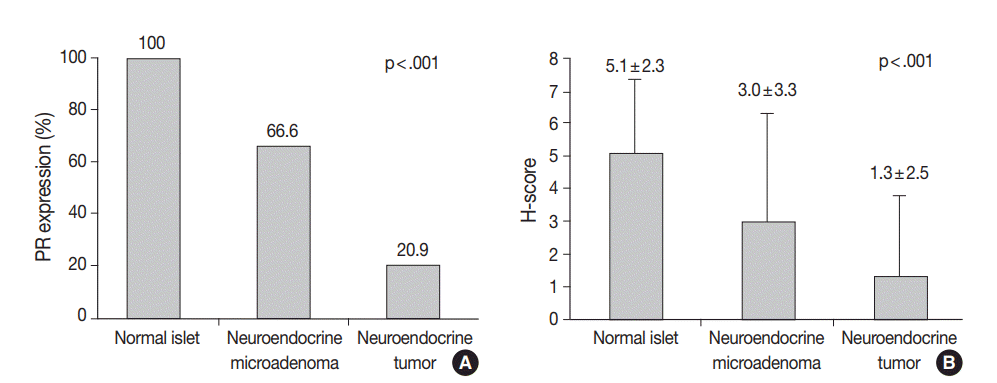
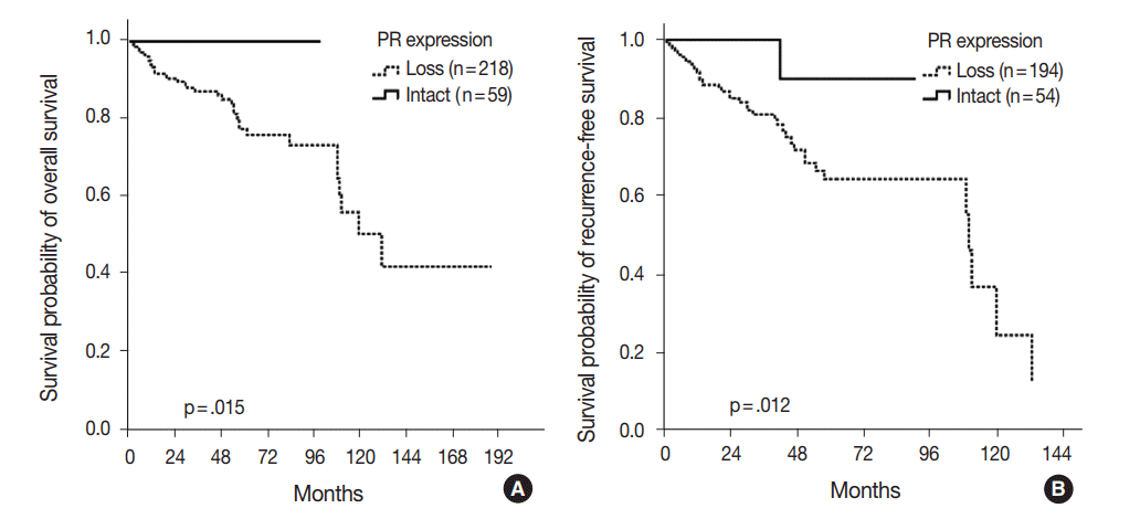


Fig. 1.
Fig. 2.
Fig. 3.
| Characteristic | PR loss | Intact PR | p-value |
|---|---|---|---|
| Age (yr) | .412 | ||
| ≤ 60 | 149 (78.0) | 42 (22) | |
| > 60 | 69 (80.2) | 17 (19.8) | |
| Sex | .380 | ||
| Male | 107 (79.9) | 27 (20.1) | |
| Female | 111 (77.6) | 32 (22.4) | |
| Tumor size (cm) | < .001 | ||
| ≤ 3 | 126 (71.2) | 51 (28.8) | |
| > 3 | 92 (86.4) | 8 (13.6) | |
| WHO grade | .001 | ||
| Grade 1 | 65 (76.5) | 20 (23.5) | |
| Grade 2 | 83 (87.4) | 12 (12.6) | |
| Grade 3 | 8 (100.0) | 0 | |
| pT classification | < .001 | ||
| pT1 | 70 (62.5) | 42 (27.5) | |
| pT2–T4 | 148 (89.7) | 17 (10.3) | |
| Lymphovascular invasion | .077 | ||
| Absent | 151 (76.3) | 47 (23.7) | |
| Present | 67 (84.8) | 12 (15.2) | |
| Perineural invasion | .028 | ||
| Absent | 180 (76.6) | 55 (23.4) | |
| Present | 38 (90.5) | 4 (9.5) | |
| Lymph node metastasis | .004 | ||
| Absent | 181 (76.1) | 57 (23.9) | |
| Present | 37 (94.9) | 2 (5.1) | |
| Distant metastasis | .639 | ||
| Absent | 210 (78.7) | 57 (21.3) | |
| Present | 8 (80.0) | 2 (20.0) | |
| Ki-67 labeling index (%) | .004 | ||
| < 3 | 137 (73.3) | 50 (26.7) | |
| ≥ 3 and < 20 | 57 (89.1) | 7 (10.9) | |
| ≥ 20 | 2 (100.0) | 0 | |
| Hormone expression | < .001 | ||
| Absent | 91 (94.8) | 5 (5.2) | |
| Present | 49 (68.1) | 23 (31.9) | |
| ALT expression | .005 | ||
| Absent | 166 (75.7) | 54 (24.5) | |
| Present | 52 (91.2) | 5 (8.8) | |
| ATRX/DAXX expression | .015 | ||
| Absent | 45 (90.0) | 5 (10.0) | |
| Present | 166 (75.5) | 54 (24.5) |
| Characteristic | Variables | Univariate analyses |
p-value | Multivariate analyses |
p-value | |
|---|---|---|---|---|---|---|
| 5-Year survival rate (%) | Hazard ratio | 95% confidence interval | ||||
| PR expression | Loss | 64.1 | .012 | 0.20 | 0.03–1.49 | .117 |
| Intact | 90 | |||||
| Age (yr) | ≤ 60 | 70.3 | .015 | 1.58 | 0.76–3.23 | .228 |
| > 60 | 62.1 | |||||
| Sex | Male | 59.1 | .609 | - | - | - |
| Female | 56.9 | |||||
| Tumor size (cm) | ≤ 3 | 74.6 | .008 | 0.94 | 0.37–2.39 | .879 |
| > 3 | 55.7 | |||||
| WHO grade | Grade 1 | 76.9 | < .001 | 1.00 | - | .001 |
| Grade 2 | 65.8 | 1.24 | 0.60–2.56 | .558 | ||
| Grade 3 | 0 | 10.74 | 3.79–30.45 | < .001 | ||
| pT classification | pT1 | 76.7 | .015 | 1.14 | 0.39–3.35 | .974 |
| pT2-T4 | 61.8 | |||||
| Lymphovascular invasion | Absent | 71.5 | .002 | 2.19 | 1.13–4.23 | .020 |
| Present | 54 | |||||
| Perineural invasion | Absent | 71.5 | < .001 | 1.12 | 0.40–3.16 | .813 |
| Present | 41 | |||||
| Lymph node metastasis | Absent | 73 | < .001 | 1.65 | 0.60–4.55 | .341 |
| Present | 27.8 | |||||
Values are presented as number (%). PR, progesterone receptor; PanNET, pancreatic neuroendocrine tumor; WHO, World Health Organization; ALT, alternative lengthening of telomeres.
PR, progesterone receptor; PanNET, pancreatic neuroendocrine tumor; WHO, World Health Organization.

