 E-submission
Articles
- Page Path
- HOME > J Pathol Transl Med > Volume 60(3); 2026 > Article
-
Newsletter
What’s new in digital and computational pathology 2026: advances in adoption, standards, AI technologies, and clinical integration -
Selim Sevim1
 , Chadi Hajar2, Snehal Sonawane3
 -
Journal of Pathology and Translational Medicine 2026;60(3):364-370.
DOI: https://doi.org/10.4132/jptm.2026.04.27
Published online: May 15, 2026
1Cancer Early Detection Advanced Research Center (CEDAR), Knight Cancer Institute, Oregon Health and Science University, Portland, OR, USA
2Department of Pathology and Laboratory Medicine, Medical University of South Carolina, Charleston, SC, USA
3Department of Pathology and Laboratory Medicine, University of Illinois at Chicago, Chicago, IL, USA
- Corresponding Author: Snehal Sonawane, MD Department of Pathology and Laboratory Medicine, University of Illinois at Chicago, Chicago, IL, USA E-mail: snehal@uic.edu
- This article has been published jointly, with consent, in both Journal of Pathology and Translational Medicine and PathologyOutlines.com.
• Received: March 27, 2026 • Accepted: April 27, 2026
© The Korean Society of Pathologists/The Korean Society for Cytopathology
This is an Open Access article distributed under the terms of the Creative Commons Attribution Non-Commercial License (https://creativecommons.org/licenses/by-nc/4.0) which permits unrestricted non-commercial use, distribution, and reproduction in any medium, provided the original work is properly cited.
- 2,734 Views
- 127 Download
- Abstract
- DIGITAL PATHOLOGY INFRASTRUCTURE – UNITED STATES AND GLOBAL
- SURGE IN DIGITAL PATHOLOGY ADOPTION
- LATEST GUIDELINES AND RECOMMENDATIONS
- INTEROPERABILITY AND STANDARDIZED FRAMEWORKS
- REGULATORY PROGRESS AND FDA APPROVALS
- MULTIMODAL DATA INTEGRATION
- BIG DATA REPOSITORIES
- DIGITAL PATHOLOGY IN TRAINING AND EDUCATION
- AI-POWERED TOOLS FOR CLINICAL USE
- VIRTUAL STAINING
- FOUNDATION MODELS
- AI COPILOTS
- DIGITAL AND COMPUTATIONAL PATHOLOGY IN CLINICAL TRIALS AND DRUG DISCOVERY
- Meet the Authors
- REFERENCES
Abstract
- Digital and computational pathology are expanding rapidly worldwide, driven by advances in whole‑slide imaging, AI algorithms, multimodal data integration, and improved digital infrastructure. Adoption continues to accelerate in the United States and internationally, supported by professional guidelines, emerging reimbursement pathways, and the growing need for remote workflows and collaborative diagnostics. Progress in interoperability standards, regulatory frameworks, and FDA approvals has strengthened the foundation for clinical deployment, while large‑scale data repositories and federated learning approaches enable more robust and privacy‑preserving model development. Foundation models, multimodal AI systems, and LLM‑based copilots are reshaping diagnostic support, prognostication, workflow efficiency, clinical trials and drug discovery.
- • The implementation of computational and digital pathology in the United States continues to develop despite the problems related to financial investment, data storage, reimbursement uncertainties, and regulatory constraints.
- • The digital pathology market is projected to exceed $2 billion by 2032 [1-3]. This increase is supported by new applications, such as biomarker quantification, large-scale image archiving, inter-institutional slide consultation, and telepathology [1].
- • Broader implementation and standardization are encouraged by guidelines and recommendations issued by entities like the College of American Pathologists (CAP) and other professional bodies, such as the Digital Pathology Association (DPA), the European Society of Digital and Integrative Pathology (ESDIP), and others.
- • On par with the United States, the primary factors that drive international adoption are based on the growing burden of cancer worldwide, understaffing, the need for personalized medicine, and collaborative diagnostic processes [3,4].
- • International adoption of digital and computational pathology remains uneven. A higher pace of adoption is mentioned in high-income countries. In contrast, developing countries face technical, financial, and workforce challenges when implementing digital initiatives [3,4].
- • A growing need for professional awareness, domain‑specific education, and broader advocacy for digital pathology is reflected in the recent establishment of several continental societies, including the Asian Society of Digital Pathology (ASDP), the African Society of Digital Pathology (AFSDP), and the Latin American Society of Digital Pathology (LASDP), alongside the longer‑standing ESDIP and DPA.
DIGITAL PATHOLOGY INFRASTRUCTURE – UNITED STATES AND GLOBAL
- • A global surge in adoption of digital pathology has been driven primarily by advances in scanning technology and speed, the expansion of AI tools, and the growing need for remote workflows (Fig. 1).
- • An international survey conducted in 2023 across 127 laboratories reported that 57% of these laboratories had implemented digital pathology for research or clinical purposes. This implementation works to improve turnaround time, case traceability within departments, and multi-site collaboration. The survey also reported challenges with laboratory information systems (LIS) integration and a lack of workforce training [5].
- • In 2024, CAP estimated that digital pathology adoption is approximately 10% in U.S. labs [6].
- • In 2023, the American Society of Cytopathology (ASC) conducted a large-scale international survey about the use of scanners in cytology, with 327 participants. The results indicated that most respondents do not routinely scan cytology slides, highlighting concerns of image quality and the cost of implementation; however, pathologists also indicated interest in the implementation of the technology for screening liquid-based Papanicolaou tests, rapid onsite evaluation, and AI-assisted screening [7].
SURGE IN DIGITAL PATHOLOGY ADOPTION
- • CAP recommends using a validation set of at least 60 cases, with consistency between whole-slide images (WSI) and glass slide reads greater than 95%, for implementation of digital pathology workflow [8].
- • CAP also advocated for the implementation of 30 new Category 3 CPT codes in 2024 to highlight the additional work involved in digitizing surgical pathology slides for primary diagnosis [9].
- • The Center for Medicare & Medicaid Services (CMS) has released their newest recommendation in 2025, which indicates that all remote review of digital cytology specimens requires a remote location with a separate CLIA certificate [10].
- • ASC proposed a structured validation process for telecytology, including training, retrospective slide review, and hardware/software testing [11].
- • Consensus-based recommendations released by the Royal College of Pathologists (RCP) (UK) [12], ESDIP [13], and European Society of Pathology (ESP) [14] reflect European approaches to the implementation of digital pathology.
LATEST GUIDELINES AND RECOMMENDATIONS
- • Interoperability remains hindered by technical issues, such as proprietary image file formats, diverse data types, and legacy systems.
- • Economic barriers to interoperability and standardized frameworks include high costs, vendor lock-in, and limited reimbursement incentives.
- • Regulatory and organizational challenges primarily arise from a fragmented health IT ecosystem, workflow variability, compliance complexities, and resistance to change.
- • Due to these reasons, standardization has accelerated through the adoption and implementation of key interoperability standards.
- o IHE Digital Pathology Image Acquisition (DPIA) profile, Health Level 7 (HL7) for metadata exchange, and Digital Imaging and Communications in Medicine (DICOM) for image encoding are the main standardization efforts.
- • These frameworks enable consistent communication between slide scanners, viewers, the LIS, and analytics systems.
- • While progress has been made and continues, substantial improvement remains necessary, requiring collaboration among governmental agencies, medical organizations, and the private sector [15-17].
INTEROPERABILITY AND STANDARDIZED FRAMEWORKS
- • The key criteria for the approval of AI and digital pathology in medical devices are based on safety and effectiveness through valid scientific evidence, and these should prove that the benefit outweighs the risk for the intended use in the target population.
- • FDA approvals have spanned all the domains of digital and computational pathology, including scanners, image management systems (IMS), and AI algorithms for clinical use [18].
- • Examples of the latest FDA approvals:
- o AISight Dx (PathAI): cloud-based digital viewing and management platform, which supports integration with several slide scanners [19].
- o PathPresenter Clinical Viewer: digital pathology image management and viewer platform used for primary diagnosis, assisting in case tracking, image archiving, and collaboration features [20].
- o Roche Digital Pathology Dx (VENTANA DP 200): automated digital slide creation, viewing, and management system intended for in vitro diagnostic use as an aid to the pathologist to review and interpret WSIs [21].
REGULATORY PROGRESS AND FDA APPROVALS
- • Progress in multimodal data integration can be seen in devices and techniques that fuse histopathological images with clinical and molecular data.
- o Rapid optical-genomic screening system DeepGlioma combines stimulated Raman histology with deep learning-based genomics to predict key glioma molecular alterations with 93% accuracy in < 90 seconds, demonstrating the potential of real-time histology and genomics integration in pathology [22].
- • Federated learning methods tailored to pathology enable multi-institutional model training by learning from local healthcare datasets and aggregating updates to build more robust and generalizable AI models while preserving data privacy (e.g., federated frameworks for WSI analysis).
- • Innovative approaches, like FedMM, address modality gaps across hospitals by training separate single-modality feature extractors, resulting in superior classification accuracy and area under the curve (AUC) on multi-institutional datasets [23].
- • To address heterogeneity across multiple pathology labs and medical institutions, PathFL recently introduced multi-level alignment strategies.
- o Those strategies are applied at three distinct levels: “style, feature, and model aggregation.”
- o This alignment involves slide scanners, organs, modalities, and sources to improve validity in pathology image segmentation [24].
MULTIMODAL DATA INTEGRATION
- • High-throughput scanners revolutionized the digital pathology field due to rapid scanning of glass slides with file sizes ranging from hundreds of megabytes to gigabytes. Traditional databases are inadequate to hold this large amount of data [25].
- • In recent years, structured data repositories have been developed to enable standardized storage, metadata annotation, and efficient data sharing. The aim is to support foundation model development, biomarker discovery, drug research, and education while accelerating AI innovation through access to large and diverse cohorts [26].
- o Examples of some big data repositories are: BIGPICTURE [26], BD4BO [27], eTOX [28], eTRANSAFE [29], MELLODDY [30], OPTIMA [31], VICT3R [32].
BIG DATA REPOSITORIES
- • Digital and computational pathology innovation has expanded its benefits in pathology education and training. It has enabled the creation of web-based image collections/libraries of various diseases. These libraries are accompanied by annotations, which may highlight the various features on WSIs.
- o Institutional and academic‑society WSI collections range from open access to closed and vary in curation rigor.
- o PathologyOutlines.com continues to expand its virtual‑slide content on textbook pages.
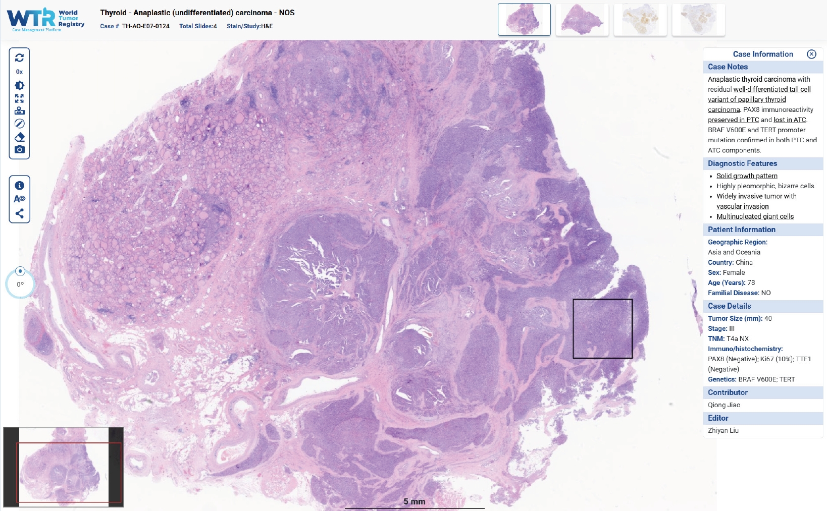
- o World Tumor Registry (WTR) is a web-based open-access collection of WSIs of tumors from every region of the world, annotated by subspecialty experts (Fig. 2). It removes geographic boundaries and serves as an educational and practical resource for cancer care and research [33].
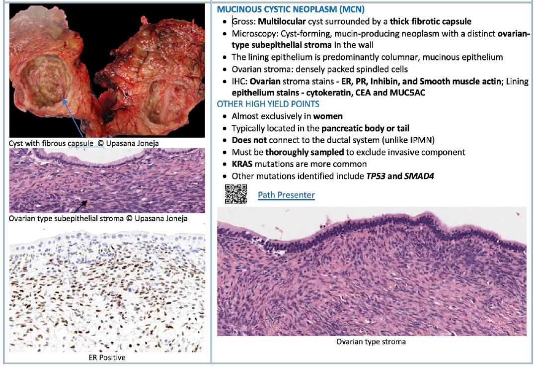
- • The innovative integration of a web-based pathology image collection with organ-based text can be supported by QR codes. These codes link each topic to its corresponding virtual slide. This approach combines the traditional hard-copy reading experience with immediate access to virtual slides, providing a dynamic and interactive learning environment for the next generation of pathologists (Fig. 3) [34].
DIGITAL PATHOLOGY IN TRAINING AND EDUCATION
- • In recent years, various AI-based tools and related technologies have demonstrated strong diagnostic and predictive/prognostic capabilities. In different studies, the effectiveness of those beneficial tools has been repeatedly verified [35].
- • Expert panels predicted that many tasks in digitized pathology laboratories could be delegated to AI by the next decade [36]. The most promising applications include cancer detection and grading in histopathology (e.g. prostate, breast, gastrointestinal) and cervical cytology screening [37].
- • AI algorithms evaluate various tissue compartments and predict disease outcomes in breast, oropharyngeal, bladder, prostate, and non‐small‐cell lung cancers. Results from those algorithms showed great promise to shape treatment modality choice and patient-specific approaches instead of tumor/disease-specific management by emphasizing risk scores of individuals [38].
- o Diagnostic tools: FDA clearance of AI-powered applications reflects their diagnostic capability [39,40], as demonstrated by the approvals of Paige Prostate and Ibex Prostate Detect for prostate cancer diagnostics [41,42].
- o Predictive/prognostic tools: predictive tools nourished by multimodal datasets, such as H&E, clinical data (age, PSA, grading, Ki67 status, etc.), and molecular data, to create a digital biomarker of indolence/aggressiveness of tumor. These algorithms can also predict treatment response. In recent years, AI‐powered tools that combine clinical data and H&E images have demonstrated their potential in the field [43-45].
- • Artera AI app uses multimodal AI to predict 5- and 10-year risk of distant metastasis and biochemical failure, as well as prostate cancer-specific and overall survival [46]. In addition, the potential benefit of androgen deprivation therapy, radiotherapy, and castration resistance prediction for patients has been effectively estimated in clinical trials [47,48].
- • RlapsRisk BC predicts 5-year metastasis-free survival in early, ER-positive, HER2-negative breast cancer [44].
- • Computational histology and AI (CHAI) is an algorithm that works on high-risk non-muscle invasive bladder cancer to predict BCG response [45]. It creates an AI-based signature to stratify these patient groups as high- and low-risk in terms of recurrence-free and progression-free survival.
AI-POWERED TOOLS FOR CLINICAL USE
- • The concept of virtual staining has emerged in recent years as a valuable complement to the diagnostic process, driven by the increasing use of high‑throughput digital pathology and deep learning methods. Virtual staining refers to the synthetic generation of images that replicate routine diagnostic stains. When properly validated and integrated into the digital pathology workflow, virtual staining can offer meaningful gains in cost‑effectiveness and time efficiency [49].
VIRTUAL STAINING
- • Foundation models (FMs) are large-scale AI models trained on vast and diverse datasets that can be adapted to a wide range of downstream tasks with minimal fine-tuning.
- • FMs can be applied to a wide range of downstream tasks, including cancer subtyping, mutation prediction, biomarker detection, spatial proteomics analysis, and pan‑cancer detection independent of tissue type.
- o Prov-GigaPath is a whole-slide pathology FM trained on 1.3 billion tiles [50]. It demonstrated robust performance in cancer subtyping and pathomics tasks.
- o Virchow model achieved a 0.95 AUC in detecting 16 types of cancers, both rare and common, using nearly 1.5 million H&E slides [51].
- o CytoFM, the first cytology FM, demonstrated the adaptability of FMs to cytology specimens and was trained on 1.4 million cytology patches [52].
- o KRONOS, a spatial proteomics FM, was trained on 8 fluorescence-based imaging platforms, 16 tissue types, 175 protein markers, and 47 million image patches [53].
- • Studies have shown that FMs can help guide computational pathology to tailor patient-specific approaches.
FOUNDATION MODELS
- • Large-language models (LLMs) are AI systems trained on massive amounts of text data to understand, generate, and respond to human language in a way that mimics human communication. The input and output of these models can be unimodal or multimodal, spanning text, images, audio, and other data types.
- • The interactive nature of LLMs and generative AI is driving the emergence of specialized AI assistants in human‑integrated workflows.
- • In pathology, generative AI has a revolutionary potential to improve diagnostic accuracy, workflow efficiency, education, and research [54].
- o PathChat is a vision-language generalist AI tool to assist pathologists in diagnostic tasks [55]. This multimodal AI copilot was generated on approximately one million pathology-related answers and questions. The performance of that algorithm was evaluated by both multiple-choice and open-ended questions.
- o Alba AI copilot has the potential to aggregate multi-sourced data, help with routine diagnostics, perform image analysis for cancer detection, screen biomarkers, and generate pathology reports with the help of voice and chat assistance [56].
- o PathAsst multimodal LLM was designed to overcome the lack of high-quality data and specialized models [57]. Experimental results showed that it outperformed existing models in visual question answering tasks in pathology.
- o TeamPath algorithm was designed to enhance multimodal pathology diagnosis with the help of reasoning AI copilots [58]. TeamPath’s framework was shaped by human-AI collaboration and showcases the power of an algorithm to correct and verify pathologist-provided answers and reasoning paths.
- • General‑purpose LLMs such as ChatGPT and Gemini are widely used by pathologists for administrative, educational, and other tasks [59]. Because they are not trained on domain‑rich pathology datasets and lack clinical‑grade validation or approval, their use for diagnostic purposes is discouraged due to limited performance and privacy risks [60].
AI COPILOTS
- • Digital and computational pathology provide multidimensional benefits in clinical trials and drug discovery by enabling centralized slide review, improving accuracy and efficiency, and supporting standardized and reproducible interpretation across participating centers. Various integrated data analytics tools allow efficient patient enrollment, randomization, stratification, and endpoint evaluation in clinical trials.
- • Improved digital pathology platforms streamline study protocols and enable real-time result evaluation, enhancing toxicopathology workflows, which involve evaluating tissue changes in non‑clinical models to assess drug safety. Digitization also facilitates navigation of complex regulatory requirements, including specimen handling, data safety, integrity, and compliance.
- • Lately, the integration of molecular/genomic data has enabled complex tissue-based experiments and strengthened global collaboration among pharmaceutical companies, technology vendors, and clinical researchers. These advances are helping to drive the continued development of precision medicine [61].
DIGITAL AND COMPUTATIONAL PATHOLOGY IN CLINICAL TRIALS AND DRUG DISCOVERY
- Dr. Selim Sevim is a pathologist with a background in digital pathology, artificial intelligence, and 3D tissue imaging. After completing his pathology residency at the Department of Pathology, Ankara University (Türkiye), he joined the Cancer Early Detection Advanced Research Center (CEDAR) at Oregon Health and Science University. His work focuses on AIassisted histopathological analysis and novel computational tools for cancer diagnosis.
- Dr. Chadi Hajar has received his medical degree from the Lebanese University School of Medicine. He completed his Anatomic and Clinical Pathology Residency in Colorado at Penrose-Saint Francis Medical Center, followed by a Fellowship in Surgical, Gastrointestinal & Liver Pathology at the Medical University of South Carolina. In addition, he completed his MBA in Health Administration at the University of Colorado, Denver, and has an interest in digital and computational pathology. He is currently an Assistant Professor in Surgical, Gastrointestinal & Liver Pathology in the Department of Pathology and Laboratory Medicine at the Medical University of South Carolina.
- Dr. Snehal Sonawane has been an author for PathologyOutlines since 2016. She has completed an Anatomic and Clinical Pathology Residency at the University of Illinois College of Medicine in Chicago, followed by a Fellowship in Surgical/GI Pathology at Loyola University Medical Center. She has also completed a Master’s in Health Informatics and has an interest in digital and computational pathology. She works as an Assistant Professor at the Department of Pathology, College of Medicine, University of Illinois at Chicago.
Meet the Authors

Fig. 2.Interface of the World Tumor Registry public platform showing WSI viewer, multiple slides representing a single case, descriptive case notes, diagnostic features annotated directly on the slides, and accompanying case details.

Fig. 3.Image demonstrating a surgical pathology book with web-based pathology image collection and organ-based text. This book uses QR codes to link each topic to the corresponding virtual slide. Gupta, Akanksha. Ace the Boards: Surgical Pathology Reimagined. 2022 (used with permission).

- 1. Bessen JL, Alexander M, Foroughi O, et al. Perspectives on reducing barriers to the adoption of digital and computational pathology technology by clinical labs. Diagnostics (Basel) 2025; 15: 794.ArticlePubMedPMC
- 2. Ardon O, Klein E, Manzo A, et al. Digital pathology operations at a tertiary cancer center: Infrastructure requirements and operational cost. J Pathol Inform 2023; 14: 100318.ArticlePubMedPMC
- 3. Ahmed MI, Spooner B, Isherwood J, Lane M, Orrock E, Dennison A. A systematic review of the barriers to the implementation of artificial intelligence in healthcare. Cureus 2023; 15: e46454.ArticlePubMedPMC
- 4. Coudry RA, Assis EACP, Frassetto FP, et al. Crossing the andes: challenges and opportunities for digital pathology in Latin America. J Pathol Inform 2024; 15: 100369.ArticlePubMedPMC
- 5. Pinto DG, Bychkov A, Tsuyama N, Fukuoka J, Eloy C. Real-world implementation of digital pathology: results from an intercontinental survey. Lab Invest 2023; 103: 100261.ArticlePubMed
- 6. Digital pathology at 10% adoption and in-lab plans [Internet]. Northfield: CAP Today, 2025 [cited 2025 Dec 20]. Available from: https://www.captodayonline.com/digital-pathology-at-10-adoption-and-in-lab-plans/.
- 7. Kim D, Thrall MJ, Michelow P, et al. The current state of digital cytology and artificial intelligence (AI): global survey results from the American Society of Cytopathology Digital Cytology Task Force. J Am Soc Cytopathol 2024; 13: 319-28. ArticlePubMed
- 8. Evans AJ, Brown RW, Bui MM, et al. Validating whole slide imaging systems for diagnostic purposes in pathology. Arch Pathol Lab Med 2022; 146: 440-50. ArticlePDF
- 9. Advocacy update: July 11, 2023 [Internet]. Northfield: College of American Pathologists, 2023 [cited 2025 Dec 20]. Available from: https://www.cap.org/advocacy/latest-news-and-practice-data/july-11-2023.
- 10. Current CAP guidelines [Internet]. Northfield: College of American Pathologists, 2025 [cited 2025 Dec 20]. Available from: https://www.cap.org/protocols-and-guidelines.
- 11. Lin O, Alperstein S, Barkan GA, et al. American Society of Cytopathology Telecytology validation recommendations for rapid on-site evaluation (ROSE). J Am Soc Cytopathol 2024; 13: 111-21. ArticlePubMed
- 12. Cross S, Furness P, Igali L, Snead D, Treanor D. Best practice recommendations for implementing digital pathology January 2018. London: The Royal College of Pathologists, 2018.
- 13. Fraggetta F, L'Imperio V, Ameisen D, et al. Best practice recommendations for the implementation of a digital pathology workflow in the anatomic pathology laboratory by the European Society of Digital and Integrative Pathology (ESDIP). Diagnostics (Basel) 2021; 11: 2167.ArticlePubMedPMC
- 14. Eloy C, Fraggetta F, van Diest PJ, et al. Digital transformation of pathology: the European Society of Pathology expert opinion paper. Virchows Arch 2025; 487: 971-81. ArticlePubMedPMCPDF
- 15. Dash RC, Jones N, Merrick R, et al. Integrating the health-care enterprise pathology and laboratory medicine guideline for digital pathology interoperability. J Pathol Inform 2021; 12: 16.ArticlePubMedPMC
- 16. Mantri M, Taran S, Sunder G. DICOM integration libraries for medical image interoperability: a technical review. IEEE Rev Biomed Eng 2022; 15: 247-59. Article
- 17. Abels E, Pantanowitz L, Aeffner F, et al. Computational pathology definitions, best practices, and recommendations for regulatory guidance: a white paper from the Digital Pathology Association. J Pathol 2019; 249: 286-94. ArticlePubMedPMCPDF
- 18. Zhang DY, Venkat A, Khasawneh H, Sali R, Zhang V, Pei Z. Implementation of digital pathology and artificial intelligence in routine pathology practice. Lab Invest 2024; 104: 102111.ArticlePubMed
- 19. 510(k) Premarket notification K243391 [Internet]. Silver Spring: U.S. Food and Drug Administration, 2024 [cited 2025 Jan 12]. Available from: https://www.accessdata.fda.gov/scripts/cdrh/cfdocs/cfpmn/pmn.cfm?ID=K243391.
- 20. 510(k) Premarket notification K250968 [Internet]. Silver Spring: U.S. Food and Drug Administration, 2025 [cited 2025 Jan 12]. Available from: https://www.accessdata.fda.gov/scripts/cdrh/cfdocs/cfpmn/pmn.cfm?ID=K250968.
- 21. 510(k) Premarket notification K232879 [Internet]. Silver Spring: U.S. Food and Drug Administration, 2024 [cited 2026 Mar 6]. Available from: https://www.accessdata.fda.gov/cdrh_docs/reviews/K232879.pdf.
- 22. Hollon T, Jiang C, Chowdury A, et al. Artificial-intelligence-based molecular classification of diffuse gliomas using rapid, label-free optical imaging. Nat Med 2023; 29: 828-32. ArticlePubMedPMCPDF
- 23. Peng Y, Bian J, Xu J. FedMM: federated multi-modal learning with modality heterogeneity in computational pathology. In: ICASSP 2024-2024 IEEE International Conference on Acoustics, Speech and Signal Processing (ICASSP); 2024 Apr 14-19; Seoul, Korea.
- 24. Lu MY, Chen RJ, Kong D, et al. Federated learning for computational pathology on gigapixel whole slide images. Med Image Anal 2022; 76: 102298.ArticlePubMedPMC
- 25. Zarella MD, Rivera Alvarez K. High-throughput whole-slide scanning to enable large-scale data repository building. J Pathol 2022; 257: 383-90. ArticlePubMedPMCPDF
- 26. Moulin P, Grunberg K, Barale-Thomas E, der Laak JV. IMI-Bigpicture: a central repository for digital pathology. Toxicol Pathol 2021; 49: 711-3. ArticlePubMedPDF
- 27. Szocska M, Jayawardana S, Smand C, Salimullah T, Reed C, Hanif S. Big data for better outcomes: supporting health care system transformation in Europe. Eurohealth 2017; 23: 7-9.
- 28. Sanz F, Pognan F, Steger-Hartmann T, et al. Legacy data sharing to improve drug safety assessment: the eTOX project. Nat Rev Drug Discov 2017; 16: 811-2. ArticlePubMedPDF
- 29. Sanz F, Pognan F, Steger-Hartmann T, et al. eTRANSAFE: data science to empower translational safety assessment. Nat Rev Drug Discov 2023; 22: 605-6. ArticlePubMed
- 30. Heyndrickx W, Mervin L, Morawietz T, et al. MELLODDY: cross-pharma federated learning at unprecedented scale unlocks benefits in QSAR without compromising proprietary information. J Chem Inf Model 2024; 64: 2331-44. ArticlePubMed
- 31. N’Dow J, Smith EJ, Polychronopoulos K, et al. 917P OPTIMA: improve care for patients with prostate, breast, and lung cancer through artificial intelligence. Ann Oncol 2022; 33(Suppl 7): S966.Article
- 32. Steger-Hartmann T, Sanz F, Bringezu F, Soininen I. IHI VICT3R: developing and implementing virtual control groups to reduce animal use in toxicology research. Toxicol Pathol 2025; 53: 230-3. ArticlePDF
- 33. The World Tumor Registry: tell all your friends [Internet]. London: The Pathologist, 2025 [cited 2025 Dec 20]. Available from: https://thepathologist.com/issues/2025/articles/june/the-world-tumor-registry-tell-all-your-friends/.
- 34. Gupta A, Sonawane S. Pathology visions 2023 overview. The path to unprecedented excellence: redefining pathology education through Ace My Path surgical pathology reimagined. J Pathol Inform 2024; 15: 100362.Article
- 35. McGenity C, Clarke EL, Jennings C, et al. Artificial intelligence in digital pathology: a systematic review and meta-analysis of diagnostic test accuracy. NPJ Digit Med 2024; 7: 114.ArticlePubMedPMCPDF
- 36. Berbis MA, McClintock DS, Bychkov A, et al. Computational pathology in 2030: a Delphi study forecasting the role of AI in pathology within the next decade. EBioMedicine 2023; 88: 104427.ArticlePubMedPMC
- 37. Chong Y, Bychkov A. Commercially available artificial intelligence solutions for gynaecologic cytology screening and their integration into clinical workflow. Cytopathology 2026; 37: 24-44. Article
- 38. Bera K, Schalper KA, Rimm DL, Velcheti V, Madabhushi A. Artificial intelligence in digital pathology: new tools for diagnosis and precision oncology. Nat Rev Clin Oncol 2019; 16: 703-15. ArticlePDF
- 39. Pantanowitz L, Quiroga-Garza GM, Bien L, et al. An artificial intelligence algorithm for prostate cancer diagnosis in whole slide images of core needle biopsies: a blinded clinical validation and deployment study. Lancet Digit Health 2020; 2: e407-16. ArticlePubMed
- 40. Raciti P, Sue J, Ceballos R, et al. Novel artificial intelligence system increases the detection of prostate cancer in whole slide images of core needle biopsies. Mod Pathol 2020; 33: 2058-66. ArticlePubMedPMCPDF
- 41. Evaluation of automatic class III designation for Paige Prostate (DEN200080) [Internet]. Silver Spring: U.S. Food and Drug Administration (FDA), 2021 [cited 2025 Dec 20]. Available from: https://www.accessdata.fda.gov/cdrh_docs/reviews/DEN200080.pdf.
- 42. Ibex Medical Analytics receives first FDA 510(k) clearance [Internet]. Tel Aviv: Ibex Medical Analytics, 2025 [cited 2025 Dec 13]. Available from: https://ibex-ai.com/fda-510k-clearance/.
- 43. Esteva A, Feng J, van der Wal D, et al. Prostate cancer therapy personalization via multi-modal deep learning on randomized phase III clinical trials. NPJ Digit Med 2022; 5: 71.ArticlePubMedPMCPDF
- 44. Garberis I, Gaury V, Saillard C, et al. Deep learning assessment of metastatic relapse risk from digitized breast cancer histological slides. Nat Commun 2025; 16: 5876.ArticlePubMedPMCPDF
- 45. Lotan Y, Krishna V, Abuzeid WM, et al. Predicting response to intravesical bacillus Calmette-Guérin in high-risk nonmuscle-invasive bladder cancer using an artificial intelligence-powered pathology assay: development and validation in an international 12-center cohort. J Urol 2025; 213: 192-204. ArticlePubMed
- 46. De Novo Classification Request for DEN240068 [Internet]. Silver Spring: U.S. Food and Drug Administration, 2025 [cited 2025 Dec 13]. Available from: https://www.accessdata.fda.gov/scripts/cdrh/cfdocs/cfpmn/denovo.cfm?id=DEN240068.
- 47. Armstrong AJ, Liu VY, Selvaraju RR, et al. Development and validation of an artificial intelligence digital pathology biomarker to predict benefit of long-term hormonal therapy and radiotherapy in men with high-risk prostate cancer across multiple phase III trials. J Clin Oncol 2025; 43: 3494-504. ArticlePubMedPMC
- 48. Feng FY, Smith MR, Saad F, et al. Digital pathology-based multimodal artificial intelligence scores and outcomes in a randomized phase III trial in men with nonmetastatic castration-resistant prostate cancer. JCO Precis Oncol 2025; 9: e2400653.ArticlePubMed
- 49. Bai B, Yang X, Li Y, Zhang Y, Pillar N, Ozcan A. Deep learning-enabled virtual histological staining of biological samples. Light Sci Appl 2023; 12: 57.ArticlePubMedPMCPDF
- 50. Xu H, Usuyama N, Bagga J, et al. A whole-slide foundation model for digital pathology from real-world data. Nature 2024; 630: 181-8. ArticlePubMedPMC
- 51. Vorontsov E, Bozkurt A, Casson A, et al. A foundation model for clinical-grade computational pathology and rare cancers detection. Nat Med 2024; 30: 2924-35. ArticlePubMedPMCPDF
- 52. Ivezic V, Radhachandran A, Redekop E, et al. CytoFM: the first cytology foundation model. In: 2025 IEEE/CVF Conference on Computer Vision and Pattern Recognition Workshops; 2025 Jun 11-12; Nashville, TN, USA.
- 53. Shaban M, Chang Y, Qiu H, et al. A foundation model for spatial proteomics. Preprint arXiv: 250603373. https://doi.org/10.48550/arXiv.2506.03373 (2025). Article
- 54. Brodsky V, Ullah E, Bychkov A, et al. Generative artificial intelligence in anatomic pathology. Arch Pathol Lab Med 2025; 149: 298-318. ArticlePubMedPDF
- 55. Lu MY, Chen B, Williamson DFK, et al. A multimodal generative AI copilot for human pathology. Nature 2024; 634: 466-73. ArticlePubMedPMCPDF
- 56. ALBA: generalist foundation model for computational pathology [Internet]. New York: Paige, 2025 [cited 2025 Dec 13]. Available from: https://www.paige.ai/al/alba.
- 57. Sun Y, Zhu C, Zheng S, et al. PathAsst: a generative foundation AI assistant towards artificial general intelligence of pathology. In: Proceedings of the 38th AAAI Conference on Artificial Intelligence; 2024 Feb 20-27; Vancouver, Canada.
- 58. Liu T, Xuan W, Wu H, et al. TeamPath: building multimodal pathology experts with reasoning AI copilots. Preprint arXiv: 251117652. https://doi.org/10.48550/arXiv.2511.17652 (2025). Article
- 59. Laohawetwanit T, Pinto DG, Bychkov A. A survey analysis of the adoption of large language models among pathologists. Am J Clin Pathol 2025; 163: 52-9. ArticlePubMedPDF
- 60. Laohawetwanit T, Apornvirat S, Asaturova A, Li H, Lami K, Bychkov A. Evaluation of general-purpose large language models as diagnostic support tools in cervical cytology. Pathol Res Pract 2025; 274: 156159.ArticlePubMed
- 61. Sebastian M, Batra H, Saini ML, et al. Applications and challenges of utilizing digital pathology and AI-enabled workflows in clinical trials. J Pathol Inform 2026; 20: 100542.ArticlePubMedPMC
REFERENCES
Figure & Data
References
Citations
Citations to this article as recorded by 
 PubReader
 ePub Link
-
 Cite this Article
- Cite this Article
-
- Close
- Download Citation
- Close
- Figure
-

- Related articles
-
- What’s new in medical renal pathology 2025: Updates on podocytopathy and immunofluorescence staining in medical kidney
- What’s new in genitourinary pathology 2023: WHO 5th edition updates for urinary tract, prostate, testis, and penis
- What’s new in hematopathology 2023: updates on mature T-cell neoplasms in the 5th edition of the WHO classification
- What’s new in bone and soft tissue pathology 2023: guidelines for molecular testing
- What’s new in neuromuscular pathology 2022: myopathy updates and gene therapies
What’s new in digital and computational pathology 2026: advances in adoption, standards, AI technologies, and clinical integration


Fig. 1. Brief overview of the advancements in digital and computational pathology.
Fig. 2. Interface of the World Tumor Registry public platform showing WSI viewer, multiple slides representing a single case, descriptive case notes, diagnostic features annotated directly on the slides, and accompanying case details.
Fig. 3. Image demonstrating a surgical pathology book with web-based pathology image collection and organ-based text. This book uses QR codes to link each topic to the corresponding virtual slide. Gupta, Akanksha. Ace the Boards: Surgical Pathology Reimagined. 2022 (used with permission).
Fig. 1.
Fig. 2.
Fig. 3.
What’s new in digital and computational pathology 2026: advances in adoption, standards, AI technologies, and clinical integration

