
 E-submission

Articles
- Page Path
- HOME > J Pathol Transl Med > Forthcoming articles > Article
-
Newsletter
What’s new in gastrointestinal pathology 2026: updates from the latest WHO classification -
Raul S. Gonzalez

-
DOI: https://doi.org/10.4132/jptm.2026.10.20
Published online: October 27, 2026
Department of Pathology and Laboratory Medicine, Emory University, Atlanta, GA, USA
- Corresponding Author: Raul S. Gonzalez, MD Department of Pathology and Laboratory Medicine, Emory University, Atlanta, GA, USA E-mail: rsgonza@emory.edu
This article has been published jointly, with consent, in both Journal of Pathology and Translational Medicine and PathologyOutlines.com.
• Received: September 20, 2026 • Accepted: October 16, 2026
© The Korean Society of Pathologists/The Korean Society for Cytopathology
This is an Open Access article distributed under the terms of the Creative Commons Attribution Non-Commercial License (http://creativecommons.org/licenses/by-nc/4.0) which permits unrestricted non-commercial use, distribution, and reproduction in any medium, provided the original work is properly cited.
- 16 Views
- 1 Download


Abstract
- The 6th edition of the World Health Organization Classification of Tumors: Digestive System Tumors was released online in a beta edition in spring 2026, with a physical print edition released in August. Compared to the 5th edition from 2019, the new edition includes several new entities, completely overhauls a few key concepts, and offers timely updates on most additional neoplasms. Most entries also include new photomicrographs and example wholeslides images. This newsletter will cover selected highlights from the new online edition.
- • Squamous papilloma is no longer regarded as an entity driven by HPV infection.
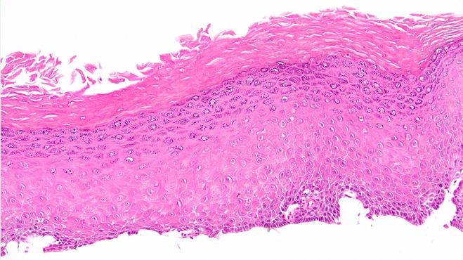
- • Epidermoid metaplasia has been added as a neoplastic precursor to squamous cell carcinoma, wherein the mucosa develops a granular layer and keratosis (Fig. 1).
- • The histopathologic descriptions of lowgrade and high-grade dysplasia in Barrett’s esophagus have been revised, and the “four layers” concept for identifying non-dysplastic Barrett’s mucosa is explained. The utility of p53 immunohistochemistry in diagnosing Barrett’srelated dysplasia is now discussed.
- • Gastroesophageal adenocarcinoma invading between two layers of duplicated muscularis mucosae, but not through the lower layer, should be staged as pT1a.
- • The entry on undifferentiated carcinoma includes information about the subtype driven by alterations in genes of the SWI/SNF chromatin remodeling complex.
ESOPHAGUS
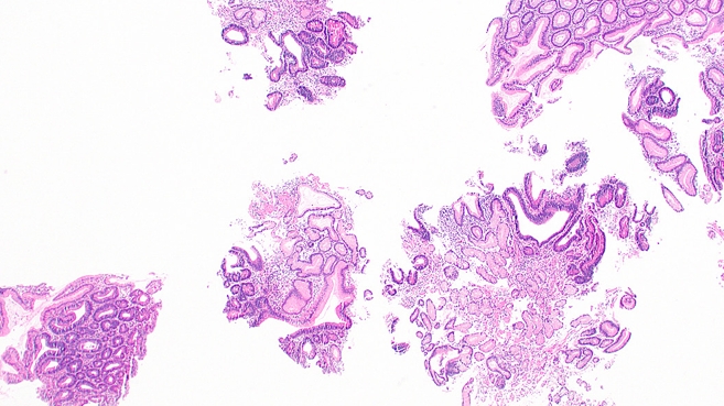
- • Gastric dysplasia is conceptually reframed as more akin to “gastritis-associated dysplasia,” particularly for cases that are endoscopically flat or poorly defined. The WHO recommends reporting raised lesions in this setting as “polypoid dysplasia/adenoma”; this includes foveolar-type adenomas and intestinal-type adenomas (Fig. 2). Polyposis patients can still develop sporadic polyps.
- • Subtypes of gastric cancer have undergone minor revisions (e.g., sarcomatoid has been added, parietal cell has been removed). The biomarker section now focuses on the utility of PD-L1 and claudin 18.2.
STOMACH
- • This has been split into two chapters, one on duodenum/ampulla and one on jejunum/ ileum. Most updates throughout are minor. There is now a specific entry on Peutz–Jeghers polyps, underscoring that they sometimes can be sporadic rather than syndromic.
SMALL BOWEL
- • Use of the terminology “traditional serrated adenoma” in appendiceal polyps is discouraged.
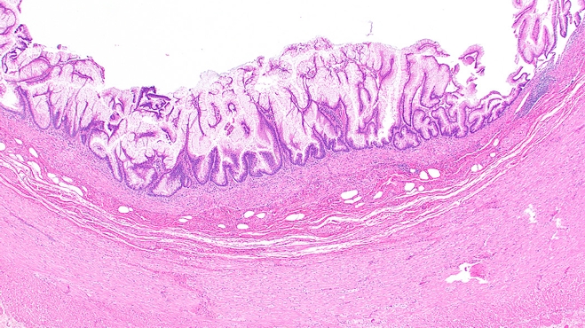
- • Mucinous adenoma has been added as a new entity in the section on benign epithelial tumors. It resembles low-grade appendiceal mucinous neoplasm, except it is completely confined to the mucosal surface and the muscularis mucosae is intact (Fig. 3). The entire appendix should be submitted and examined histologically to distinguish a mucinous adenoma from low- or high-grade appendiceal mucinous neoplasms.
- • A topic on conventional appendiceal adenomas (e.g., tubular adenoma) has been added. These are largely analogous to conventional adenomas of the colon.
- • A new table has been added that clarifies and streamlines reporting and definitions for appendiceal mucinous neoplasms and associated peritoneal metastases (clinically pseudomyxoma peritonei). The pathologic term “mucinous carcinoma peritonei” is officially preferred for the latter.
- • High-grade appendiceal mucinous neoplasm now requires 10% high-grade dysplasia.
- • Goblet cell adenocarcinoma grading is similar to the 5th edition, though the descriptive wording of low-grade and high-grade features is revamped. The minimum diagnostic criterion is no longer a conventional low-grade focus; instead, the tumor’s amphicrine nature must be established (see neuroendocrine section below).
APPENDIX
- • Hyperplastic polyps have been separated out into their own entry.
- • The WHO recommends reporting mixed polyps with features of both tubulovillous adenoma and traditional serrated adenoma (TSA) as TSA with low-grade or high-grade dysplasia on the basis of nuclear cytology.
- • Serrated polyposis has been moved to this chapter to emphasize that it is not really a genetic syndrome.
- • Non-syndromic juvenile polyps are now discussed in this chapter.
- • Architectural changes are emphasized as a key component of recognizing high-grade dysplasia in colorectal adenomas.
- • The term “pseudoinvasion” should no longer be used to describe epithelial misplacement in adenomas.
- • Colitis-associated dysplasia in the setting of inflammatory bowel disease has been expanded, and the histologic descriptions have been overhauled. Subtypes include conventional (adenomatous) dysplasia, serrated dysplasia, hypermucinous dysplasia, goblet celldeficient dysplasia, and crypt cell dysplasia. Immunohistochemistry for p53 may be helpful in diagnosing these lesions.
- • The section on colorectal cancer has been expanded. Key updates include the approach to grading, new details regarding molecular alterations, and an expanded roster of subtypes, which are now summarized in a table.
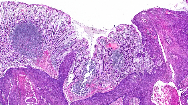
- o New subtypes include squamous cell carcinoma (Fig. 4) and lymphoglandular complex–like adenocarcinoma.
- o Carcinomas meeting criteria for both mucinous adenocarcinoma and signet-ring cell carcinoma may be called “mucin-rich signet-ring cell carcinomas.”
- o “Carcinoma with sarcomatoid components” is now termed “carcinoma with mesenchymal differentiation.”
- o Intramucosal carcinoma has been formally recognized as a rare phenomenon distinct from high-grade dysplasia and often linked to hereditary syndromes.
COLON AND RECTUM
- • Giant condyloma (Buschke–Löwenstein tumor) is clearly recognized as a neoplasm linked to low-oncogenic-risk HPV infection and distinct from verrucous carcinoma, which is not related to HPV infection.
- • The section on HPV-related anal dysplasia now more closely aligns with the widely adopted Lower Anogenital Squamous Terminology (LAST) classification.
- • HPV-independent forms of anal dysplasia, such as differentiated intraepithelial neoplasia, are briefly discussed.
- • Terminology has been updated for anal adenocarcinoma (including the rare “perianal adenocarcinoma”), and the role of HPV in driving a subset of cases has been added. Rare examples resemble cervical stratified mucinproducing carcinomas.
ANAL CANAL
- • The lack of molecular abnormalities is emphasized in focal nodular hyperplasia.
- • Hepatocellular adenoma is now defined as arising in a non-cirrhotic liver. The sonic hedgehog subtype has been added to the classification, and additional details are provided regarding ancillary testing for difficult cases.
- • Updates to the section on hepatocellular carcinoma include the addition of sarcomatoid carcinoma as a subtype and the utility of albumin ISH (which is sensitive but not specific) for diagnosis. Cases showing a “vessels encapsulating tumor clusters” pattern appear to have a worse prognosis.
- • Hepatocellular neoplasm, NOS has been added under the hemangioblastoma topic, and other terminology here has been slightly revised.
- • Now by definition, mucinous cystic neoplasm of the liver cannot occur in men.
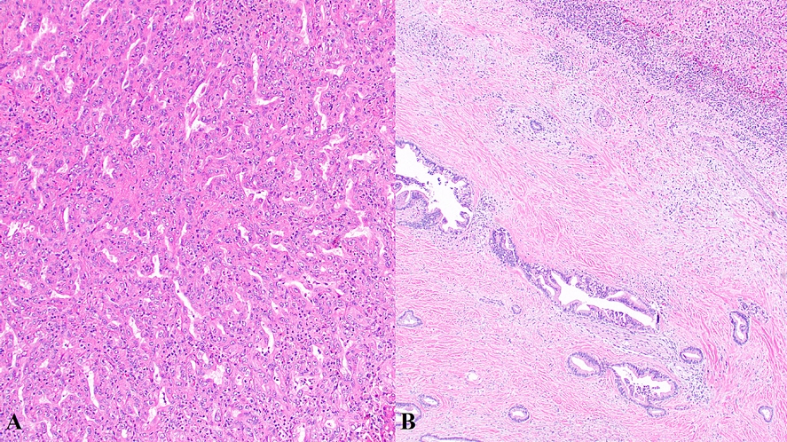
- • Intrahepatic cholangiocarcinoma has been formally split into small duct type (Fig. 5A) and large duct type (Fig. 5B), with clinical, morphologic, molecular, and prognostic differences. Most of this information is summarized in a table. The former often arises in the setting of chronic liver disease, and the latter bears similarities to extrahepatic cholangiocarcinoma and has a poor prognosis. The recently described solid tubulocystic subtype of small duct intrahepatic cholangiocarcinoma (also known as “cholangioblastic” carcinoma) demonstrates NIPBL::NACC1 fusion.
- • Immunohistochemistry is now mentioned as often helpful/necessary to confirm a diagnosis of combined hepatocellular carcinomacholangiocarcinoma. Therapy for these lesions should be based on the predominant component.
LIVER AND INTRAHEPATIC BILE DUCTS
- • Intraductal tubulopapillary neoplasm and intraductal oncocytic papillary neoplasm have been separated out into standalone topics, in addition to intraductal papillary neoplasm of the bile ducts.
- • The concept of intracholecystic neoplasms now includes intracholecystic papillary neoplasm (which is often associated with adenocarcinoma), intracholecystic tubular neoplasm (which is generally indolent), and mural intracholecystic neoplasm (which is quite rare). “Pyloric gland adenoma” has been removed as acceptable terminology, as it appears to represent polypoid pyloric gland metaplasia.
- • The sections on extrahepatic cholangiocarcinoma and carcinoma of the gallbladder have undergone minor revisions.
GALLBLADDER AND EXTRAHEPATIC BILE DUCTS
- • Acinar cystic transformation, a non-neoplastic process, was included in the 5th edition but has been removed from this edition.
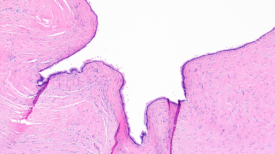
- • Simple mucinous cyst has been added as a lowgrade neoplasm with KRAS mutations that does not connect with the main pancreatic duct and can rarely progress to malignancy (Fig. 6).
- • The typical size of intraductal papillary mucinous neoplasm has been updated; it was previously > 0.5 cm and is now > 1.0 cm.
- • Intraductal oncocytic papillary neoplasm of the pancreas is now known to harbor PRKACA or PRKACB fusions.
- • Key updates to pancreatic ductal adenocarcinoma include expanded information on grading and targetable molecular alterations. Histologic information now includes the transcriptomics-derived subtyping of basal-like and classic forms.
- • Accepted synonyms are pared down for solid pseudopapillary neoplasm, and extrapancreatic examples are no longer discussed. Expanded pathogenetic information implicates CTNNB1 mutations as causing the tumor’s unique morphology. The utility of LEF1 immunohistochemistry for the diagnosis is mentioned.
PANCREAS
- • The introduction to this chapter discourages the practice of counting mast cells outside the setting of systemic mastocytosis.
- • Two entities have been added: EBV-positive mucocutaneous ulcer and indolent NKcell lymphoproliferative disorder of the gastrointestinal tract.
- • A handful of entities have been removed (including post-transplant lymphoproliferative disorders), and the remaining entities have mostly received minor updates. Notably, follicular lymphomas should not be graded.
HEMATOLYMPHOID TUMORS
- • Descriptions of next-generation immunohistochemistry have been added for some lesions, such as CAMTA1 for epithelioid hemangioendothelioma and SS18-SSX for synovial sarcoma.
- • The section on gastrointestinal stromal tumors has been expanded, and the succinate dehydrogenase-deficient variety has been pulled out into a separate entry.
- • Liposarcoma has been added; it most often presents as a giant fibrovascular polyp of the esophagus and is usually well differentiated but may be dedifferentiated.
- • Calcifying fibrous tumor, a benign hypocellular lesion most commonly involving the stomach or small intestine, has been added.
- • Leiomyoma and leiomyosarcoma have been compiled into a single entry on smooth muscle tumors. This entry also includes EBV-associated smooth muscle tumor, a hypercellular proliferation typically seen in immunocompromised patients.
- • Terminology for vascular and lymphatic lesions has been updated based on that of the International Society for the Study of Vascular Anomalies (ISSVA).
- • Potential malignancy criteria for glomus tumor have been revised; they now include size > 5 cm, cytologic atypia, and ≥ 2 mitotic figures per 10 high-power fields.
- • Schwann cell hamartoma, a bland spindled polyp of the colorectum, has been added.
- • Angiomyolipoma has been reclassified as a subtype of PEComa, rather than being roughly synonymous.
- • Additional new entries include GLI1-altered neoplasms (including gastroblastoma), NUT-rearranged sarcoma, and undifferentiated sarcoma.
- • A few lesions have been removed, most notably solitary fibrous tumor.
MESENCHYMAL TUMORS
- • This new chapter compiles neuroendocrine neoplasms previously scattered throughout the book. INSM1 is included as a useful immunohistochemical marker of neuroendocrine differentiation.
- • The WHO encourages reporting presence or absence of tumor necrosis in well-differentiated neuroendocrine tumors, and it raises the possibility of subdividing grade 2 tumors into 2a (Ki67 of 3% to < 10%) and 2b (Ki67 of 10% to 20%). It also stresses that Ki67 is a continuous variable and an actual value should be reported.
- • The term “carcinoid” is expressly no longer recommended. Neuroendocrine tumors at all sites now include SSTR2 immunohistochemical expression as a desirable diagnostic criterion.
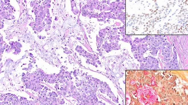
- • Amphicrine-like carcinoma is introduced as a homogeneous neoplasm composed of an intimate admixture of neuroendocrine and nonneuroendocrine cells (Fig. 7).
- o The neuroendocrine component must represent at least 30% of the lesion and express at least 2 neuroendocrine markers.
- o The non-endocrine component may be adenocarcinoma, squamous cell carcinoma, etc.
- o This is in contrast to mixed neuroendocrine– non-neuroendocrine neoplasms, which consist of a discrete neuroendocrine component and a separate discrete nonneuroendocrine component.
- • Patient symptoms should inform the terminology used for a neuroendocrine tumor.
- o For instance, a duodenal lesion showing classic “somatostatinoma” morphology that is positive for somatostatin by immunohistochemistry but does not cause systemic symptoms of somatostatin hypersecretion (i.e., is nonfunctional) should instead be termed “somatostatin cell neuroendocrine tumor.” If appropriate clinical symptoms are present, the term “somatostatinoma” can be used.
- • A minimum size cutoff of 0.5 cm is now included for gastric neuroendocrine tumors.
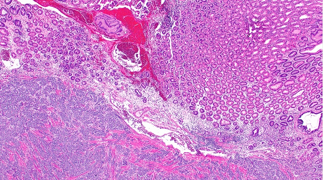
- • Two new subtypes of gastric neuroendocrine tumor have been added. Type 4 is rare and linked to ATP4A germline mutations. Type 5 is increasingly common, indolent, and linked to long-term proton pump inhibitor use (Fig. 8).
- • An entry on paraganglioma has been added, and gangliocytic paraganglioma has been renamed “composite gangliocytoma/neuroma and neuroendocrine tumor”.
NEUROENDOCRINE NEOPLASMS
- • This expanded section now includes extensive details on liver metastases, peritoneal metastases of appendiceal origin, and peritoneal metastases of non-appendiceal origin.
- • A thorough entry on carcinoma of unknown primary provides information on workup and classification of such lesions. A small percentage are deemed favorable based on certain clinicopathologic characteristics, though most are considered unfavorable.
OTHER TUMORS
- • The section on Lynch syndrome has been comprehensively updated. The somewhat related entity of constitutional mismatch repair deficiency syndrome is now given its own full topic.
- • SMAD4 and BMPR1A mutations are emphasized in the definition of juvenile polyposis syndrome, and similarly STK11 mutations are emphasized in the definition of Peutz–Jeghers syndrome, though diagnostic criteria are essentially unchanged.
- • Updated diagnostic criteria for the renamed “hereditary gastric and breast cancer syndrome” are provided; most patients harbor a germline CDH1 or CTNNA1 mutation.
- • Cowden syndrome has been formally renamed PTEN hamartoma tumor syndrome.
- • A few rare syndromes now have entire topics devoted to them, including glucagon cell hyperplasia and neoplasia, MAFA-related familial insulinomatosis, and gastrointestinal stromal tumor predisposition syndrome.
GENETIC TUMOR SYNDROMES
- Raul S. Gonzalez, MD is Professor of Pathology and Laboratory Medicine at Emory University Hospital. He has written more than 180 topics for PathologyOutlines. com and previously served as the website’s Editor in Chief. He was a coauthor for three entries in the WHO classification discussed in this manuscript.
Meet the Authors
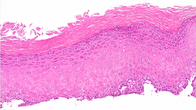
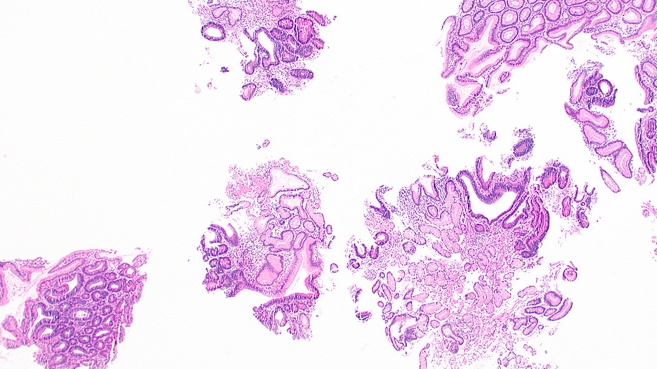
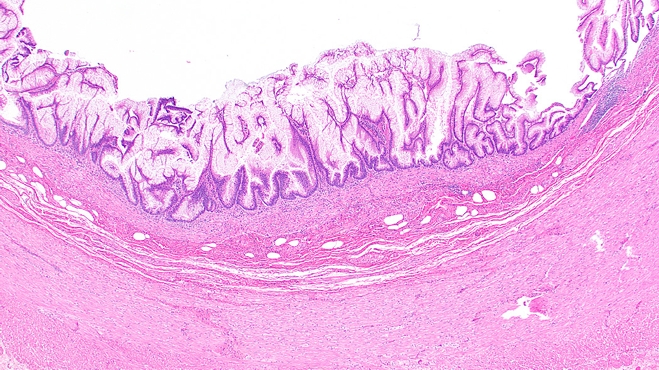
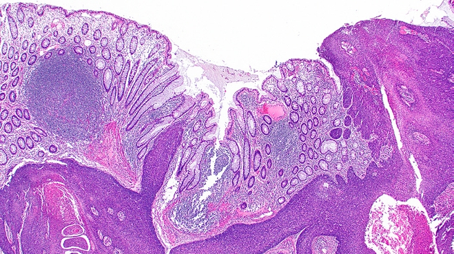
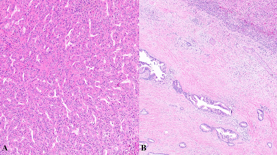
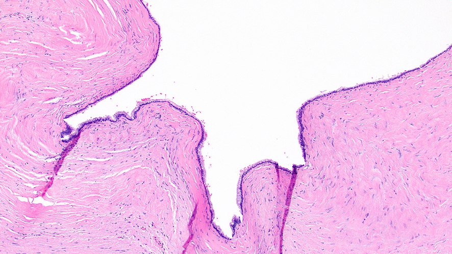
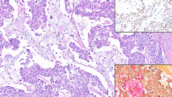
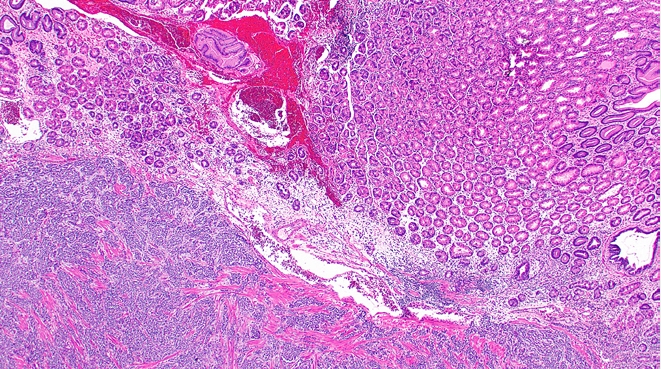
Figure & Data
References
Citations
Citations to this article as recorded by 
 PubReader
 ePub Link
-
 Cite this Article
- Cite this Article
-
- Close
- Download Citation
- Close
 XML Download
- Figure
-
- Related articles
-
- What’s new in neoplastic pulmonary pathology 2026: updates on grading, staging and molecular testing
- What’s new in digital and computational pathology 2026: advances in adoption, standards, AI technologies, and clinical integration
- What's new in molecular genetic pathology 2026: emerging biomarkers for personalized cancer therapies
- What’s new in hematopathology 2025: myeloid neoplasms in the WHO 5th edition and ICC
- What’s new in medical renal pathology 2025: Updates on podocytopathy and immunofluorescence staining in medical kidney
What’s new in gastrointestinal pathology 2026: updates from the latest WHO classification
Fig. 1. Epidermoid metaplasia of the esophagus.
Fig. 2. Polypoid intestinal-type gastric dysplasia.
Fig. 3. Mucinous adenoma of the appendix.
Fig. 4. Squamous cell carcinoma of the colon.
Fig. 5. Small duct (A) and large duct (B) intrahepatic cholangiocarcinoma.
Fig. 6. Simple mucinous cyst of the pancreas.
Fig. 7. Amphicrine-like adenocarcinoma. Inset stains are INSM1 and mucicarmine.
Fig. 8. Type V gastric neuroendocrine tumor.
Fig. 1.
Fig. 2.
Fig. 3.
Fig. 4.
Fig. 5.
Fig. 6.
Fig. 7.
Fig. 8.
What’s new in gastrointestinal pathology 2026: updates from the latest WHO classification


