E-submission
Articles
- Page Path
- HOME > J Pathol Transl Med > Volume 47(2); 2013 > Article
-
Brief Case Report
Mucinous Non-neoplastic Cyst of the Pancreas - Jae Do Yang, Ji Soo Song1, Sang Jae Noh2, Woo Sung Moon2
-
Korean Journal of Pathology 2013;47(2):188-190.
DOI: https://doi.org/10.4132/KoreanJPathol.2013.47.2.188
Published online: April 24, 2013
Department of Surgery, Research Institute of Clinical Medicine and Research Institute for Endocrine Sciences, Chonbuk National University Medical School, Jeonju, Korea.
1Department of Radiology, Research Institute of Clinical Medicine and Research Institute for Endocrine Sciences, Chonbuk National University Medical School, Jeonju, Korea.
2Department of Pathology, Research Institute of Clinical Medicine and Research Institute for Endocrine Sciences, Chonbuk National University Medical School, Jeonju, Korea.
- Corresponding Author: Woo Sung Moon, M.D. Department of Pathology, Chonbuk National University Medical School, 567 Baekje-daero, Deokjin-gu, Jeonju 561-756, Korea. Tel: +82-63-270-3086, Fax: +82-63-270-3135, mws@chonbuk.ac.kr
© 2013 The Korean Society of Pathologists/The Korean Society for Cytopathology
This is an Open Access article distributed under the terms of the Creative Commons Attribution Non-Commercial License (http://creativecommons.org/licenses/by-nc/3.0/) which permits unrestricted non-commercial use, distribution, and reproduction in any medium, provided the original work is properly cited.
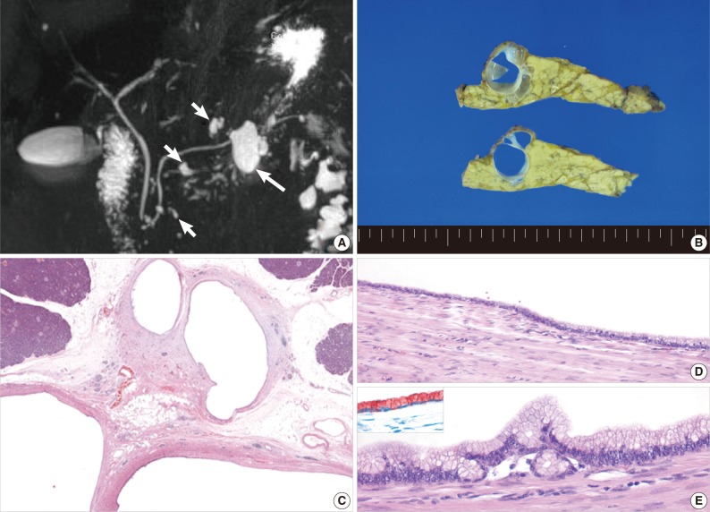
- A 69-year-old woman was admitted to our hospital for evaluation of an asymptomatic cystic mass of the pancreas detected incidentally during a health checkup. The patient did not have a history of smoking or alcohol abuse. The serum amylase level was 60 IU/L (normal range, 54 to 168 IU/L), lipase level was 39 IU/L (normal range, 15 to 60 IU/L), and clinical signs suggesting pancreatitis were not observed. Routine laboratory values, including tumor markers and serological tests for hepatitis B and hepatitis C virus, were negative. Contrast-enhanced abdominal computed tomography demonstrated a multilocular low density cystic lesion measuring approximately 3.0 cm in the tail of the pancreas. The diameter of the main pancreatic duct was normal, and there was no evidence of an enhanced solid portion. Three-dimensional magnetic resonance cholangiopancreatography imaging revealed a multilocular cystic mass in the pancreatic tail with multiple cysts in the head and body of the pancreas (Fig. 1A). Surgical exploration revealed a 3×2.5 cm-sized, multilocular cyst in the tail of the pancreas. The lesion consisted of variable-sized (0.3-1.5 cm) multiple macrocysts separated by thin septa. Mucinous material was observed in several cysts. There was no communication with the main or large branch pancreatic ducts (Fig. 1B). The cysts were lined by a monolayer of cuboidal to columnar epithelial cells with focal pseudostratification (Fig. 1C-E). The cyst walls consisted of paucicellular fibrous tissue. There was no ovarian-like stroma in the cyst walls. There was no evidence of nuclear pleomorphism or mitotic figure, suggesting a malignant tumor. The remaining pancreatic tissue was histologically unremarkable. Immunohistochemically, the duct lining epithelial cells were diffusely strongly positive for keratin 7, 19, and Muc6 and showed weak Muc1 expression, whereas the cells were negative for Muc2 and Muc5Ac (Fig. 1E, inset). On the basis of histological and immunohistochemical findings, a final diagnosis of MNCP was made.
CASE REPORT
- In 2002, Kosmahl et al.1 reported a novel non-neoplastic cystic lesion of the pancreas in five patients designated as MNCP. Later, 21 more cases were reported.2-5 The incidence of MNCP is 2.1-3.4% in resected pancreatic lesions.2,3 MNCP is found over a wide age range (20-88 years) but tends to be more common in females over 50 years of age.1-3 MNCP is found most commonly in the pancreas head (65%, 13/20) and may present as an unilocular or multilocular cyst. The average lesion size is 2.6±2.5 cm (n=20) but can range from 1-12 cm. Most MNCPs present as a single cyst (70%, 14/20), with only 30% presenting with multiple lesions, as in our case. The clinical presentations of MNCP patients are usually non-specific and rarely present as obstructive jaundice due to external compression of the common bile duct. The pathogenesis of MNCP is unclear. Cao et al.2 proposed that MNCPs might be related to acinar-ductal mucinous metaplasia. The key clinicopathological features of MNCP include 1) mucinous differentiation of the lining epithelium; 2) lack of cellular atypia or increased proliferation; 3) a thin rim of supporting, almost acellular stroma; 4) lack of communication with the duct or biliary system; and 5) preferential localization in the pancreatic head.1 Histologically, the differential diagnosis of a MNCP includes intraductal papillary mucinous tumor (IPMT), mucinous cystic neoplasm (MCN), and retention cyst.1-3 One of the most common neoplastic mucinous cystic lesions in the pancreas is IPMT, which has malignant potential.6 MNCP can easily be distinguished from IPMT by its lack of communication with the duct system and papillary proliferation of neoplastic cells.1 In previous studies on mucin expression in MNCP, expression of Muc5Ac and Muc1 was reported in 14 of 20 (70%) and five of 20 (25%) cases, respectively. Expression of Muc2 was not observed in any of these cases.1,2 In the present case, staining for Muc1 and Muc6 was positive, whereas that for Muc2 and Muc5Ac was negative. Absence of Muc2 expression in MNCP is also useful in the differential diagnosis of MNCP and IPMT, because Muc2 is expressed in 71% cases of IPMT.2 MCN should always be included in the differential diagnosis because MNCP and MCN share several features.1 Lining epithelial cells of both lesions are positive for pancreatic ductal cell markers keratin 7, 19, and Muc5Ac.1,7 Although MNCP and MCN have similar epithelial phenotypes, they differ in stromal components and in potential for malignant transformation. We excluded MCN because there was no ovarian-like stroma or dysplastic mucinous lining epithelium, which are necessary for the diagnosis of MCN. Although a retention cyst may share similar mucinous lining epithelium as MNCP, a retention cyst can be excluded by the absence of potential causes for ductal obstruction, such as calculi, chronic pancreatitis, and lack of communication with the duct system. Moreover, retention cysts are usually smaller than 1 cm.1
- The clinical course of MNCP is known as benign.1-3 In the present case, after eight months of follow-up, the patient remained in good health with no evidence of recurrence. Recognition of the clinicopathological features of this lesion is essential for a differential diagnosis and treatment guidance.
DISCUSSION
Acknowledgments
Acknowledgments
- 1. Kosmahl M, Egawa N, Schröder S, Carneiro F, Lüttges J, Klöppel G. Mucinous nonneoplastic cyst of the pancreas: a novel nonneoplastic cystic change? Mod Pathol 2002; 15: 154-158. ArticlePubMedPDF
- 2. Cao W, Adley BP, Liao J, et al. Mucinous nonneoplastic cyst of the pancreas: apomucin phenotype distinguishes this entity from intraductal papillary mucinous neoplasm. Hum Pathol 2010; 41: 513-521. ArticlePubMed
- 3. Kosmahl M, Pauser U, Peters K, et al. Cystic neoplasms of the pancreas and tumor-like lesions with cystic features: a review of 418 cases and a classification proposal. Virchows Arch 2004; 445: 168-178. ArticlePubMedPDF
- 4. Brunner A, Ladurner R, Kosmahl M, Mikuz G, Tzankov A. Mucinous non-neoplastic cyst of the pancreas accompanied by non-parasitic asymptomatic liver cysts. Virchows Arch 2004; 444: 482-484. ArticlePubMedPDF
- 5. Goh BK, Tan YM, Tan PH, Ooi LL. Mucinous nonneoplastic cyst of the pancreas: a truly novel pathological entity? World J Gastroenterol 2005; 11: 2045-2047. ArticlePubMedPMC
- 6. Adsay NV. Cystic neoplasia of the pancreas: pathology and biology. J Gastrointest Surg 2008; 12: 401-404. ArticlePubMedPDF
- 7. Osborn M, van Lessen G, Weber K, Klöppel G, Altmannsberger M. Differential diagnosis of gastrointestinal carcinomas by using monoclonal antibodies specific for individual keratin polypeptides. Lab Invest 1986; 55: 497-504. PubMed
REFERENCES

Figure & Data
References
Citations
- Molecular Characterization of Pancreatic Simple Mucinous Cysts With GNAS Mutation: A Case Report and Literature Review
Shoichiro Mizukami, Koji Imai, Hiroyuki Takahashi, Shingo Shimada, Nobue Tamamura, Miyuki Mori, Koji Nishikawa, Yusuke Ono, Mishie Tanino, Yusuke Mizukami, Hideki Yokoo
Pancreas.2026; 55(1): e75. CrossRef - Simple mucinous cyst: another potential cancer precursor in the pancreas? Case report with molecular characterization and systematic review of the literature
Anna Caterina Milanetto, Alice Sabrina Tonello, Giovanni Valotto, Giada Munari, Claudio Luchini, Matteo Fassan, Claudio Pasquali
Virchows Archiv.2021; 479(1): 179. CrossRef - Mucinous Non-neoplastic Cyst of the Pancreas
Jun Hyung Kim, Dong Eun Park, Keum Ha Choi
The Korean Journal of Gastroenterology.2019; 73(4): 235. CrossRef - Mucinous nonneoplastic cyst of the pancreas: CT and MRI appearances
Kousei Ishigami, Akihiro Nishie, Naoki Mochidome, Yoshiki Asayama, Yasuhiro Ushijima, Daisuke Kakihara, Daisuke Okamoto, Nobuhiro Fujita, Takao Ohtsuka, Yoshihiro Miyasaka, Tomoyuki Hida, Tomoharu Yoshizumi, Hiroshi Honda
Abdominal Radiology.2017; 42(12): 2827. CrossRef - Pathologic Evaluation and Reporting of Intraductal Papillary Mucinous Neoplasms of the Pancreas and Other Tumoral Intraepithelial Neoplasms of Pancreatobiliary Tract
Volkan Adsay, Mari Mino-Kenudson, Toru Furukawa, Olca Basturk, Giuseppe Zamboni, Giovanni Marchegiani, Claudio Bassi, Roberto Salvia, Giuseppe Malleo, Salvatore Paiella, Christopher L. Wolfgang, Hanno Matthaei, G. Johan Offerhaus, Mustapha Adham, Marco J.
Annals of Surgery.2016; 263(1): 162. CrossRef - Rare Nonneoplastic Cysts of Pancreas
Yeon Suk Kim, Jae Hee Cho
Clinical Endoscopy.2015; 48(1): 31. CrossRef
 PubReader
 ePub Link
-
 Cite this Article
- Cite this Article
-
- Close
- Download Citation
- Close
- Figure
-