 E-submission
Articles
- Page Path
- HOME > J Pathol Transl Med > Volume 47(5); 2013 > Article
-
Letter to the Editor
Perineural Involvement in Benign Mixed Tumor - Se Jin Jung, Jong Cheol Lee1, Hye Jeong Choi, Hee Jeong Cha, Young Min Kim, Young Wha Koh, Jae Hee Suh
-
Korean Journal of Pathology 2013;47(5):403-404.
DOI: https://doi.org/10.4132/KoreanJPathol.2013.47.5.403
Published online: October 25, 2013
Department of Pathology, Ulsan University Hospital, Ulsan University College of Medicine, Ulsan, Korea.
1Department of Otorhinolaryngology-Head and Neck Surgery, Ulsan University Hospital, Ulsan University College of Medicine, Ulsan, Korea.
- Corresponding Author: Jae Hee Suh, M.D. Department of Pathology, Ulsan University Hospital, University of Ulsan College of Medicine, 877 Bangeojinsunhwan-doro, Dong-gu, Ulsan 682-714, Korea. Tel: +82-52-250-7263, Fax: +82-52-252-3024, drjhs@uuh.ulsan.kr
• Received: July 4, 2013 • Revised: September 13, 2013 • Accepted: September 24, 2013
© 2013 The Korean Society of Pathologists/The Korean Society for Cytopathology
This is an Open Access article distributed under the terms of the Creative Commons Attribution Non-Commercial License (http://creativecommons.org/licenses/by-nc/3.0/) which permits unrestricted non-commercial use, distribution, and reproduction in any medium, provided the original work is properly cited.
- 1. Ali TZ, Epstein JI. Perineural involvement by benign prostatic glands on needle biopsy. Am J Surg Pathol 2005; 29: 1159-1163. ArticlePubMed
- 2. Skidmore RA, Woosley JT, Tomsick RS. Renaut bodies: benign disease process mimicking neurotropic tumor infiltration. Dermatol Surg 1996; 22: 969-971. PubMed
REFERENCES
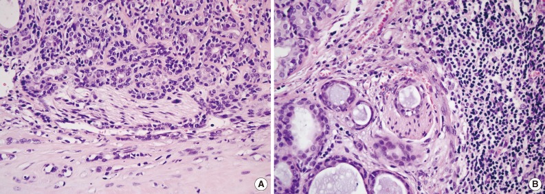
Figure & Data
References
Citations
Citations to this article as recorded by 
- Sclerosing Odontogenic Carcinoma: A Review of a Challenging Entity in Malignant Odontogenic Classification With a Focus on Perineural Invasion and Cellular Proliferation
Merva Soluk‐Tekkesin, Keith David Hunter, Ronell Bologna‐Molina, Kelly Magliocca, Liam Robinson, Akinyele Olumuyiwa Adisa, Jiang Li, Elizabeth Ann Bilodeau, Haizal Mohd Hussaini, Madhu Shrestha, Ricardo Santiago Gomez, Willie van Heerden, Wanninayake Mudi
Journal of Oral Pathology & Medicine.2026;[Epub] CrossRef - Benign Pleomorphic Adenoma in the Facial Nerve
Gemma E. Hogg, Richard A. Steven, Patrick M. Spielmann
Ear, Nose & Throat Journal.2020; 99(6): 361. CrossRef - Benign pleomorphic adenoma of minor salivary gland showing perineural invasion: a rare entity
Rahul Jayaram, Dipen Patel, Vijay Santhanam
British Journal of Oral and Maxillofacial Surgery.2015; 53(1): 81. CrossRef
 PubReader
 ePub Link
-
 Cite this Article
- Cite this Article
-
- Close
- Download Citation
- Close
- Figure
-

Perineural Involvement in Benign Mixed Tumor

Fig. 1 Histologic findings. Two peripheral nerve twigs with perineural (A) and neural (B) tumor involvement in the subcapsular space and the fibrous tumor capsule.
Fig. 1
Perineural Involvement in Benign Mixed Tumor

