Articles
- Page Path
- HOME > J Pathol Transl Med > Volume 49(1); 2015 > Article
-
Brief Case Report
A Rare Case of Mixed Type A Thymoma and Micronodular Thymoma with Lymphoid Stroma - Yoon Jin Cha, Joungho Han, Jimin Kim, Kyung Soo Lee1, Young Mog Shim2
-
Journal of Pathology and Translational Medicine 2015;49(1):75-77.
DOI: https://doi.org/10.4132/jptm.2014.10.27
Published online: January 15, 2015
Department of Pathology, Samsung Medical Center, Sungkyunkwan University School of Medicine, Seoul, Korea
1Department of Radiology, Samsung Medical Center, Sungkyunkwan University School of Medicine, Seoul, Korea
2Department of Thoracic Surgery, Samsung Medical Center, Sungkyunkwan University School of Medicine, Seoul, Korea
- Corresponding Author: Joungho Han, M.D. Department of Pathology, Samsung Medical Center, Sungkyunkwan University School of Medicine, 81 Irwon-ro, Gangnam-gu, Seoul 135-710, Korea Tel: +82-2-3410-2800, Fax: +82-2-3410-0025, E-mail: hanjho@skku.edu
• Received: September 2, 2014 • Accepted: October 24, 2014
© 2015 The Korean Society of Pathologists/The Korean Society for Cytopathology
This is an Open Access article distributed under the terms of the Creative Commons Attribution Non-Commercial License (http://creativecommons.org/licenses/by-nc/3.0/) which permits unrestricted noncommercial use, distribution, and reproduction in any medium, provided the original work is properly cited.
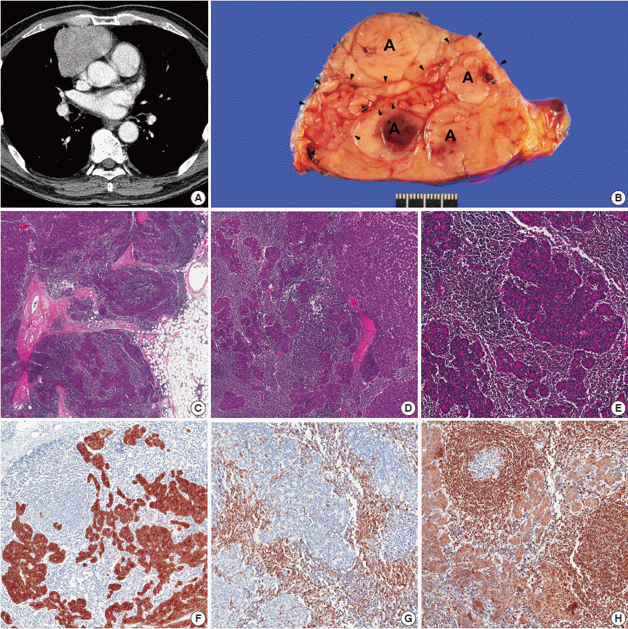
- A 63-year-old man presented with a mediastinal mass incidentally found on routine chest radiograph during a regular health check-up. He had a history of hypertension and diabetes treated with medication. There was no evidence of myasthenia gravis. On further evaluation by chest computed tomography (CT), a 7.3-cm mediastinal mass was identified in the right anterior mediastinum (Fig. 1A). The mass had a lobulated contour, and intratumoral septation suggested a thymic epithelial tumor. No enlargement of mediastinal lymph nodes or pleural seeding was found on CT. He received extended thymectomy via median sternotomy. During the operation, there was no pleural adhesion or invasion into surrounding structures.
- On cut sections, the tumor measured 7×6 cm and was enclosed by a thin fibrous capsule. Vague, tan-colored nodules averaging 1.5 cm in size were identified. These were partly separated by thin fibrous septa and areas of numerous tiny cobblestone-like micronodules (Fig. 1B). On histological examination, two different types of thymoma were noted, showing partly infiltrative growth into adjacent fat tissue (Fig. 1C). Large nodules seen were composed of epithelial cells with sparse lymphocytes. Spindled tumor cells formed an organoid pattern, arranged in short fascicles and a solid sheet. The tumor cells had elongated, bland nuclei with fine chromatin and inconspicuous small nucleoli. These characteristics, together with positivity for cytokeratin (CK) and Bcl-2, were compatible with World Health Organization (WHO) type A thymoma. The type A area was ill-defined, showing gradual transition into the rest of the tumor, which harbored a mixture of epithelial and lymphoid components (Fig. 1D). Multiple small epithelial nodules were embedded in the lymphoid stroma. The tumor cells from the smaller nodules were spindled, bland-looking and formed pseudorosettes that had elongated nuclei with fine chromatin and inconspicuous nucleoli, similar to type A thymoma. Epithelial tumor nodules were segregated by lymphoid stroma that occasionally harbored germinal centers (Fig. 1E). Tumor cells were strongly positive for CK and moderately positive for Bcl-2 (Fig. 1F, G). Lymphoid stroma was positive for CD5 (Fig. 1G). Bcl-2 was diffusely positive throughout the lymphoid stroma (Fig. 1H) but was negative in germinal centers. Additional CD20 and CD3 immunohistochemical staining showed reactive B- and T-cells in the lymphoid stroma. The patient was diagnosed ultimately with mixed type A thymoma and MNTLS.
CASE REPORT
- MNTLS, first coined by Suster and Moran [1], is a rare subtype of thymoma. A micronodular pattern of epithelial components is found in approximately 10% of type A and type AB thymoma [2]. Cases accompanied by thymic cyst [3], heart myxoma [4], and ectopic lesions arising in the salivary gland [5] have been reported. Characteristically, MNTLS demonstrates small nodules of epithelial components resembling those of type A thymoma within lymphoid stroma that is predominantly composed of B-cells with occasional germinal centers. Being devoid of epithelial components is helpful in excluding type AB thymoma, which also contains epithelial components and lymphoid follicles. With its rich B-cells, occasional germinal centers, and nodules of epithelial cells, MNTLS could be misdiagnosed as metastatic carcinoma in a small biopsy specimen [4]. Although the histogenesis of MNTLS is not yet clarified, its medullary epithelial cells suggest MNTLS may be a variant of type A thymoma in the setting of thymic B-cell hyperplasia [1]. Bcl-2, a B-cell marker, is expressed in type A thymoma and thymic carcinoma [6]. We observed Bcl-2 expression in both type A thymoma and MNTLS. These findings support the hypothesis that MNTLS originates from type A thymoma. Stobel et al. [7] found that 33% of MNTLS had lymphoid stroma of a monoclonal B-cell population whereas thymic lymphoid hyperplasia or other types of thymoma had a polyclonal population of lymphocytes. Because of the monoclonal lymphoid stroma, they suggested that MNTLS might be a precursor lesion of mediastinal lymphoma [7]. In the present case, the lymphoid stroma was positive for both CD20 and CD3, reflecting a polyclonal lymphocyte nature. Although patient outcomes of MNTLS have not been established because of its rarity, MNTLS has reported cases that have shown excellent prognosis so far [1,8]. Ishikawa et al. [9] explained that an immune response induced by intratumoral MNTLS Langerhans cells would contribute to improved patient outcomes.
- We report a case of mixed thymoma containing type A thymoma and MNTLS. In the present case, epithelial components of type A thymoma and MNTLS showed similar histologic and immunohistochemical profiles with areas of gradual transition, suggesting type A thymoma and MNTLS share a common histogenesis. Although this tumor showed focal infiltrative growth of MNTLS, it was postulated that this tumor would have a good clinical course.
DISCUSSION
Fig. 1.Mixed Type A Thymoma and Micronodular Thymoma with Lymphoid Stroma. (A) Chest computed tomography imaging demonstrates a lobulated mass in the right anterior mediastinum. (B) Grossly, the tumor is encapsulated with a thin fibrous capsule, and is composed of vague nodules with tiny internodular micronodules, which matched with both type A thymoma (capital A on figure) and micronodular thymoma (arrowheads). (C) A microscopic area of infiltrative growth into fat tissue is noted. (D) A gradual transition is found between type A thymoma and micronodular thymoma with lymphoid stroma (MNTLS). (E) The MNTLS tissue has epithelial micronodules arranged in pseudorosettes that are separated by lymphoid stroma. (F) The tumor cells are strongly positive for cytokeratin, whereas lymphoid stroma lacks any epithelial component. (G) Lymphoid stroma is diffusely positive for CD5. (H) Together with type A thymoma, the epithelial component of MNTLS is positive for Bcl-2. Lymphoid stroma, except for within germinal centers, also is positive for Bcl-2.


- 1. Suster S, Moran CA. Micronodular thymoma with lymphoid B-cell hyperplasia: clinicopathologic and immunohistochemical study of eighteen cases of a distinctive morphologic variant of thymic epithelial neoplasm. Am J Surg Pathol 1999; 23: 955-62. PubMed
- 2. Pan CC, Chen WY, Chiang H. Spindle cell and mixed spindle/lymphocytic thymomas: an integrated clinicopathologic and immunohistochemical study of 81 cases. Am J Surg Pathol 2001; 25: 111-20. PubMed
- 3. Kim NR, Lee JI, Ha SY. Micronodular thymoma with lymphoid stroma in a multilocular thymic cyst: a case study. Korean J Pathol 2013; 47: 392-4. ArticlePubMedPMC
- 4. Song HJ, Han J, Kim TS. Micronodular thymoma with lymphoid stroma: a case report. J Lung Cancer 2011; 10: 56-8. Article
- 5. Mende S, Moschopulos M, Marx A, Laeng RH. Ectopic micronodular thymoma with lymphoid stroma. Virchows Arch 2004; 444: 397-9. ArticlePubMedPDF
- 6. Hiroshima K, Iyoda A, Toyozaki T, et al. Proliferative activity and apoptosis in thymic epithelial neoplasms. Mod Pathol 2002; 15: 1326-32. ArticlePubMed
- 7. Strobel P, Marino M, Feuchtenberger M, et al. Micronodular thymoma: an epithelial tumour with abnormal chemokine expression setting the stage for lymphoma development. J Pathol 2005; 207: 72-82. ArticlePubMed
- 8. Tateyama H, Saito Y, Fujii Y, et al. The spectrum of micronodular thymic epithelial tumours with lymphoid B-cell hyperplasia. Histopathology 2001; 38: 519-27. ArticlePubMedPDF
- 9. Ishikawa Y, Tateyama H, Yoshida M, et al. Micronodular thymoma with lymphoid stroma: an immunohistochemical study of the distribution of Langerhans cells and mature dendritic cells in six patients. Histopathology 2014 Apr 4 [Epub]. http://dx.doi.org/10.1111/his.12428. Article
REFERENCES
Figure & Data
References
Citations
Citations to this article as recorded by 

-
GTF2I mutation in micronodular thymoma with lymphoid stroma
Andrea Bille, Katherine Fryer, Andrew Wallace, Daisuke Nonaka
Journal of Clinical Pathology.2024; 77(2): 125. CrossRef - Minimally invasive thoracoscopic resection of a micronodular thymoma with lymphoid stroma via a subxiphoid single-incision approach: A case report
Qiang Wu, Kun Qiao, Xiaoming Zhang, Zizi Zhou
Medicine.2024; 103(36): e39637. CrossRef - Constant p.L424H Mutation in GTF2I in Micronodular Thymomas With Lymphoid Stroma: Evidence Supporting Close Relationship With Type A and AB Thymomas
Min-Shu Hsieh, Hua-Lin Kao, Wen-Chang Huang, Shu-Ying Wang, Shin-Ying Lin, Ping-Yuan Chu, Chin-Chen Pan, Teh-Ying Chou, Hsiang-Ling Ho, Yi-Chen Yeh
Modern Pathology.2023; 36(2): 100008. CrossRef - Comparative clinicopathological and immunohistochemical study of micronodular thymoma and micronodular thymic carcinoma with lymphoid stroma
Ping Ping Liu, Yun Chao Su, Yun Niu, Yan Fen Shi, Jie Luo, Ding Rong Zhong
Journal of Clinical Pathology.2022; 75(10): 702. CrossRef - Micronodular Thymoma With Lymphoid Stroma: A Trio of Cases, With Diverse-associated Histological Features
Neha Bakshi, Shashi Dhawan, Seema Rao, Kishan Singh Rawat
International Journal of Surgical Pathology.2021; 29(6): 693. CrossRef - A case of thymoma with type A and micronodular thymoma with lymphoid stroma elements
Clayton E. Kibler, Matthew J. Cecchini, Marie-Christine Aubry, Said F. Yassin, Julie K. Harrington
Human Pathology: Case Reports.2021; 23: 200487. CrossRef - Micronodular thymoma with lymphoid stroma: a clinicopathological study of five cases
Katherine R. Hulme, Annabelle Mahar, Christopher Cao, Brian McCaughan, Wendy A. Cooper
Pathology.2021; 53(7): 930. CrossRef - Mixed Type A Thymoma and Micronodular Thymoma With Lymphoid Stroma
Renqing Wang, Ling Nie
International Journal of Surgical Pathology.2018; 26(4): 336. CrossRef - Cystic Micronodular Thymoma. Report of a Case
Mlika M
Journal of Clinical, Medical and Experimental Images.2017; 1(1): 001. CrossRef - Two Invasive Thymomas Incidentally Found during Coronary Artery Bypass Graft Surgery
Navid Omidifar, Maral Mokhtari, Mansoureh Shokripour
Case Reports in Pathology.2016; 2016: 1. CrossRef
 PubReader
 ePub Link
-
 Cite this Article
- Cite this Article
-
- Close
- Download Citation
- Close
- Figure
A Rare Case of Mixed Type A Thymoma and Micronodular Thymoma with Lymphoid Stroma

Fig. 1. Mixed Type A Thymoma and Micronodular Thymoma with Lymphoid Stroma. (A) Chest computed tomography imaging demonstrates a lobulated mass in the right anterior mediastinum. (B) Grossly, the tumor is encapsulated with a thin fibrous capsule, and is composed of vague nodules with tiny internodular micronodules, which matched with both type A thymoma (capital A on figure) and micronodular thymoma (arrowheads). (C) A microscopic area of infiltrative growth into fat tissue is noted. (D) A gradual transition is found between type A thymoma and micronodular thymoma with lymphoid stroma (MNTLS). (E) The MNTLS tissue has epithelial micronodules arranged in pseudorosettes that are separated by lymphoid stroma. (F) The tumor cells are strongly positive for cytokeratin, whereas lymphoid stroma lacks any epithelial component. (G) Lymphoid stroma is diffusely positive for CD5. (H) Together with type A thymoma, the epithelial component of MNTLS is positive for Bcl-2. Lymphoid stroma, except for within germinal centers, also is positive for Bcl-2.
Fig. 1.
A Rare Case of Mixed Type A Thymoma and Micronodular Thymoma with Lymphoid Stroma

 E-submission


