- Primary carcinoid tumor in the external auditory canal
-
Dong Hae Chung, Gyu Cheol Han, Na Rae Kim
-
J Pathol Transl Med. 2020;54(2):184-187. Published online November 13, 2019
-
DOI: https://doi.org/10.4132/jptm.2019.11.07
-
-
7,770
View
-
166
Download
-
4
Web of Science
-
3
Crossref
-
 Abstract Abstract
 PDF PDF
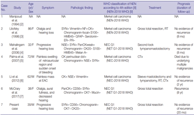
- A 39-year-old man visited the department of otolaryngology due to an ongoing hearing disturbance that had lasted for 1 year. Temporal bone computed tomography revealed soft tissue density nearly obliterating the left external auditory canal (EAC). The mass was composed of sheets of round tumor cells containing moderate amounts of fine granular cytoplasm and salt and pepper chromatin. Neither mitosis nor necrosis was found. The Ki-67 proliferation index was less than 2%. Cells were positive for CD56 and synaptophysin but negative for chromogranin, cytokeratin (CK) 20, and CK7. Based on these findings, the tumor was diagnosed as a carcinoid tumor, well differentiated neuroendocrine carcinoma, grade 1 (G1) according to current World Health Organization (WHO) classification of head and neck tumors; and a neuroendocrine tumor, G1 according to neuroendocrine neoplasm (NEN)-2018 WHO standard classification. He remained free of local recurrence and metastasis after 20 months of follow up. To date, only six cases of primary NENs in the EAC have been reported. Metastatic tumor should be included in the differential diagnoses. Because of its rarity, the prognosis and treatment have not yet been clarified.
-
Citations
Citations to this article as recorded by  - First Report on a Rare Poorly Differentiated Neuroendocrine Tumour of the External Auditory Canal Involving Pinna
Akash Varshney, Amit Kumar Tyagi, Prashant Durgapal, Kajal Mahto, Akhilesh Chandra Yadav, Ankita Semwal
Indian Journal of Otolaryngology and Head & Neck Surgery.2025; 77(4): 1922. CrossRef - Incidental finding of a neuroendocrine neoplasm in a suspected ear canal exostosis
Alexander Wieck Fjaeldstad, Gerda Elisabeth Villadsen, Gitte Dam, Stephen Jacques Hamilton-Dutoit, Thomas Winther Frederiksen
Otolaryngology Case Reports.2022; 22: 100394. CrossRef - 68Ga-DOTATATE Uptake in Well-Differentiated Neuroendocrine Tumor of the External Auditory Canal
Özge Erol Fenercioğlu, Ediz Beyhan, Rahime Şahin, Mehmet Can Baloğlu, Tevfik Fikret Çermik
Clinical Nuclear Medicine.2022; 47(8): e552. CrossRef
- Contribution of cytologic examination to diagnosis of poorly differentiated thyroid carcinoma
-
Na Rae Kim, Jae Yeon Seok, Yoo Seung Chung, Joon Hyop Lee, Dong Hae Chung
-
J Pathol Transl Med. 2020;54(2):171-178. Published online February 5, 2020
-
DOI: https://doi.org/10.4132/jptm.2019.12.03
-
-
7,173
View
-
201
Download
-
3
Web of Science
-
5
Crossref
-
Abstract
PDF
- Background
The cytologic diagnosis of poorly differentiated thyroid carcinoma (PDTC) is difficult because it lacks salient cytologic findings and shares cytologic features with more commonly encountered neoplasms. Due to diverse cytologic findings and paucicellularity of PDTC, standardization of cytologic diagnostic criteria is limited. The purpose of this study is to investigate and recognize diverse thyroid findings of fine needle aspiration (FNA) cytology and frozen smear cytology in diagnosis of this rare but aggressive carcinoma.
Methods
The present study included six cases of FNA cytology and frozen smears of histologically diagnosed PDTCs.
Results
PDTC showed cytologic overlap with well-differentiated thyroid carcinomas (WDTCs). Five of six cases showed dedifferentiation arising from well differentiated thyroid carcinomas. Only one de novo PDTC showed highly cellular smears composed of discohesive small cells, high nuclear/cytoplasmic (N/C) ratio, prominent micronucleoli, and irregular nuclei. Retrospectively reviewed, these findings are highly suspicious for PDTC. Cytologic findings of nuclear atypia, pleomorphism, and irregularity were frequently found, whereas scattered small cells were seen only in the de novo case.
Conclusions
Heterogeneous cytologic findings of PDTCs are shared with those of WDTCs and contribute to difficult preoperative cytologic diagnoses. Most PDTCs show dedifferentiation from WDTCs. Albeit rare, de novo PDTC should be considered with cytology showing discohesive small cells with high N/C ratio. This will enable precise diagnosis and prompt treatment of this aggressive malignancy
-
Citations
Citations to this article as recorded by - Non-papillary thyroid carcinoma diagnoses in The Bethesda System for Reporting Thyroid Cytopathology categories V and VI: An institutional experience
Myunghee Kang, Na Rae Kim, Jae Yeon Seok
Annals of Diagnostic Pathology.2024; 71: 152263. CrossRef - Cytologic features of differentiated high‐grade thyroid carcinoma: A multi‐institutional study of 40 cases
Vanda F. Torous, Tikamporn Jitpasutham, Zubair Baloch, Richard L. Cantley, Darcy A. Kerr, Xiaoying Liu, Zahra Maleki, Ross Merkin, Vania Nosé, Liron Pantanowitz, Isabella Tondi Resta, Esther D. Rossi, William C. Faquin
Cancer Cytopathology.2024; 132(8): 525. CrossRef - An Unexpected Finding of Poorly Differentiated Thyroid Carcinoma in a Toxic Thyroid Nodule
Kimberly Yuang, Huda Al-Bahadili, Alan Chang
JCEM Case Reports.2023;[Epub] CrossRef - Revisiting the cytomorphological features of poorly differentiated thyroid carcinoma: a comparative analysis with indeterminate thyroid fine-needle aspiration samples
Yazeed Alwelaie, Ali Howaidi, Mohammed Tashkandi, Ahmad Almotairi, Hisham Saied, Moammar Muzzaffar, Doaa Alghamdi
Journal of the American Society of Cytopathology.2023; 12(5): 331. CrossRef - Characterization of the genomic alterations in poorly differentiated thyroid cancer
Yeeun Lee, SeongRyeol Moon, Jae Yeon Seok, Joon-Hyop Lee, Seungyoon Nam, Yoo Seung Chung
Scientific Reports.2023;[Epub] CrossRef
- Comparison of papanicolaou smear and human papillomavirus (HPV) test as cervical screening tools: can we rely on HPV test alone as a screening method? An 11-year retrospective experience at a single institution
-
Myunghee Kang, Seung Yeon Ha, Hyun Yee Cho, Dong Hae Chung, Na Rae Kim, Jungsuk An, Sangho Lee, Jae Yeon Seok, Juhyeon Jeong
-
J Pathol Transl Med. 2020;54(1):112-118. Published online January 15, 2020
-
DOI: https://doi.org/10.4132/jptm.2019.11.29
-
-
10,496
View
-
251
Download
-
19
Web of Science
-
20
Crossref
-
Abstract
PDF
- Background
The decrease in incidence of cervical dysplasia and carcinoma has not been as dramatic as expected with the development of improved research tools and test methods. The human papillomavirus (HPV) test alone has been suggested for screening in some countries. The National Cancer Screening Project in Korea has applied Papanicolaou smears (Pap smears) as the screening method for cervical dysplasia and carcinoma. We evaluated the value of Pap smear and HPV testing as diagnostic screening tools in a single institution.
Methods
Patients co-tested with HPV test and Pap smear simultaneously or within one month of each other were included in this study. Patients with only punch biopsy results were excluded because of sampling errors. A total of 999 cases were included, and the collected reports encompassed results of smear cytology, HPV subtypes, and histologic examinations.
Results
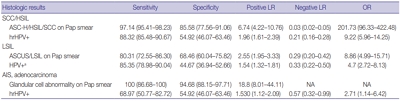
Sensitivity and specificity of detecting high-grade squamous intraepithelial lesion (HSIL) and squamous cell carcinoma (SCC) were higher for Pap smears than for HPV tests (sensitivity, 97.14%; specificity, 85.58% for Pap smears; sensitivity, 88.32%; specificity, 54.92% for HPV tests). HPV tests and Pap smears did not differ greatly in detection of low-grade squamous intraepithelial lesion (85.35% for HPV test, 80.31% for Pap smears). When atypical glandular cells were noted on Pap smears, the likelihood for histologic diagnosis of adenocarcinoma following Pap smear was higher than that of high-risk HPV test results (18.8 and 1.53, respectively).
Conclusions
Pap smears were more useful than HPV tests in the diagnosis of HSIL, SCC, and glandular lesions.
-
Citations
Citations to this article as recorded by - Detection of cervical precancerous lesions and cancer by small-scale RT-qPCR analysis of oppositely deregulated mRNAs pairs in cytological smears
Anastasia A. Artyukh, Mikhail K. Ivanov, Sergei E. Titov, Victoria V. Dzyubenko, Sergey E. Krasilnikov, Anastasia O. Shumeikina, Nikita A. Afanasev, Anastasia V. Malek, Sergei A. Glushkov, Eduard F. Agletdinov
Frontiers in Oncology.2025;[Epub] CrossRef - High burden of abnormal cervical smears in South African primary health care: health programmes implications
Olufemi B Omole, Joel M Francis, John M Musonda, Pumla P Sodo, Elizabeth Reji, Nyundu S J Phukuta, Honey L M Mabuza, Joyce S Musonda, Jimmy Akii, John V Ndimande, Olalekan A Ayo-Yusuf
Health Promotion International.2025;[Epub] CrossRef - Bibliometric analysis: a study of the microenvironment in cervical cancer (2000-2024)
Yun-Tao Zhang, Yan-Ni Wei, Chen-Chen Liu, Mai-Qing Yang
Frontiers in Oncology.2025;[Epub] CrossRef - Challenges in the diagmosis of cervical pathologies
D. Y. Chernov, O. A. Tikhonovskaya, S. V. Logvinov, I. A. Petrov, Y. S. Yuriev, A. A. Zhdankina, A. V. Gerasimov, I. V. Zingalyuk, G. A. Mikheenko
Bulletin of Siberian Medicine.2024; 22(4): 201. CrossRef - “Barriers and Advantages of Self-Sampling Tests, for HPV Diagnosis: A Qualitative Field Experience Before Implementation in a Rural Community in Ecuador”
Bernardo Vega-Crespo, Vivian Alejandra Neira, Ruth Maldonado - Rengel, Diana López, Dayanara Delgado-López, Gabriela Guerra Astudillo, Veronique Verhoeven
International Journal of Women's Health.2024; Volume 16: 947. CrossRef - Cervical Human Papillomavirus Testing
Carol N. Rizkalla, Eric C. Huang
Surgical Pathology Clinics.2024; 17(3): 431. CrossRef - Segmentation of Overlapping Cells in Cervical Cytology Images: A Survey
E Chen, Hua-Nong Ting, Joon Huang Chuah, Jun Zhao
IEEE Access.2024; 12: 114170. CrossRef - Knowledge and awareness regarding pap test and HPV typing for cervical cancer screening in Edo North, Nigeria
Amina Momodu, Johnsolomon Eghosa Ohenhen, Godfrey Innocent Iyare, Musa Abidemi Muhibi, Godwin Avwioro
Discover Public Health.2024;[Epub] CrossRef - Colposcopy Value in Young Child-bearing Women: Is New Recommendations Necessary?
Fahimeh Sabet, Avishan Aminizad, Fariba Behnamfar, Tajossadat Allameh, Seyedeh Ghazal Shahrokh, Rostami Koushan, Amirmohammad Taravati, Leila Mousavi Seresht
Advanced Biomedical Research.2024;[Epub] CrossRef - Selection of endogenous control and identification of significant microRNA deregulations in cervical cancer
T. Stverakova, I. Baranova, P. Mikyskova, B. Gajdosova, H. Vosmikova, J. Laco, V. Palicka, H. Parova
Frontiers in Oncology.2023;[Epub] CrossRef - Cytology Versus Molecular Diagnosis of HPV for Cervical Cancer Screening. Comparison of the Diagnostic Properties of Four Tests in a Rural Community of Cuenca Ecuador
Bernardo Vega Crespo, Vivian Alejandra Neira, Rocío Murillo, Cristina Ochoa Avilés
ESPOCH Congresses: The Ecuadorian Journal of S.T.E.A.M..2023; 3(1): 139. CrossRef - Attitudes towards prevention of cervical cancer and early diagnosis among female academicians
Nurhan Doğan, Gamze Fışkın
Journal of Obstetrics and Gynaecology Research.2022; 48(6): 1433. CrossRef - Role of Self-Sampling for Cervical Cancer Screening: Diagnostic Test Properties of Three Tests for the Diagnosis of HPV in Rural Communities of Cuenca, Ecuador
Bernardo Vega Crespo, Vivian Alejandra Neira, José Ortíz Segarra, Ruth Maldonado Rengel, Diana López, María Paz Orellana, Andrea Gómez, María José Vicuña, Jorge Mejía, Ina Benoy, Tesifón Parrón Carreño, Veronique Verhoeven
International Journal of Environmental Research and Public Health.2022; 19(8): 4619. CrossRef - Utility of Scoring System for Screening and Early Warning of Cervical Cancer Based on Big Data Analysis
Dan Hou, Binjie Yang, Yangdan Li, Ming Sun
Frontiers in Public Health.2022;[Epub] CrossRef - Evaluation of Urine and Vaginal Self-Sampling versus Clinician-Based Sampling for Cervical Cancer Screening: A Field Comparison of the Acceptability of Three Sampling Tests in a Rural Community of Cuenca, Ecuador
Bernardo Vega Crespo, Vivian Alejandra Neira, José Ortíz S, Ruth Maldonado-Rengel, Diana López, Andrea Gómez, María José Vicuña, Jorge Mejía, Ina Benoy, Tesifón Parrón Carreño, Veronique Verhoeven
Healthcare.2022; 10(9): 1614. CrossRef - Diagnostic distribution and pitfalls of glandular abnormalities in cervical cytology: a 25-year single-center study
Jung-A Sung, Ilias P. Nikas, Haeryoung Kim, Han Suk Ryu, Cheol Lee
Journal of Pathology and Translational Medicine.2022; 56(6): 354. CrossRef - Primary screening of cervical cancer by Pap smear in women of reproductive age group
Ruchi Mishra, Dakshina Bisht, Manisha Gupta
Journal of Family Medicine and Primary Care.2022; 11(9): 5327. CrossRef - Comparison of Learning Transfer Using Simulation Problem-Based Learning and Demonstration: An Application of Papanicolaou Smear Nursing Education
Jeongim Lee, Hae Kyoung Son
International Journal of Environmental Research and Public Health.2021; 18(4): 1765. CrossRef - Investigating host-virus interaction mechanism and phylogenetic analysis of viral proteins involved in the pathogenesis
Ahmad Abu Turab Naqvi, Farah Anjum, Alaa Shafie, Sufian Badar, Abdelbaset Mohamed Elasbali, Dharmendra Kumar Yadav, Md. Imtaiyaz Hassan, Timir Tripathi
PLOS ONE.2021; 16(12): e0261497. CrossRef - Utility of Human Papillomavirus Testing for Cervical Cancer Screening in Korea
Mee-seon Kim, Eun Hee Lee, Moon-il Park, Jae Seok Lee, Kisu Kim, Mee Sook Roh, Hyoun Wook Lee
International Journal of Environmental Research and Public Health.2020; 17(5): 1726. CrossRef
- Primary Necrobiotic Xanthogranulomatous Sialadenitis with Submandibular Gland Localization without Skin Involvement
-
Myunghee Kang, Na Rae Kim, Dong Hae Chung, Jae Yeon Seok, Dong Young Kim
-
J Pathol Transl Med. 2019;53(4):261-265. Published online January 16, 2019
-
DOI: https://doi.org/10.4132/jptm.2019.01.08
-
-
7,188
View
-
166
Download
-
1
Web of Science
-
5
Crossref
-
Abstract
PDF
- Necrobiotic xanthogranulomatous reaction is a multiorgan, non-Langerhans cell histiocytosis with an unknown etiology. Occurrence in the salivary gland is extremely rare. We recently identified a case of necrobiotic xanthogranulomatous sialadenitis in a 73-year-old Korean woman who presented with a painless palpable lesion in the chin. There was no accompanying cutaneous lesion. Partial resection and subsequent wide excision with neck dissection were performed. Pathological examination showed a severe inflammatory lesion that included foamy macrophages centrally admixed with neutrophils, eosinophils, lymphocytes, plasma cells, and scattered giant cells, as well as necrobiosis. During the 12-month postoperative period, no grossly remarkable change in size was noted. Necrobiotic xanthogranulomatous inflammation may be preceded by or combined with hematologic malignancy. Although rare, clinicians and radiologists should be aware that an adhesive necrobiotic xanthogranuloma in the salivary gland may present with a mass-like lesion. Further evaluation for hematologic disease and close follow-up are needed when a pathologic diagnosis is made.
-
Citations
Citations to this article as recorded by - Five Cases of Xanthogranulomatous Sialadenitis
Satoshi Kiyama, Hiroyuki Iuchi, Kotoko Ito, Kengo Nishimoto, Tsutomu Matsuzaki, Masaru Yamashita
Practica Oto-Rhino-Laryngologica.2022; 115(4): 315. CrossRef - Xanthogranulomatous change in a pleomorphic adenoma: An extremely rare variant/degenerative change. Is it fine needle aspiration induced?
Mukta Pujani, Dipti Sidam, Kanika Singh, Aparna Khandelwal, Khushbu Katarya
Diagnostic Cytopathology.2021;[Epub] CrossRef - A Case of Xanthogranulomatous Sialadenitis with Facial Palsy Mimicking Malignancy
Sang Hyun Kim, Sun Woo Kim, Sang Hyuk Lee
Korean Journal of Otorhinolaryngology-Head and Neck Surgery.2021; 64(6): 422. CrossRef - Xanthogranulomatous Sialadenitis, an Uncommon Reactive Change is Often Associated with Warthin’s Tumor
Lihong Bu, Hui Zhu, Emilian Racila, Sobia Khaja, David Hamlar, Faqian Li
Head and Neck Pathology.2020; 14(2): 525. CrossRef - A Case of Xanthogranulomatous Sialadenitis of the Sublingual Gland:A Review of Literature
Naoya KITAMURA, Seiji OHNO, Tetsuya YAMAMOTO
Journal of Japanese Society of Oral Medicine.2019; 25(1): 20. CrossRef
- Frozen Cytology of Meningeal Malignant Solitary Fibrous Tumor/Hemangiopericytoma
-
Myunghee Kang, Na Rae Kim, Dong Hae Chung, Gie-Taek Yie
-
J Pathol Transl Med. 2019;53(3):192-197. Published online April 11, 2019
-
DOI: https://doi.org/10.4132/jptm.2019.03.20
-
-
6,721
View
-
158
Download
-
6
Web of Science
-
7
Crossref
-
Abstract
PDF
- A 51-year-old woman presented with severe dizziness. The brain magnetic resonance image revealed a 5.5 cm multiloculated mass with a thick rim in the left temporal lobe. Cytological examination of frozen diagnosis of the mass showed hypercellular sheets of round and rhabdoid cells in a hemorrhagic background, and two mitotic figures were observed. Histologically, the excised dura-based mass consisted of predominantly round cells with small foci of rhabdoid tumor cells in a pseudoalveolar pattern in a hemorrhagic background, and the cells showed nuclear positivity for signal transducer and activator of transcription 6 as well as frequent mitosis. The mass was diagnosed as a grade 3 solitary fibrous tumor (SFT)/hemangiopericytoma (HPC). The cytological diagnosis of SFT/HPC is challenging because of the heterogeneous cytological findings, such as histological heterogeneity, and because there are no standardized cytological criteria for malignant SFT/HPC. Cytological findings, such as singly scattered small cells, hypercellularity, rare ropy collagen, and round and rhabdoid cells with pseudoalveolar pattern, may assist in the diagnosis of malignant SFT/HPC.
-
Citations
Citations to this article as recorded by - A Rare Case of Cervical Solitary Fibrous Tumor in a Pediatric Patient: Case Report and Literature Review
Eleonora Becattini, Lorenzo Sgarbanti, Giuseppina Bevacqua, Valentina Grespi, Carlo Conti
NeuroSci.2025; 6(2): 49. CrossRef - Cytologic features of mesenchymal, melanocytic and haematolymphoid tumours of the central nervous system and metastases
Carmen Bárcena, José A. Jiménez‐Heffernan
Cytopathology.2024; 35(5): 590. CrossRef - A Hemangiopericytoma in the External Auditory Canal: A Rare Clinical Presentation and Management
Vaibhavi Patil, Prasad Deshmukh, Sagar S Gaurkar , Ayushi Ghosh Moulic, Jasleen Kaur
Cureus.2024;[Epub] CrossRef - Scoring system for intraoperative diagnosis of intracranial schwannoma by squash cytology
Hirotaka Fujita, Takuma Tajiri, Tomohisa Machida, Nozomi Nomura, Suguru Toguchi, Hitoshi Itoh, Shinichiro Hiraiwa, Tomoko Sugiyama, Chie Inomoto, Masaaki Imai, Shinri Oda, Masami Shimoda, Naoya Nakamura
Cytopathology.2022; 33(2): 196. CrossRef - Occurrence of a solitary fibrous tumor adjacent to the resection bed of a high-grade meningioma: A case report
Coby Cunningham, Rocco Dabecco, Justin Davanzo
Interdisciplinary Neurosurgery.2021; 25: 101277. CrossRef - A case of solitary fibrous tumor arising in the meninge
Saori NAKANISHI, Naoto KURODA, Toshiko TAKAI, Mari KOJIMA, Misato OONOGI
The Journal of the Japanese Society of Clinical Cytology.2021; 60(4): 224. CrossRef - Intraoperative frozen cytology of intraosseous cystic meningioma in the sphenoid bone
Na Rae Kim, Gie-Taek Yie
Journal of Pathology and Translational Medicine.2020; 54(6): 508. CrossRef
- Squamous Cell Carcinoma of the Extrahepatic Common Hepatic Duct
-
Myunghee Kang, Na Rae Kim, Dong Hae Chung, Hyun Yee Cho, Yeon Ho Park
-
J Pathol Transl Med. 2019;53(2):112-118. Published online October 1, 2018
-
DOI: https://doi.org/10.4132/jptm.2018.09.03
-
-
7,806
View
-
172
Download
-
9
Web of Science
-
9
Crossref
-
Abstract
PDF
- We report a rare case of hilar squamous cell carcinoma. A 62-year-old Korean woman complaining of nausea was referred to our hospital. Her biliary computed tomography revealed a 28 mm-sized protruding solid mass in the proximal common bile duct. The patient underwent left hemihepatectomy with S1 segmentectomy and segmental excision of the common bile duct. Microscopically, the tumor was a moderately differentiated squamous cell carcinoma of the extrahepatic bile duct, without any component of adenocarcinoma or metaplastic portion in the biliary epithelium. Immunohistochemically, the tumor was positive for cytokeratin (CK) 5/6, CK19, p40, and p63. Squamous cell carcinoma of the extrahepatic bile duct is rare. To date, only 24 cases of biliary squamous cell carcinomas have been reported. Here, we provide a clinicopathologic review of previously reported extrahepatic bile duct squamous cell carcinomas.
-
Citations
Citations to this article as recorded by - Cholangiocarcinoma With Liver Metastasis in Squamous Cell Carcinoma Type: A Case Report
Jane Chiang
Journal of Diagnostic Medical Sonography.2024; 40(6): 609. CrossRef - A Rare Case of Squamous Cell Carcinoma of the Bile Duct
Julianna Tantum, Rachael Schneider, Stefanie Gallagher, Kyley Leroy, Jared Lander, Patricia Wong
ACG Case Reports Journal.2023; 10(8): e01119. CrossRef - Metastatic Anal Squamous Cell Carcinoma Presenting as an Indeterminate Biliary Stricture Diagnosed By Cholangioscopy
Ritu Nahar, Ian Holmes, Jeffrey Baliff, Austin Chiang, Thomas Kowalski
ACG Case Reports Journal.2022; 9(6): e00785. CrossRef - Temporal Changes in Cholangiocarcinoma Incidence and Mortality in the United States from 2001 to 2017
Milind Javle, Sunyoung Lee, Nilofer S Azad, Mitesh J Borad, Robin Kate Kelley, Smitha Sivaraman, Anna Teschemaker, Ishveen Chopra, Nora Janjan, Shreekant Parasuraman, Tanios S Bekaii-Saab
The Oncologist.2022; 27(10): 874. CrossRef - PRIMARY SQUAMOUS CELL CARCINOMA OF THE COMMON BILE DUCT WITH LIVER METASTASES
Dhouha BACHA, Mohamed HAJRI, Wael FERJAOUI, Ghofrane TALBI, Lasaad GHARBI, Mohamed Taher KHALFALLAH, Sana ben SLAMA, Ahlem LAHMAR
ABCD. Arquivos Brasileiros de Cirurgia Digestiva (São Paulo).2021;[Epub] CrossRef - S1510 A Rare Case of Squamous Cell Carcinoma of the Bile Duct
Stefanie Gallagher, Kyley Leroy, Julianna Tantum, Babak Etemad
American Journal of Gastroenterology.2021; 116(1): S688. CrossRef - Heparin
Reactions Weekly.2019; 1752(1): 184. CrossRef - Carcinoma primario de células escamosas del conducto hepático común: a propósito de un caso
Ana Delgado Maroto, Andrés Barrientos Delgado, Marta Lázaro Sáez, Samia Hallouch Toutouh, Enrique Práxedes González
Gastroenterología y Hepatología.2019; 42(7): 436. CrossRef - Primary squamous cell carcinoma of the extrahepatic bile duct: A case report
Ana Delgado Maroto, Andrés Barrientos Delgado, Marta Lázaro Sáez, Samia Hallouch Toutouh, Enrique Práxedes González
Gastroenterología y Hepatología (English Edition).2019; 42(7): 436. CrossRef
- Intraoperative Frozen Cytology of Central Nervous System Neoplasms: An Ancillary Tool for Frozen Diagnosis
-
Myunghee Kang, Dong Hae Chung, Na Rae Kim, Hyun Yee Cho, Seung Yeon Ha, Sangho Lee, Jungsuk An, Jae Yeon Seok, Gie-Taek Yie, Chan Jong Yoo, Sang Gu Lee, Eun Young Kim, Woo Kyung Kim, Seong Son, Sun Jin Sym, Dong Bok Shin, Hee Young Hwang, Eung Yeop Kim, Kyu Chan Lee
-
J Pathol Transl Med. 2019;53(2):104-111. Published online January 14, 2019
-
DOI: https://doi.org/10.4132/jptm.2018.11.10
-
-
11,694
View
-
662
Download
-
8
Web of Science
-
8
Crossref
-
Abstract
PDF
- Background
Pathologic diagnosis of central nervous system (CNS) neoplasms is made by comparing light microscopic, immunohistochemical, and molecular cytogenetic findings with clinicoradiologic observations. Intraoperative frozen cytology smears can improve the diagnostic accuracy for CNS neoplasms. Here, we evaluate the diagnostic value of cytology in frozen diagnoses of CNS neoplasms.
Methods
Cases were selected from patients undergoing both frozen cytology and frozen sections. Diagnostic accuracy was evaluated.
Results
Four hundred and fifty-four cases were included in this retrospective single-center review study covering a span of 10 years. Five discrepant cases (1.1%) were found after excluding 53 deferred cases (31 cases of tentative diagnosis, 22 cases of inadequate frozen sampling). A total of 346 cases of complete concordance and 50 cases of partial concordance were classified as not discordant cases in the present study. Diagnostic accuracy of intraoperative frozen diagnosis was 87.2%, and the accuracy was 98.8% after excluding deferred cases. Discrepancies between frozen and permanent diagnoses (n = 5, 1.1%) were found in cases of nonrepresentative sampling (n = 2) and misinterpretation (n = 3). High concordance was observed more frequently in meningeal tumors (97/98, 99%), metastatic brain tumors (51/52, 98.1%), pituitary adenomas (86/89, 96.6%), schwannomas (45/47, 95.8%), high-grade astrocytic tumors (47/58, 81%), low grade astrocytic tumors (10/13, 76.9%), non-neoplastic lesions (23/36, 63.9%), in decreasing frequency.
Conclusions
Using intraoperative cytology and frozen sections of CNS tumors is a highly accurate diagnostic ancillary method, providing subtyping of CNS neoplasms, especially in frequently encountered entities.
-
Citations
Citations to this article as recorded by - Intraoperative Integrated Diagnostic System for Malignant Central Nervous System Tumors
Takahiro Hayashi, Kensuke Tateishi, Shinichiro Matsuyama, Hiromichi Iwashita, Yohei Miyake, Akito Oshima, Hirokuni Honma, Jo Sasame, Katsuhiro Takabayashi, Kyoka Sugino, Emi Hirata, Naoko Udaka, Yuko Matsushita, Ikuma Kato, Hiroaki Hayashi, Taishi Nakamur
Clinical Cancer Research.2024; 30(1): 116. CrossRef - A multicenter proof-of-concept study on deep learning-based intraoperative discrimination of primary central nervous system lymphoma
Xinke Zhang, Zihan Zhao, Ruixuan Wang, Haohua Chen, Xueyi Zheng, Lili Liu, Lilong Lan, Peng Li, Shuyang Wu, Qinghua Cao, Rongzhen Luo, Wanming Hu, Shanshan lyu, Zhengyu Zhang, Dan Xie, Yaping Ye, Yu Wang, Muyan Cai
Nature Communications.2024;[Epub] CrossRef - Advancements in Neurosurgical Intraoperative Histology
Ali A. Mohamed, Emma Sargent, Cooper Williams, Zev Karve, Karthik Nair, Brandon Lucke-Wold
Tomography.2024; 10(5): 693. CrossRef - Unveiling the potential application of intraoperative brain smear for brain tumor diagnosis in low-middle-income countries: A comprehensive systematic review
Muhammad Shakir, Ahmed Altaf, Hawra Hussain, Syed Muhammad Aqeel Abidi, Zoey Petitt, Mahnoor Tariq, Ahmed Gilani, S. Ather Enam
Surgical Neurology International.2023; 14: 325. CrossRef - A Comparative Study of Squash Smear Cytology Diagnosis and Radiological Diagnosis with Histopathology in Central Nervous System Lesions
B N Kumarguru, G Santhipriya, S Kranthi Kumar, R Ramesh Kumar, A S Ramaswamy, P Janakiraman
Journal of Cytology.2022; 39(1): 1. CrossRef - Intraoperative squash cytology provides a qualitative intraoperative diagnosis for cases in which frozen section yields a diagnosis of equivocal brain tumour
Hirotaka Fujita, Takuma Tajiri, Tomohisa Machida, Nozomi Nomura, Suguru Toguchi, Hitoshi Itoh, Shinichiro Hiraiwa, Tomoko Sugiyama, Masaaki Imai, Shinri Oda, Masami Shimoda, Naoya Nakamura
Cytopathology.2020; 31(2): 106. CrossRef - Intraoperative frozen cytology of intraosseous cystic meningioma in the sphenoid bone
Na Rae Kim, Gie-Taek Yie
Journal of Pathology and Translational Medicine.2020; 54(6): 508. CrossRef - Use of 5-Aminolevulinic Acid for Confirmation of Lesional Biopsy Sample in Presumed High-Grade Glioma
Victoria L. Watson, Jeffrey W. Cozzens
World Neurosurgery.2019; 132: 21. CrossRef
- Post-transplant Amputation Traumatic Neuroma of the Hilum and Extrahepatic Duct in a Liver Donor
-
Na Rae Kim, Hyun Yee Cho, Dong Hae Chung, Keon Kuk Kim, Jae Hee Cho, Seung Joon Choi
-
J Pathol Transl Med. 2018;52(3):191-194. Published online August 4, 2017
-
DOI: https://doi.org/10.4132/jptm.2017.01.20
-
-
6,242
View
-
112
Download
-
1
Web of Science
-
2
Crossref
-
PDF
-
Citations
Citations to this article as recorded by - Biliary tree traumatic neuroma following laparoscopic cholecystectomy: A case report and literature review
Hemn Kaka Ali, Dana Gharib, Marwan Hassan, Ari Abdullah, Deari Ismaeil, Omar Ghalib Hawramy, Dlshad Ahmed, Dilan Hiwa, Berun Abdalla, Fahmi Kakamad
Medicine International.2023;[Epub] CrossRef - Hilar Biliary Amputation Neuroma Following Liver Transplant: A Case Report and Review of the Literature for this Diagnostic and Therapeutic Challenge
Sarang Thaker, Najib Nassani, Bartlomiej Lukasz Radzik, Christine Chan, Wadih Chacra, Sean Koppe, Grace Guzman, Adam E. Mikolajczyk
Transplantation Direct.2022; 8(12): e1405. CrossRef
- Rare Case of Anal Canal Signet Ring Cell Carcinoma Associated with Perianal and Vulvar Pagetoid Spread
-
Na Rae Kim, Hyun Yee Cho, Jeong-Heum Baek, Juhyeon Jeong, Seung Yeon Ha, Jae Yeon Seok, Sung Won Park, Sun Jin Sym, Kyu Chan Lee, Dong Hae Chung
-
J Pathol Transl Med. 2016;50(3):231-237. Published online October 8, 2015
-
DOI: https://doi.org/10.4132/jptm.2015.08.08
-
-
12,289
View
-
141
Download
-
4
Web of Science
-
4
Crossref
-
Abstract
PDF
- A 61-year-old woman was referred to surgery for incidentally found colonic polyps during a health examination. Physical examination revealed widespread eczematous skin lesion without pruritus in the perianal and vulvar area. Abdominopelvic computed tomography showed an approximately 4-cm-sized, soft tissue lesion in the right perianal area. Inguinal lymph node dissection and Mils’ operation extended to perianal and perivulvar skin was performed. Histologically, the anal canal lesion was composed of mucin-containing signet ring cells, which were similar to those found in Pagetoid skin lesions. It was diagnosed as an anal canal signet ring cell carcinoma (SRCC) with perianal and vulvar Pagetoid spread and bilateral inguinal lymph node metastasis. Anal canal SRCC is rare, and the current case is the third reported case in the English literature. Seven additional cases were retrieved from the world literature. Here, we describe this rare case of anal canal SRCC with perianal Pagetoid spread and provide a literature review.
-
Citations
Citations to this article as recorded by - Primary Carcinomas of the Episiotomy Scar Site: A Systematic Literature Review
Andrea Palicelli, Federica Torricelli, Gabriele Tonni, Alessandra Bisagni, Eleonora Zanetti, Magda Zanelli, Venus Damaris Medina-Illueca, Beatrice Melli, Maurizio Zizzo, Andrea Morini, Maria Paola Bonasoni, Giacomo Santandrea, Giuseppe Broggi, Rosario Cal
Current Oncology.2025; 32(2): 65. CrossRef - A Case of Prostatic Signet-Ring Cell-like Carcinoma with Pagetoid Spread and Intraductal Carcinoma and Long-Term Survival: PD-L1 and Mismatch Repair System Proteins (MMR) Immunohistochemical Evaluation with Systematic Literature Review
Nektarios Koufopoulos, Argyro-Ioanna Ieronimaki, Andriani Zacharatou, Alina Roxana Gouloumis, Danai Leventakou, Ioannis Boutas, Dionysios T. Dimas, Adamantia Kontogeorgi, Kyparissia Sitara, Lubna Khaldi, Magda Zanelli, Andrea Palicelli
Journal of Personalized Medicine.2023; 13(6): 1016. CrossRef - Anal canal adenocarcinoma with neuroendocrine features accompanying secondary extramammary Paget disease, successfully treated with modified FOLFOX6: a case report
Masamichi Yamaura, Takeshi Yamada, Rei Watanabe, Hitomi Kawai, Suguru Hirose, Hiroki Tajima, Masashi Sato, Yuichi Uchida, Daisuke Suganuma, Yoshiyuki Yamamoto, Toshikazu Moriwaki, Ichinosuke Hyodo
BMC Cancer.2018;[Epub] CrossRef - Solitary left axillary lymph node metastasis after curative resection of carcinoma at the colostomy site: a case report
Ken Imaizumi, Shigenori Homma, Tadashi Yoshida, Tatsushi Shimokuni, Hideyasu Sakihama, Norihiko Takahashi, Hideki Kawamura, Emi Takakuwa, Akinobu Taketomi
Surgical Case Reports.2016;[Epub] CrossRef
- Morphologic Analysis of Cytomegalovirus Infected Cells in Bronchial Washing Cytology: Comparison of Liquid-Based Preparation and Conventional Smear
-
Jae Yeon Seok, Jungsuk An, Seung Yeon Ha, Dong Hae Chung, Sangho Lee, Hyunchul Kim
-
J Pathol Transl Med. 2016;50(2):147-154. Published online February 15, 2016
-
DOI: https://doi.org/10.4132/jptm.2015.12.25
-
-
10,651
View
-
89
Download
-
3
Web of Science
-
2
Crossref
-
Abstract
PDF
- Background
The cytopathic effects of cytomegalovirus (CMV) infection have been well described since the virus was first reported; however, the morphology of CMV infection has not been clearly studied. We examined the difference in detailed cytologic findings in bronchial washing cytology between liquid-based and conventionally prepared smears. Methods: Bronchial washing cytology was processed using either the conventional preparation (CP) or liquid-based preparation (LBP). Sixty-nine cells with typical cytopathic effects of CMV infection were detected on CP slides and 18 cells on LBP slides. Using the image analyzer, area, circumference, major axis, and minor axis of the cytoplasm, nucleus, and intranuclear inclusion were measured in singly scattered CMV-infected cells, and histiocytes were used as a control. Results: The mean cytoplasmic area of CMV-infected cells was 1.47 times larger than that of histiocytes in CP and 2.92 times larger in LBP (p<.05). The mean nuclear area of CMV-infected cells was 2.61 times larger than that of histiocytes in CP and 4.25 times larger in LBP (p<.05). The nucleus to cytoplasm ratio and intranuclear inclusion to cytoplasm ratio of the mean area, circumference, major axis, and minor axis in CP were larger than those in LBP (p<.05). Conclusions: The sizes of cytoplasm, nucleus, and intranuclear inclusion were larger in LBP than in CP, indicating that CMV-infected cells are easily detectable in LBP. However, the nucleus-to-cytoplasm ratio was larger in CP, suggesting that differentiation from malignancy or regenerative atypia requires caution in CP.
-
Citations
Citations to this article as recorded by - Tissue Pathogens and Cancers: A Review of Commonly Seen Manifestations in Histo- and Cytopathology
Tzy Harn Chua, Lavisha S Punjabi, Li Yan Khor
Pathogens.2021; 10(11): 1410. CrossRef - Diagnosis of Infectious Diseases in the Lower Respiratory Tract: A Cytopathologist's Perspective
Rebecca J. Baldassarri, Deepika Kumar, Stephen Baldassarri, Guoping Cai
Archives of Pathology & Laboratory Medicine.2019; 143(6): 683. CrossRef
- Comparison of Cytologic Characteristics between Adenoid Cystic Carcinoma and Adenoid Basal Carcinoma in the Uterine Cervix
-
Juhyeon Jeong, Seung Yeon Ha, Hyun Yee Cho, Dong Hae Chung, Jungsuk An
-
J Pathol Transl Med. 2015;49(5):396-402. Published online August 17, 2015
-
DOI: https://doi.org/10.4132/jptm.2015.07.08
-
-
9,714
View
-
95
Download
-
1
Web of Science
-
2
Crossref
-
Abstract
PDF
- Background
Adenoid cystic carcinoma (ACC) and adenoid basal carcinoma (ABC) are rare in the uterine cervix. ACC is more aggressive than ABC, thus accurate differential diagnosis is important. In this study, we identified cytologic features useful in distinguishing these two tumors for diagnosis. Methods: Three cases of ACC and five cases of ABC were selected for this study. Cervicovaginal smear slides were reviewed retrospectively, and the area, circumference, major axis, and minor axis of nuclei were measured using an image analyzer. Results: ACC displayed three-dimensional clusters with a small acini pattern. ABC displayed peripheral palisading without an acini pattern. The nuclei of ACC were more irregular and angulated than those of ABC, and the former showed a coarsely granular chromatin pattern. The nucleic area, circumference, major axis, and minor axis were 18.556±8.665 µm2, 23.320±11.412 µm, 5.664±1.537 µm, and 4.127±1.107 µm in ACC and 11.017±4.440 µm2, 15.920±5.664 µm, 4.612±1.025 µm, and 3.088±0.762 µm in the cases of ABC. All measured values showed statistically significant difference (p < .001). Conclusions: Although the nuclei of both of these tumor types were oval shaped, inferred from the ratio of minor axis to major axis (0.728 in ACC and 0.669 in ABC), the area of nuclei was approximately 1.7 times larger in ACC than in ABC. Distinguishing nucleic features, including area, morphology, and chromatin pattern, may be helpful in making a correct diagnosis.
-
Citations
Citations to this article as recorded by - Adenoid basal carcinoma of the uterine cervix
Anas Mohamed, Tesfalem Korga, Ahlam Ali, Javier Laurini
International Journal of Gynecological Cancer.2025; : 101873. CrossRef - Adenoid Basal Carcinoma of the Uterine Cervix: A Case Report
Tatsuya Kanuma, Keiko Kigure, Tosio Nishimura, Yuji Ibuki, Shigeru Tsuchida, Harumi Kamiyama, Misa Iijima, Kazuto Nakamura
The KITAKANTO Medical Journal.2016; 66(1): 11. CrossRef
- Alveolar Rhabdomyosarcoma of the Lip in an Adult with Clear Cell Features
-
Jae Yeon Seok, Juhyeon Jeong, Young Woo Cheon, Hyun Yee Cho, Seung Yeon Ha, Dong Hae Chung
-
J Pathol Transl Med. 2015;49(1):81-84. Published online January 15, 2015
-
DOI: https://doi.org/10.4132/jptm.2014.06.03
-
-
PDF
- Cystic Brunner’s Gland Hamartoma in the Gastric Body: A Case Report
-
Dong Hae Chung, Na Rae Kim, Hyun Yee Cho, Yoon Jae Kim
-
Korean J Pathol. 2014;48(5):371-374. Published online October 27, 2014
-
DOI: https://doi.org/10.4132/KoreanJPathol.2014.48.5.371
-
-
9,433
View
-
60
Download
-
1
Crossref
-
PDF
-
Citations
Citations to this article as recorded by - An Unusual Gastric Polyp: Brunneroma
Jad Mhanna, Fadi F. Francis, Bassel Zein Sabatto, Ayman Tawil, Jana G. Hashash
ACG Case Reports Journal.2021; 8(11): e00681. CrossRef
- Bilateral Stafne Bone Cavity in the Anterior Mandible with Heterotopic Salivary Gland Tissue: A Case Report
-
Hyunchul Kim, Jae Yeon Seok, Sangho Lee, Jungsuk An, Na Rae Kim, Dong Hae Chung, Hyun Yee Cho, Seung Yeon Ha
-
Korean J Pathol. 2014;48(3):248-249. Published online June 26, 2014
-
DOI: https://doi.org/10.4132/KoreanJPathol.2014.48.3.248
-
-
13,805
View
-
98
Download
-
11
Crossref
-
PDF
-
Citations
Citations to this article as recorded by - Diagnostic approach for the rare anterior variant of mandibular bone depression often misdiagnosed as tumorous lesions
Hak-Sun Kim
Journal of Dental Sciences.2025; 20(1): 502. CrossRef - Static bone cavity occurred in the buccal side of the mandible: A case report and review of literature
Hideki Hojo, Takanori Eguchi, Yumi Ito, Yoshiki Hamada
Journal of Oral and Maxillofacial Surgery, Medicine, and Pathology.2025; 37(4): 698. CrossRef - Bilateral Stafne Bone Cavity in the Body of the Mandible: An Unusual Case Report and Literature Review
Mayank Pahadia, Rutvi Vyas
Cureus.2023;[Epub] CrossRef - Effect of Stafne bone defect on the adjacent tooth: A review of the literature
Mahdi Niknami, Azin Parsa, Zahra Khodadadi
Imaging Science in Dentistry.2022; 52(2): 165. CrossRef - Assessment of prevalence and volumetric estimation of possible Stafne bone concavities on cone beam computed tomography images
Alaettin Koç, Cennet Neslihan Eroğlu, Ersen Bilgili
Oral Radiology.2020; 36(3): 254. CrossRef - Stafne’s bone cyst revisited and renamed: the benign mandibular concavity
Johan K.M. Aps, Natasha Koelmeyer, Cina Yaqub
Dentomaxillofacial Radiology.2020; 49(4): 20190475. CrossRef - Cone‐beam computed tomography analysis of lingual mandibular bone depression in the premolar region: A case report
Saeed Asgary, Naghmeh Emadi
Clinical Case Reports.2020; 8(3): 523. CrossRef - Letters to the Editor
Ariyan S Araghi, Richard M Graham
Dental Update.2019; 46(8): 792. CrossRef - Radiographic features of lingual mandibular bone depression using dental cone beam computed tomography
Liu Liu, Byung Cheol Kang, Suk Ja Yoon, Jae Seo Lee, Sel Ae Hwang
Dentomaxillofacial Radiology.2018; 47(6): 20170383. CrossRef - Stafne's bone cavity – unusual presentation in the anterior mandible
Ioan Davies, Holly Boyes, James Wykes, Graham Smith
Dental Update.2018; 45(4): 340. CrossRef - Anterior stafne bone cyst mimicking periapical cyst: a case report
Ji-Young Song
Journal of Dental Rehabilitation and Applied Science.2016; 32(3): 209. CrossRef
- Uncommon and Rare Human Papillomavirus Genotypes Relating to Cervical Carcinomas
-
Na Rae Kim, Myunghee Kang, Soon Pyo Lee, Hyunchul Kim, Jungsuk An, Dong Hae Chung, Seung Yeon Ha, Hyun Yee Cho
-
Korean J Pathol. 2014;48(1):43-49. Published online February 25, 2014
-
DOI: https://doi.org/10.4132/KoreanJPathol.2014.48.1.43
-
-
8,337
View
-
53
Download
-
10
Crossref
-
Abstract
PDF
- Background
Human papillomavirus (HPV) is an oncogenic virus in cervical cancer and most invasive carcinomas (ICs) are caused by HPV16 and 18. However, the roles and contributions of other uncommon and rare genotypes remain uncertain. MethodsHPV genotypes were retrospectively assessed using an HPV DNA chip that can specify up to 32 HPV genotypes. We arbitrarily regarded genotypes accounting for less than 6% of the total as uncommon and rare genotypes. ResultsA total of 3,164 HPV-positive cases were enrolled. In groups 2A, 2B, 3, and unclassified HPV genotypes, 2.4% of cases with uncommon HPV genotypes (68, 26, 34, 53, 66, 69, 70, 73, 40, 42, 43, 44, 54, 55, 61, 62, 6, and 11) showed high grade squamous intraepithelial lesions and ICs. There were no HPV32- and 57-infected cases. ConclusionsWe found that the uncommon and rare HPV genotypes may provide incremental etiologic contributions in cervical carcinogenesis, especially HPV68, 70, and 53. Further studies on these uncommon and rare HPV genotypes will be of importance in establishing the significance of genotypes in different regions, especially in planning a strategy for further vaccine development as well as follow-up on the effectiveness of the currently used vaccines.
-
Citations
Citations to this article as recorded by - High-risk human papillomavirus diversity among indigenous women of western Botswana with normal cervical cytology and dysplasia
Patricia S. Rantshabeng, Billy M. Tsima, Andrew K. Ndlovu, Keneilwe Motlhatlhedi, Kirthana Sharma, Carol B. Masole, Natasha O. Moraka, Kesego Motsumi, Angela K. T. Maoto-Mokote, Alemayehu B. Eshetu, Leabaneng Tawe, Tendani Gaolathe, Sikhulile Moyo, Lynnet
BMC Infectious Diseases.2024;[Epub] CrossRef - Human Papillomavirus (HPV69/HPV73) Coinfection associated with Simultaneous Squamous Cell Carcinoma of the Anus and Presumed Lung Metastasis
Stephanie Shea, Marina Muñoz, Stephen C. Ward, Mary B. Beasley, Melissa R Gitman, Michael D Nowak, Jane Houldsworth, Emilia Mia Sordillo, Juan David Ramirez, Alberto E. Paniz Mondolfi
Viruses.2020; 12(3): 349. CrossRef - Human Papillomavirus Selected Properties and Related Cervical Cancer Prevention Issues
Saule Balmagambetova, Andrea Tinelli, Ospan A. Mynbaev, Arip Koyshybaev, Olzhas Urazayev, Nurgul Kereyeva, Elnara Ismagulova
Current Pharmaceutical Design.2020; 26(18): 2073. CrossRef - Periungual Bowen's disease with a narrow longitudinal melanonychia mimicking periungual warts
Taiyo HITAKA, Michiko HASEGAWA, Akira SHIMIZU, Yuko KURIYAMA, Atsushi TAMURA
Skin Cancer.2019; 33(3): 211. CrossRef - Detection of HPV RNA molecules in stratified mucin-producing intraepithelial lesion (SMILE) with concurrent cervical intraepithelial lesion: a case report
Shiho Fukui, Kazunori Nagasaka, Naoko Iimura, Ranka Kanda, Takayuki Ichinose, Takeru Sugihara, Haruko Hiraike, Shunsuke Nakagawa, Yuko Sasajima, Takuya Ayabe
Virology Journal.2019;[Epub] CrossRef - Pitfalls of commercially available HPV tests in HPV68a detection
Hana Jaworek, Katerina Kubanova, Vladimira Koudelakova, Rastislav Slavkovsky, Jiri Drabek, Marian Hajduch, Craig Meyers
PLOS ONE.2019; 14(8): e0220373. CrossRef - Overall accuracy of cervical cytology and clinicopathological significance of LSIL cells in ASC‐H cytology
S. H. Kim, J. M. Lee, H. G. Yun, U. S. Park, S. U. Hwang, J.‐S. Pyo, J. H. Sohn
Cytopathology.2017; 28(1): 16. CrossRef - Human papillomavirus genotyping by Linear Array and Next-Generation Sequencing in cervical samples from Western Mexico
María Guadalupe Flores-Miramontes, Luis Alberto Torres-Reyes, Liliana Alvarado-Ruíz, Salvador Angel Romero-Martínez, Verenice Ramírez-Rodríguez, Luz María Adriana Balderas-Peña, Verónica Vallejo-Ruíz, Patricia Piña-Sánchez, Elva Irene Cortés-Gutiérrez, Lu
Virology Journal.2015;[Epub] CrossRef - Impact of human papillomavirus coinfections on the risk of high-grade squamous intraepithelial lesion and cervical cancer
Adela Carrillo-García, Sergio Ponce-de-León-Rosales, David Cantú-de-León, Verónica Fragoso-Ontiveros, Imelda Martínez-Ramírez, Asunción Orozco-Colín, Alejandro Mohar, Marcela Lizano
Gynecologic Oncology.2014; 134(3): 534. CrossRef - Human papillomavirus 66‐associated subungual squamous cell carcinoma
Jin Hee Kang, Hwa young Ahn, Miri Kim, Shin Taek Oh, Baik Kee Cho, Hyun Jeong Park
The Journal of Dermatology.2014; 41(12): 1119. CrossRef
- Adenocarcinoma Arising in a Colonic Duplication Cyst: A Case Report and Review of the Literature
-
Myunghee Kang, Jungsuk An, Dong Hae Chung, Hyun Yee Cho
-
Korean J Pathol. 2014;48(1):62-65. Published online February 25, 2014
-
DOI: https://doi.org/10.4132/KoreanJPathol.2014.48.1.62
-
-
6,899
View
-
59
Download
-
12
Crossref
-
PDF
-
Citations
Citations to this article as recorded by - Low-grade mucinous neoplasm originating from intestinal duplication: a case report and review of the literature
Huihui Yin, Jie Yu, Yunzhao Chen
World Journal of Surgical Oncology.2025;[Epub] CrossRef - Adenocarcinoma originating from a colonic duplication cyst: A case report
Jeehye Lee, Jung Wook Suh
World Journal of Gastrointestinal Surgery.2025;[Epub] CrossRef - Low-Grade Mucinous Neoplasm Arising in an Enteric Duplication Cyst of Pancreas: A Case Report and Literature Review
Mengjing Fan, Fang Yang
International Journal of Surgical Pathology.2024; 32(2): 422. CrossRef - Colonic duplication in an adult with chronic constipation: a case report and review of its surgical management
Muhammad Ash-Shafhawi Adznan, Hizami Amin Tai, Aras Emre Canda, Nevra Elmas, Mustafa Cem Terzi
Annals of Coloproctology.2024; 40(Suppl 1): S6. CrossRef - Tubular adenoma arising in tubular colonic duplication: a case report
Heonwoo Lee, Hyeong Rok An, Chan Wook Kim, Young Soo Park
Journal of Pathology and Translational Medicine.2024; 58(4): 198. CrossRef - Right-Sided Colonic Duplication Cyst with a Malignant Twist in a Young Adult — a Case Report
Laxmi Radhakrishnan, Joseph George, Latha K. Abraham
Journal of Gastrointestinal Cancer.2022; 53(3): 805. CrossRef - Endoscopic resection of a duodenal duplication cyst: A case report
Sayumi Kurita, Kazuo Kitagawa, Naoki Toya, Masahiko Kawamura, Muneo Kawamura, Ken Eto
DEN Open.2022;[Epub] CrossRef - Complete colonic duplication presenting as hip fistula in an adult with pelvic malformation: A case report
Xuan Cai, Jing-Tao Bi, Zhi-Xue Zheng, Ya-Qi Liu
World Journal of Clinical Cases.2022; 10(30): 11037. CrossRef - Sigmoid colon duplication seen as a rare cause of ileus in adult: case report
Barış BAYRAKTAR, Salih BOLUK, Sümeyra Emine BÖLÜK
Anatolian Current Medical Journal.2022; 4(3): 323. CrossRef - Successful management of tubular colonic duplication using a laparoscopic approach: A case report and review of the literature
Gan-Bin Li, Jia-Gang Han, Zhen-Jun Wang, Zhi-Wei Zhai, Yu Tao
World Journal of Clinical Cases.2020; 8(15): 3291. CrossRef - Sigmoid colon duplication with ectopic immature renal tissue in an adult: A case report
Hwan Namgung
World Journal of Clinical Cases.2020; 8(24): 6346. CrossRef - Retroperitoneal Mucinous Neoplasm Arising from Colonic Duplication Cyst
María M. Rojas-Rojas, Marcela Mejiah, Martha Mora, Jorge Otero, Fernando Arias-Amézquita, Eduardo Londoño-Schimmer, Paula A. Rodríguez-Urrego
Journal of Gastrointestinal Cancer.2019; 50(3): 583. CrossRef
- Crush Cytology of Microcystic Meningioma with Extensive Sclerosis
-
Jae Yeon Seok, Na Rae Kim, Hyun Yee Cho, Dong Hae Chung, Gi-Taek Yee, Eung Yeop Kim
-
Korean J Pathol. 2014;48(1):77-80. Published online February 25, 2014
-
DOI: https://doi.org/10.4132/KoreanJPathol.2014.48.1.77
-
-
10,250
View
-
57
Download
-
6
Crossref
-
PDF
-
Citations
Citations to this article as recorded by - Cytologic features of meningioma: An analysis of common and uncommon subtypes and diagnostic difficulties during intraoperative procedures
Ana M. Rodríguez‐García, Isabel Esteban‐Rodríguez, José A. Jiménez‐Heffernan, Carmen Bárcena, Samuel López‐Muñoz, Pilar López‐Ferrer
Cytopathology.2024; 35(5): 581. CrossRef - Exploring the role of epidermal growth factor receptor variant III in meningeal tumors
Rashmi Rana, Vaishnavi Rathi, Kirti Chauhan, Kriti Jain, Satnam Singh Chhabra, Rajesh Acharya, Samir Kumar Kalra, Anshul Gupta, Sunila Jain, Nirmal Kumar Ganguly, Dharmendra Kumar Yadav, Timir Tripathi
PLOS ONE.2021; 16(9): e0255133. CrossRef - Intraoperative frozen cytology of intraosseous cystic meningioma in the sphenoid bone
Na Rae Kim, Gie-Taek Yie
Journal of Pathology and Translational Medicine.2020; 54(6): 508. CrossRef - Can amide proton transfer–weighted imaging differentiate tumor grade and predict Ki-67 proliferation status of meningioma?
Hao Yu, Xinrui Wen, Pingping Wu, Yueqin Chen, Tianyu Zou, Xianlong Wang, Shanshan Jiang, Jinyuan Zhou, Zhibo Wen
European Radiology.2019; 29(10): 5298. CrossRef - Intraoperative Frozen Cytology of Central Nervous System Neoplasms: An Ancillary Tool for Frozen Diagnosis
Myunghee Kang, Dong Hae Chung, Na Rae Kim, Hyun Yee Cho, Seung Yeon Ha, Sangho Lee, Jungsuk An, Jae Yeon Seok, Gie-Taek Yie, Chan Jong Yoo, Sang Gu Lee, Eun Young Kim, Woo Kyung Kim, Seong Son, Sun Jin Sym, Dong Bok Shin, Hee Young Hwang, Eung Yeop Kim, K
Journal of Pathology and Translational Medicine.2019; 53(2): 104. CrossRef - Crush Cytology of Secretory Meningioma: A Case Report
Na Rae Kim, Gie-Taek Yee, Hyun Yee Cho
Brain Tumor Research and Treatment.2015; 3(2): 147. CrossRef
- Primary Gastric Melanoma with Rhabdoid Features: A Case Report
-
Na Rae Kim, Woon Kee Lee, Dong Hae Chung
-
Korean J Pathol. 2013;47(6):606-609. Published online December 24, 2013
-
DOI: https://doi.org/10.4132/KoreanJPathol.2013.47.6.606
-
-
6,403
View
-
49
Download
-
6
Crossref
-
PDF
-
Citations
Citations to this article as recorded by - Primary Cutaneous Rhabdomyosarcomatous Melanomas—A Report of Two Cases and Literature Review
Andreea Iliesiu, Victor Nimigean, Dana Antonia Tapoi, Mariana Costache
Diagnostics.2025; 15(11): 1357. CrossRef - Primary gastric melanoma in adult population: a systematic review of the literature
Dimitrios Schizas, Nefeli Tomara, Ioannis Katsaros, Stratigoula Sakellariou, Nikolaos Machairas, Anna Paspala, Diamantis I. Tsilimigras, Ioannis S. Papanikolaou, Dimitrios Mantas
ANZ Journal of Surgery.2021; 91(3): 269. CrossRef - A Rare Case of Primary Gastric Melanoma
Maciej Wiewiora, Katrzyna Steplewska, Jerzy Z. Piecuch, Jerzy Piecuch
Indian Journal of Surgery.2020; 82(3): 442. CrossRef - Primary malignant melanoma of the stomach: A rare neoplasm
Samreen Zaheer, Divya Khosla, Kannan Periasamy, Sakshi Rana, Renu Madan, Geethanjali Gude, RakeshK Vasishta, Rakesh Kapoor
Clinical Cancer Investigation Journal.2020; 9(5): 216. CrossRef - The Challenge of Primary Gastric Melanoma: a Systematic Review
Gregory S Mellotte, Diya Sabu, Mary O’Reilly, Ray McDermott, Anthony O’Connor, Barbara M Ryan
Melanoma Management.2020;[Epub] CrossRef - Primary Gastric Melanoma: Case Report of a Rare Malignancy
Alexander Augustyn, Emma Diaz de Leon, Adam C. Yopp
Rare Tumors.2015; 7(1): 46. CrossRef
- Fine Needle Aspiration Cytology of Postoperative Spindle Cell Nodule in Neck after Thyroidectomy: A Case Report
-
Myunghee Kang, Seung Yeon Ha, Hyun Yee Cho, Jungsuk An, Dong Hae Chung, Yoo Seung Chung
-
Korean J Pathol. 2013;47(1):89-91. Published online February 25, 2013
-
DOI: https://doi.org/10.4132/KoreanJPathol.2013.47.1.89
-
-
7,823
View
-
44
Download
-
3
Crossref
-
PDF
-
Citations
Citations to this article as recorded by - Post-surgical thyroid bed myofibroma simulating a recurrent papillary thyroid carcinoma: A case report and review of the literature
Jun Hyeon Park, Kyung Sik Yi, Chi-Hoon Choi, Yook Kim, Jisun Lee, Yeongtae Park, Ok-Jun Lee
Medicine.2024; 103(2): e36945. CrossRef - USP6‐associated neoplasm as a tentative subset of postoperative spindle cell nodule
Lili Sun, Zehua Zhao, Yanmei Zhu
Histopathology.2023; 82(4): 587. CrossRef - Diagnostic Performance of Core Needle Biopsy for Characterizing Thyroidectomy Bed Lesions
So Yeong Jeong, Jung Hwan Baek, Sae Rom Chung, Young Jun Choi, Dong Eun Song, Ki-Wook Chung, Won Woong Kim, Jeong Hyun Lee
Korean Journal of Radiology.2022; 23(10): 1019. CrossRef
- Cervical Lymphadenopathy Mimicking Angioimmunoblastic T-Cell Lymphoma after Dapsone-Induced Hypersensitivity Syndrome
-
Min Young Rim, Junshik Hong, Inku Yo, Hyeonsu Park, Dong Hae Chung, Jeong Yeal Ahn, Sanghui Park, Jinny Park, Yun Soo Kim, Jae Hoon Lee
-
Korean J Pathol. 2012;46(6):606-610. Published online December 26, 2012
-
DOI: https://doi.org/10.4132/KoreanJPathol.2012.46.6.606
-
-
9,221
View
-
62
Download
-
6
Crossref
-
Abstract
PDF
A 36-year-old woman presented with erythematous confluent macules on her whole body with fever and chills associated with jaundice after 8 months of dapsone therapy. Her symptoms had developed progressively, and a physical examination revealed bilateral cervical lymphadenopathy and splenomegaly. Excisional biopsy of a cervical lymph node showed effacement of the normal architecture with atypical lymphoid hyperplasia and proliferation of high endothelial venules compatible with angioimmunoblastic T-cell lymphoma. However, it was assumed that the cervical lymphadenopathy was a clinical manifestation of a systemic hypersensitivity reaction because her clinical course was reminiscent of dapsone-induced hypersensitivity syndrome. A liver biopsy revealed drug-induced hepatitis with no evidence of lymphomatous involvement. Intravenous glucocorticoid was immediately initiated and her symptoms and clinical disease dramatically improved. The authors present an unusual case of cervical lymphadenopathy mimicking angioimmunoblastic T-cell lymphoma as an adverse reaction to dapsone. -
Citations
Citations to this article as recorded by - Morphologic Spectrum of Lymphadenopathy in Drug Reaction With Eosinophilia and Systemic Symptoms Syndrome
Hui-Chun Chen, Ren Ching Wang, Huey-Pin Tsai, L. Jeffrey Medeiros, Kung-Chao Chang
Archives of Pathology & Laboratory Medicine.2022; 146(9): 1084. CrossRef - Antibacterial antibiotic-induced drug reaction with eosinophilia and systemic symptoms (DRESS) syndrome: a literature review
Shiva Sharifzadeh, Amir Hooshang Mohammadpour, Ashraf Tavanaee, Sepideh Elyasi
European Journal of Clinical Pharmacology.2021; 77(3): 275. CrossRef - Drug-Induced Hypersensitivity Syndrome: A Clinical, Radiologic, and Histologic Mimic of Lymphoma
Faaria Gowani, Bradley Gehrs, Teresa Scordino
Case Reports in Hematology.2018; 2018: 1. CrossRef - In vitrotesting for diagnosis of idiosyncratic adverse drug reactions: Implications for pathophysiology
Abdelbaset A. Elzagallaai, Michael J. Rieder
British Journal of Clinical Pharmacology.2015; 80(4): 889. CrossRef - Dapsone-induced drug reaction with eosinophilia and systemic symptoms syndrome, misdiagnosed as lymphoma
Bomi Shin, So Young Park, Sun-Young Yoon, Eun-Hye Shin, Young-Joo Yang, Hyung-Jin Cho, Il-Young Jang, Dong-Uk Kang, Tae-Bum Kim, You Sook Cho, Hee-Bom Moon, Hyouk-Soo Kwon
Allergy, Asthma & Respiratory Disease.2013; 1(4): 400. CrossRef - T-cell lymphoma presenting as drug rash with eosinophilia and systemic symptoms syndrome
Mi-Ae Kim, Hye-Soo Yoo, Sun Hyuk Hwang, Yoo Seob Shin, Dong-Ho Nahm, Hae-Sim Park
Allergy Asthma & Respiratory Disease.2013; 1(3): 280. CrossRef
|
 E-submission