 E-submission
Search
- Page Path
- HOME > Search
Original Article
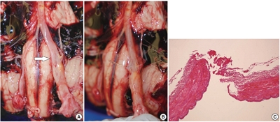
- Single umbilical artery and associated birth defects in perinatal autopsies: prenatal diagnosis and management
- Manushree Saxena, Bhagyashri Hungund
- J Pathol Transl Med. 2024;58(5):214-218. Published online July 9, 2024
- DOI: https://doi.org/10.4132/jptm.2024.07.03

- 25,980 View
- 496 Download
- 2 Web of Science
- 3 Crossref
-
 Abstract
 PDF
PDF - Background
The umbilical cord forms the connection between the fetus and the placenta at the feto-maternal interface and normally comprises two umbilical arteries and one umbilical vein. In some cases, only a single umbilical artery (SUA) is present. This study was conducted to evaluate associations between SUA and other congenital malformations discovered in perinatal autopsies and to ascertain the existence of preferential associations between SUA and certain anomalies.
Methods
We evaluated records of all fetuses sent for autopsy to the Department of Pathology during the 10-year period from 2013 through 2022 (n = 1,277). The data were obtained from the hospital’s pathology laboratory records. The congenital anomalies were grouped by organ or system for analysis and included cardiovascular, urinary tract, nervous system, gastrointestinal tract, musculoskeletal, and lung anomalies.
Results
A SUA was present in 8.61% of the autopsies. The gestational age of the affected fetuses ranged between 13 to 40 weeks. An SUA presented as an isolated single anomaly in 44 cases (3.4%). Of the 110 SUA cases, 60% had other congenital anomalies. There was a significant association between birth defects and SUAs (p < .001). Strong associations between SUA and urinary tract, lung, and musculoskeletal anomalies were observed.
Conclusions
A SUA is usually seen in association with other congenital malformations rather than as an isolated defect. Therefore, examination for associated anomalies when an SUA is detected either antenatally or postnatally is imperative. The findings of this study should be helpful in counseling expectant mothers and their families in cases of SUA. -
Citations
Citations to this article as recorded by
- Prevalence of Congenital Abnormalities in Abortuses and Stillbirths in India: A Systematic Review and Meta‐Analysis
Sushant Swaroop Das, Shalika Sharma, Reeha Mahajan, Sushruti Kaushal, Manupriya Sharma, Harsimranjit Singh, M. Ramkumar
Congenital Anomalies.2026;[Epub] CrossRef - Single Umbilical Artery with Symmetrical IUGR and Multiple Fetal Anomalies - An Interesting Case Report
Amulya Choudary Kotapati, Bhargavi Khandru, Vijayasree M.
Journal of Evolution of Medical and Dental Sciences.2025; : 10. CrossRef - Epidemiological and Histopathological Characteristics of Fetuses with Congenital Disorders: A Study in Greece
Despoina Nteli, Maria Nteli, Konstantinos Konstantinidis, Maria Ouzounidou, Paschalis Theotokis, Maria-Eleni Manthou, Iasonas Dermitzakis, Xeni Miliara, Chrysoula Gouta, Stamatia Angelidou, Dimosthenis Miliaras, Soultana Meditskou
Biology.2025; 14(6): 626. CrossRef
- Prevalence of Congenital Abnormalities in Abortuses and Stillbirths in India: A Systematic Review and Meta‐Analysis
Case Study
- Inconspicuous longitudinal tears of the intracranial vertebral artery in traumatic basal subarachnoid hemorrhage
- Seongho Kim
- J Pathol Transl Med. 2020;54(2):179-183. Published online November 8, 2019
- DOI: https://doi.org/10.4132/jptm.2019.10.15
- 11,511 View
- 211 Download
- 1 Web of Science
- 2 Crossref
-
Abstract
PDF
- Blunt force trauma to the head or neck region can cause traumatic basal subarachnoid hemorrhage (TBSAH), which can result in rapid loss of consciousness and death; however, detecting such a vascular injury is difficult. Posterior neck dissection was performed to investigate the bleeding focus in TBSAH cases 2018 and 2019. In all four cases, autopsies revealed a longitudinal tear in the midsection of the vertebral artery’s intracranial portion. The midportion of the intracranial vertebral artery appears to be most vulnerable to TBSAH. Interestingly, three of the cases showed only a vaguely visible longitudinal fissure in the artery without a grossly apparent tear; rupture was confirmed by microscopic examination. Longitudinal fissures of the intracranial vertebral artery, which are difficult to identify without detailed examination, may be overlooked in some cases of TBSAH. Thus, careful gross and microscopic examination of the vertebral artery is recommended in cases of TBSAH.
-
Citations
Citations to this article as recorded by- Traumatic vertebrobasilar pseudoaneurysms: diagnostic pitfalls on CT angiography with forensic implications — two case reports
Numfon Tweeatsani, Kana Unuma, Yukiko Uemura, Hirotaro Iwase, Yohsuke Makino
Emergency Radiology.2025; 33(1): 189. CrossRef - Effect of Ginseng Extract Ginsenoside Rg1 on Mice with Intracerebral Injury
Zixin Zhuang, Jinman Chen, Hao Xu, Yongjun Wang, Qianqian Liang
Chinese Medicine and Culture.2023;[Epub] CrossRef
- Traumatic vertebrobasilar pseudoaneurysms: diagnostic pitfalls on CT angiography with forensic implications — two case reports
Review
- Acute Atherosis of the Uterine Spiral Arteries: Clinicopathologic Implications
- Joo-Yeon Kim, Yeon Mee Kim
- J Pathol Transl Med. 2015;49(6):462-471. Published online November 4, 2015
- DOI: https://doi.org/10.4132/jptm.2015.10.23
- 22,587 View
- 246 Download
- 36 Web of Science
- 40 Crossref
-
Abstract
PDF
- Acute atherosis is unique vascular changes of the placenta associated with poor placentation. It is characterized by subendothelial lipid-filled foam cells, fibrinoid necrosis of the arterial wall, perivascular lymphocytic infiltration, and it is histologically similar to early-stage atherosclerosis. Acute atherosis is rare in normal pregnancies, but is frequently observed in non- transformed spiral arteries in abnormal pregnancies, such as preeclampsia, small for gestational age (SGA), fetal death, spontaneous preterm labor and preterm premature rupture of membranes. In preeclampsia, spiral arteries fail to develop physiologic transformation and retain thick walls and a narrow lumen. Failure of physiologic transformation of spiral arteries is believed to be the main cause of uteroplacental ischemia, which can lead to the production of anti-angiogenic factors and induce endothelial dysfunction and eventually predispose the pregnancy to preeclampsia. Acute atherosis is more frequently observed in the spiral arteries of the decidua of the placenta (parietalis or basalis) than in the decidual or myometrial segments of the placental bed. The presence and deeper location of acute atherosis is associated with poorer pregnancy outcomes, more severe disease, earlier onset of preeclampsia, and a greater frequency of SGA neonates in patients with preeclampsia. Moreover, the idea that the presence of acute atherosis in the placenta may increase the risk of future cardiovascular disease in women with a history of preeclampsia is of growing concern. Therefore, placental examination is crucial for retrospective investigation of pregnancy complications and outcomes, and accurate placental pathology based on universal diagnostic criteria in patients with abnormal pregnancies is essential for clinicopathologic correlation.
-
Citations
Citations to this article as recorded by- Placental vascular remodeling in preeclampsia: A three-dimensional analysis of microvascular alterations across disease severity
Mingqun Li, Xiaoqiang Han, Yao Peng, Yang He, Qiangqiang You, Jiaqi Zhang
Placenta.2026; 174: 96. CrossRef - Placental aberrant inflammation and spatial-specific lipid metabolism contribute to hypertensive disorder of pregnancy susceptibility in preeclampsia offspring
Pei-ran Hu, Jing-hui Xu, Yan Shi, Ying Zhu, Gao-chen Zhang, Jie-ru Yang, Yue Xu, Ming-hao Li, Xian-hua Lin, Yu Zhang, He-feng Huang
Biochimica et Biophysica Acta (BBA) - Molecular Basis of Disease.2026; 1872(4): 168168. CrossRef - Cardiac Implications of Preeclampsia: A Review
Beani J. Forst, Linda R. Chambliss, David S. Majdalany
Journal of Personalized Medicine.2026; 16(5): 265. CrossRef - Circulating vascular endothelial growth factor receptor‐3, a pro‐lymphangiogenic and pro‐angiogenic mediator, is decreased in pre‐eclampsia
Ana C. Palei, Julyane N. S. Kaihara, Ricardo C. Cavalli, Valeria C. Sandrim
International Journal of Gynecology & Obstetrics.2025; 168(1): 210. CrossRef - ECHS1 as a Lipid Metabolism Biomarker for Pediatric Focal Segmental Glomerulosclerosis
Chao He, Wei Peng, Sheng Li, Can Xu, Xiuping Chen, Yuanhan Qin, Nasar Alwahaibi
PLOS ONE.2025; 20(3): e0319049. CrossRef - PlacEntal Acute atherosis RefLecting Subclinical systemic atherosclerosis in women up to 20 years after pre-eclampsia (PEARLS): research protocol for a cohort study
Gwyneth Jansen, Robert-Jan Alers, Emma BNJ Janssen, Laura M Jorissen, Eri Morina - Shijaku, Carmen Severens-Rijvers, Arnoud van ’t Hof, J van Drongelen, Ralph R Scholten, Salwan Al-Nasiry, Droima Stevens, Wessel Ganzevoort, Sanne Gordijn, Jérôme Cornette,
BMJ Open.2025; 15(5): e100542. CrossRef - Understanding Preeclampsia: Cardiovascular Pathophysiology, Histopathological Insights and Molecular Biomarkers
Kaltrina Kutllovci Hasani, Nurxhan Ajeti, Nandu Goswami
Medical Sciences.2025; 13(3): 154. CrossRef - Evidence that atherosis of the spiral artery represents atherosclerotic lesions similar to those of native and transplant-induced atherosclerosis: implications for understanding the pathophysiology of obstetrical syndromes and long-term cardiovascular ris
Carlos A. Labarrere, Roberto Romero, Hector L. DiCarlo, James W. Hardin, Yeon Mee Kim, Arun Meyyazhagan, Offer Erez, Piya Chaemsaithong, Liliana Voto, Awoniyi Awonuga, Tinnakorn Chaiworapongsa, Ghassan S. Kassab
American Journal of Obstetrics and Gynecology.2025;[Epub] CrossRef - Human Placenta and Evolving Insights into Pathological Changes of Preeclampsia: A Comprehensive Review of the Last Decade
Diana Maria Chiorean, Esra Cobankent Aytekin, Melinda-Ildiko Mitranovici, Sabin Gligore Turdean, Mirpooya Salehi Moharer, Ovidiu Simion Cotoi, Havva Serap Toru
Fetal and Pediatric Pathology.2024; 43(1): 33. CrossRef - Effects of hypertensive disorders of pregnancy on the complications in very low birth weight neonates
Baoquan Zhang, Xiujuan Chen, Changyi Yang, Huiying Shi, Wenlong Xiu
Hypertension in Pregnancy.2024;[Epub] CrossRef - Prevention of Pregnancy Complications Using a Multimodal Lifestyle, Screening, and Medical Model
Jim Parker, Pierre Hofstee, Shaun Brennecke
Journal of Clinical Medicine.2024; 13(15): 4344. CrossRef - Placental growth factor mediates pathological uterine angiogenesis by activating the NFAT5-SGK1 signaling axis in the endometrium: implications for preeclampsia development
Janet P. Raja Xavier, Toshiyuki Okumura, Melina Apweiler, Nirzari A. Chacko, Yogesh Singh, Sara Y Brucker, Satoru Takeda, Florian Lang, Madhuri S Salker
Biological Research.2024;[Epub] CrossRef - Genome-wide DNA methylation and gene expression in human placentas derived from assisted reproductive technology
Pauliina Auvinen, Jussi Vehviläinen, Karita Rämö, Ida Laukkanen, Heidi Marjonen-Lindblad, Essi Wallén, Viveca Söderström-Anttila, Hanna Kahila, Christel Hydén-Granskog, Timo Tuuri, Aila Tiitinen, Nina Kaminen-Ahola
Communications Medicine.2024;[Epub] CrossRef - Missing links in preeclampsia cell model systems of endothelial dysfunction
Sarah Viana-Mattioli, Miriam Helena Fonseca-Alaniz, Iguaracy Pinheiro-de-Sousa, José Eduardo Krieger, Valéria Cristina Sandrim
Trends in Molecular Medicine.2023; 29(7): 541. CrossRef - Roles of maternal HDL during pregnancy
Laura A. Woollett, Janet M. Catov, Helen N. Jones
Biochimica et Biophysica Acta (BBA) - Molecular and Cell Biology of Lipids.2022; 1867(3): 159106. CrossRef - The role of the placenta in spontaneous preterm labor and delivery with intact membranes
Sunil Jaiman, Roberto Romero, Gaurav Bhatti, Eunjung Jung, Francesca Gotsch, Manaphat Suksai, Dahiana M. Gallo, Tinnakorn Chaiworapongsa, Nicholas Kadar
Journal of Perinatal Medicine.2022; 50(5): 553. CrossRef - Gestational Antibodies to C. pneumoniae, H. pylori and CMV in Women with Preeclampsia and in Matched Controls
Abdul Wajid, David Todem, Mark R. Schleiss, David F. Colombo, Nigel S. Paneth
Maternal and Child Health Journal.2022; 26(10): 2040. CrossRef - Toward a new taxonomy of obstetrical disease: improved performance of maternal blood biomarkers for the great obstetrical syndromes when classified according to placental pathology
Roberto Romero, Eunjung Jung, Tinnakorn Chaiworapongsa, Offer Erez, Dereje W. Gudicha, Yeon Mee Kim, Jung-Sun Kim, Bomi Kim, Juan Pedro Kusanovic, Francesca Gotsch, Andreea B. Taran, Bo Hyun Yoon, Sonia S. Hassan, Chaur-Dong Hsu, Piya Chaemsaithong, Nardh
American Journal of Obstetrics and Gynecology.2022; 227(4): 615.e1. CrossRef - Preeclampsia and Fetal Growth Restriction as Risk Factors of Future Maternal Cardiovascular Disease—A Review
Sylwia Sławek-Szmyt, Katarzyna Kawka-Paciorkowska, Aleksandra Ciepłucha, Maciej Lesiak, Mariola Ropacka-Lesiak
Journal of Clinical Medicine.2022; 11(20): 6048. CrossRef - The Role of NF-κB in Uterine Spiral Arteries Remodeling, Insight into the Cornerstone of Preeclampsia
Maciej W. Socha, Bartosz Malinowski, Oskar Puk, Mateusz Wartęga, Martyna Stankiewicz, Anita Kazdepka-Ziemińska, Michał Wiciński
International Journal of Molecular Sciences.2021; 22(2): 704. CrossRef - Pathogenesis of uteroplacental acute atherosis: An update on current research
Shu Li, Yan‐Wei Hu
American Journal of Reproductive Immunology.2021;[Epub] CrossRef - Disorders of placental villous maturation are present in one-third of cases with spontaneous preterm labor
Sunil Jaiman, Roberto Romero, Percy Pacora, Offer Erez, Eunjung Jung, Adi L. Tarca, Gaurav Bhatti, Lami Yeo, Yeon Mee Kim, Chong Jai Kim, Jung-Sun Kim, Faisal Qureshi, Suzanne M. Jacques, Nardhy Gomez-Lopez, Chaur-Dong Hsu
Journal of Perinatal Medicine.2021; 49(4): 412. CrossRef - The COVID-19 Pandemic: an Appraisal of its Impact on Human Immunodeficiency Virus Infection and Pre-Eclampsia
Rowen Govender, Jagidesa Moodley, Thajasvarie Naicker
Current Hypertension Reports.2021;[Epub] CrossRef - Acute Atherosis Lesions at the Fetal-Maternal Border: Current Knowledge and Implications for Maternal Cardiovascular Health
Daniel Pitz Jacobsen, Heidi Elisabeth Fjeldstad, Guro Mørk Johnsen, Ingrid Knutsdotter Fosheim, Kjartan Moe, Patji Alnæs-Katjavivi, Ralf Dechend, Meryam Sugulle, Anne Cathrine Staff
Frontiers in Immunology.2021;[Epub] CrossRef - Aetiology, prophylaxis and management of preeclampsia
Karolina Gronkowska
Acta Universitatis Lodziensis. Folia Biologica et Oecologica.2021; 17: 111. CrossRef - HMOX1 is partly responsible for phenotypic and functional abnormalities in mesenchymal stem cells/stromal cells from placenta of preeclampsia (PE) patients
Yasser S. Basmaeil, Dana Algudiri, Reem Alenzi, Abdullah Al Subayyil, Ayodele Alaiya, Tanvir Khatlani
Stem Cell Research & Therapy.2020;[Epub] CrossRef - Analyzing Preeclampsia as the Tip of the Iceberg Represented by Women with Long-Term Cardiovascular Disease, Atherosclerosis, and Inflammation
Angélica Lemos Debs Diniz, Maria Marta Bini Martins Paes, Aline Debs Diniz
Current Atherosclerosis Reports.2020;[Epub] CrossRef - Lipids in preeclampsia: pathogenic parallels to atherosclerosis
V. I. Shcherbakov, Ya. V. Polonskaya, E. V. Kashtanova, A. V. Shirinskaya
"Arterial’naya Gipertenziya" ("Arterial Hypertension").2020; 26(2): 163. CrossRef - Transthyretin increases migration and invasion of rat placental trophoblast cells
Xiao‐Peng Ma, Chong‐Dong Liu, Guang‐Ming Cao, Zhen‐Yu Zhang
FEBS Open Bio.2020; 10(8): 1568. CrossRef - Early Onset Preeclampsia Is Associated With Glycocalyx Degradation and Reduced Microvascular Perfusion
Tracey L. Weissgerber, Oscar Garcia‐Valencia, Natasa M. Milic, Elizabeth Codsi, Hajrunisa Cubro, Meryl C. Nath, Wendy M. White, Karl A. Nath, Vesna D. Garovic
Journal of the American Heart Association.2019;[Epub] CrossRef - The immunophenotype of decidual macrophages in acute atherosis
Navleen Gill, Yaozhu Leng, Roberto Romero, Yi Xu, Bogdan Panaitescu, Derek Miller, Afrah Arif, Salma Mumuni, Faisal Qureshi, Chaur‐Dong Hsu, Sonia S. Hassan, Anne Cathrine Staff, Nardhy Gomez‐Lopez
American Journal of Reproductive Immunology.2019;[Epub] CrossRef - The potential effects of pomegranate peel extract and bee venom in improving the diabetes induced damaging of spiral artery
HIH El-Sayyad, HA El-Ghawet, AMA El-Sayed
Studies on Stem Cells Research and Therapy.2019; 5(1): 007. CrossRef - Race and risk of maternal vascular malperfusion lesions in the placenta
Vanessa Assibey-Mensah, W. Tony Parks, Alison D. Gernand, Janet M. Catov
Placenta.2018; 69: 102. CrossRef - Preclinical atherosclerosis at the time of pre‐eclamptic pregnancy and up to 10 years postpartum: systematic review and meta‐analysis
N. M. Milic, J. Milin‐Lazovic, T. L. Weissgerber, G. Trajkovic, W. M. White, V. D. Garovic
Ultrasound in Obstetrics & Gynecology.2017; 49(1): 110. CrossRef - Is an episode of suspected preterm labor that subsequently leads to a term delivery benign?
Roberto Romero, Offer Erez, Eli Maymon, Percy Pacora
American Journal of Obstetrics and Gynecology.2017; 216(2): 89. CrossRef - Establishment of the Human Uteroplacental Circulation: A Historical Perspective
Kenna Degner, Ronald R. Magness, Dinesh M. Shah
Reproductive Sciences.2017; 24(5): 753. CrossRef - Preeclampsia and coronary plaque erosion: Manifestations of endothelial dysfunction resulting in cardiovascular events in women
Saskia C.A. de Jager, John A.L. Meeuwsen, Freeke M. van Pijpen, Gerbrand A. Zoet, Arjan D. Barendrecht, Arie Franx, Gerard Pasterkamp, Bas B. van Rijn, Marie-José Goumans, Hester M. den Ruijter
European Journal of Pharmacology.2017; 816: 129. CrossRef - Placental histopathology lesions and pregnancy outcome in pregnancies complicated with symptomatic vs. non-symptomatic placenta previa
Eran Weiner, Hadas Miremberg, Ehud Grinstein, Letizia Schreiber, Shimon Ginath, Jacob Bar, Michal Kovo
Early Human Development.2016; 101: 85. CrossRef - Porphyromonas gingivalis within Placental Villous Mesenchyme and Umbilical Cord Stroma Is Associated with Adverse Pregnancy Outcome
Sizzle F. Vanterpool, Jasper V. Been, Michiel L. Houben, Peter G. J. Nikkels, Ronald R. De Krijger, Luc J. I. Zimmermann, Boris W. Kramer, Ann Progulske-Fox, Leticia Reyes, Motohiro Komaki
PLOS ONE.2016; 11(1): e0146157. CrossRef - Pregnant women with heart disease: Placental characteristics and their association with fetal adverse events
Fabio V. Lima, Paraskevi Koutrolou-Sotiropoulou, Puja B. Parikh, Cecilia Avila, Javed Butler, Kathleen Stergiopoulos
Acute Cardiac Care.2016; 18(3): 56. CrossRef
- Placental vascular remodeling in preeclampsia: A three-dimensional analysis of microvascular alterations across disease severity
Original Article
- Therapeutic Effects of Umbilical Cord Blood Derived Mesenchymal Stem Cell-Conditioned Medium on Pulmonary Arterial Hypertension in Rats
- Jae Chul Lee, Choong Ik Cha, Dong-Sik Kim, Soo Young Choe
- J Pathol Transl Med. 2015;49(6):472-480. Published online October 16, 2015
- DOI: https://doi.org/10.4132/jptm.2015.09.11
- Retraction in: J Pathol Transl Med 2016;50(4):325
- 18,239 View
- 83 Download
- 7 Web of Science
- 8 Crossref
Case Report
- A Case of Intimal Sarcoma Arising in the Left Common Iliac Artery.
- Ji Young Park, Kun Young Kwon, Hyoung Tae Kim, Sang Sook Lee
- Korean J Pathol. 2011;45(3):311-314.
- DOI: https://doi.org/10.4132/KoreanJPathol.2011.45.3.311
- 4,244 View
- 28 Download
-
Abstract
PDF
- Primary tumors of the great vessels are rare. Most encountered cases are sarcomas which most commonly develop in the aorta, pulmonary artery, and inferior vena cava. We experienced an intimal sarcoma arising in the left common iliac artery in a 68-year-old male, who suffered from claudication in his left lower extremity for a year and was diagnosed as arteriosclerosis obliterans, clinically. Bypass surgery was performed on the obstructive lesion. Grossly, the vascular lumen was filled with dark hemorrhagic materials. Microscopically, the lesion showed proliferation of anaplastic spindle cells with a marked nuclear atypia, arranged haphazardly. There were numerous mitotic figures. Foci of cholesterol clefts were also found in the intima. Immunohistochemically, the tumor cells were positive for vimentin, smooth muscle actin, and cytokeratin in certain areas. Stains for CD34, desmin, myosin heavy chain, caldesmon, and S-100 protein were negative. A pathologic diagnosis was made as intimal sarcoma with myofibroblastic differentiation.
Original Article
- Pulmonary Vascular Sarcomas: Clinicopathologic Analysis of 14 Cases.
- Na Rae Kim, Jhingook Kim, Seung Yeon Ha, Joungho Han
- Korean J Pathol. 2011;45(2):132-138.
- DOI: https://doi.org/10.4132/KoreanJPathol.2011.45.2.132
- 5,416 View
- 34 Download
- 4 Crossref
-
Abstract
PDF
- BACKGROUND
Pulmonary vessel sarcomas are rare, and their pathogenesis is still unclear.
METHODS
We focus on the pathologic findings of fourteen pulmonary artery and/or vein sarcomas along with clinical prognosis.
RESULTS
Nine patients were male and five were female, and they ranged in age from 26 to 72 years (mean, 47 years). There were ten cases of pulmonary artery sarcoma, three cases of pulmonary artery and vein sarcoma, and one case of pure pulmonary vein sarcoma. Ten out of the fourteen cases were associated with pulmonary thromboembolism. Microscopically, all the tumors showed an undifferentiated sarcomatous portion. There were leiomyosarcoma portions in 8 cases, malignant fibrous histiocytomatous portions in 7 cases, angiosarcomatous differentiation in 3 cases, and osteosarcomatous portion in 1 case. All but two patients died during the follow up period (range, 1 to 78 months). The mean survival time of the patients who died was 14 months and the longest survival time was 78 months after surgical resection.
CONCLUSIONS
The current study is one of the largest single institutional reviews of pulmonary artery and/or vein sarcoma. Regardless of the histological components and macroscopic growth patterns, these rare tumors have a grave prognosis. -
Citations
Citations to this article as recorded by- Therapeutic Use of Bee Venom and Potential Applications in Veterinary Medicine
Roberto Bava, Fabio Castagna, Vincenzo Musella, Carmine Lupia, Ernesto Palma, Domenico Britti
Veterinary Sciences.2023; 10(2): 119. CrossRef - Intimal Sarcoma of the Great Vessels
Alan M. Ropp, Allen P. Burke, Seth J. Kligerman, Jay S. Leb, Aletta A. Frazier
RadioGraphics.2021; 41(2): 361. CrossRef - Incidence of pulmonary non-epithelial tumors: 18 years’ experience at a single institute
In Ho Choi, Dae Hyun Song, Kang Min Han, Yong Soo Choi, Joungho Han
Pathology - Research and Practice.2014; 210(4): 210. CrossRef - Pleomorphic Malignant Histiocytoma of Pulmonary Arteries Presenting as Pulmonary Aneurysms
Gustavo Armando De La Cerda Belmont, Carlos Alberto Lezama Urtecho
The Annals of Thoracic Surgery.2013; 95(3): 1091. CrossRef
- Therapeutic Use of Bee Venom and Potential Applications in Veterinary Medicine
Case Reports
- Cystic Adventitial Disease of the Popliteal Artery: A case report.
- Soo Min Kang, Kyeong Cheon Jung, Je G Chi
- Korean J Pathol. 1993;27(4):418-420.
- 2,220 View
- 15 Download
-
Abstract
PDF
- Localized cystic degeneration of peripheral arteries represents and unusual cause of arterial insufficiency. It frequently occurs in patient without generalized arteriosclerosis. It has been reported in patients from age 11 to 62 years. Cystic adventitial disease is most common in the popliteal artery. At least 115 cases have been reported worldwide, but none in Korea. We report a case of cystic adventitial disease involving the left popliteal artery. This 64-year-old man presented with an 18-month history of cramping pain of sudden onset in the left calf and claudication. Angiographic findings showed a 6 cm length of luminal obliteration of the popliteal artery. Segmentally resected popliteal artery showed two longitudinally directed cystic masses measuring 3.5x1.5 cm and 2.5x1.5 cm in the adventitia. Microscopic examination revealed cystic space in the arterial adventitia compressing arterial lumen. There were a number of foamy histiocytes collected along the cystic lumen.
- Multiple Fibromuscular Dysplasia of Arteries with Aneurysm Formation: A case report.
- Soo Min Kang, Sang Yong Song, Yong Il Kim, Sung Kwon Kim, Jung Sang Lee, Sang Joon Kim
- Korean J Pathol. 1992;26(3):288-292.
- 2,104 View
- 13 Download
-
Abstract
PDF
- Multiple involvement of arterial fibromuscular dysplasia is unusal and such a case is quite limited in the literature. We present a case of multiple arterial fibromuscular dysplasia with aneurysm formation in a 38-year-old non-hypertensive man. Angiographic study showed multiple aneurysmal dilatations of the right renal, superior mesenteric, celiac axis, and left colic arteries. The resected right kidney demonstrated an aneurysm of the main renal artery and extensive infarction in the midportion of the renal parenchyma. The segmentally resected superior mesenteric artery disclosed a similar nature of aneurysm occluded by recent thrombi. Microscopically, all the resected arterial walls showed both intimal and medial fibroplasia together with periadventitial fibrosis resulting in marked narrowing of the lumen and subsequent thrombus formation.
 First
First Prev
Prev


