 E-submission
Search
- Page Path
- HOME > Search
- A comprehensive review of ossifying fibromyxoid tumor: insights into its clinical, pathological, and molecular landscape
- Kyriakos Chatzopoulos, Antonia Syrnioti, Mohamed Yakoub, Konstantinos Linos
- J Pathol Transl Med. 2026;60(1):6-19. Published online January 14, 2026
- DOI: https://doi.org/10.4132/jptm.2025.10.02
- 4,723 View
- 209 Download
-
 Abstract
 PDF
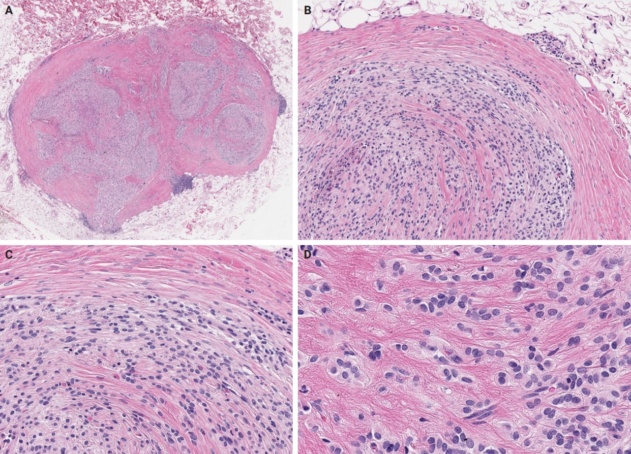
PDF - Ossifying fibromyxoid tumor (OFMT) is a rare mesenchymal neoplasm first described in 1989. It typically arises in the superficial soft tissues of the extremities as a slow-growing, painless mass. Histologically, it is commonly characterized by a multilobular architecture composed of uniform epithelioid cells embedded in a fibromyxoid matrix, often surrounded by a rim of metaplastic bone. While classic cases are readily identifiable, the tumor's histopathological heterogeneity can mimic a range of benign and malignant neoplasms, posing significant diagnostic challenges. Molecularly, most OFMTs harbor PHF1 rearrangements, commonly involving fusion partners such as EP400, MEAF6, or TFE3. This review underscores the importance of an integrated diagnostic approach- incorporating histopathological, immunohistochemical, and molecular data- to accurately classify OFMT and distinguish it from its mimics. Expanding awareness of its morphologic and molecular spectrum is essential for precise diagnosis, optimal patient management, and a deeper understanding of this enigmatic neoplasm.
- Juxtacortical chondromyxoid fibroma in the small bones: two cases with unusual location and a literature review
- Sun-Ju Oh, So Hak Chung
- J Pathol Transl Med. 2022;56(3):157-160. Published online January 21, 2022
- DOI: https://doi.org/10.4132/jptm.2021.12.15

- 7,227 View
- 198 Download
- 3 Web of Science
- 1 Crossref
-
Abstract
PDF
- Chondromyxoid fibroma is a rare bone tumor of cartilaginous origin, representing less than 1% of all bone tumors. It preferentially arises in the eccentric location of the metaphysis of a long tubular bone. Juxtacortical locations are reported infrequently in the long bones and even more rarely in short tubular bones, with only three cases documented. Here we present two new cases of juxtacortical chondromyxoid fibroma in the small bones. One was an intracortical osteolytic lesion of the metatarsal bone of the foot with degenerative atypia that histologically should be differentiated from chondrosarcoma. The other was a phalangeal mass protruding into the interphalangeal joint of the hand, which had been labeled mistakenly as a soft tissue mass preoperatively. These cases illustrated that chondromyxoid fibromas have various the manifestations and should be included in the differential diagnosis of an osteolytic lesion or an exophytic mass in the small bones.
-
Citations
Citations to this article as recorded by
- Cartilage Forming Tumors of the Skeleton
Julio A. Diaz-Perez, Andrew E. Rosenberg
Advances in Anatomic Pathology.2025; 32(2): 132. CrossRef
- Cartilage Forming Tumors of the Skeleton
- A Case of Mixed Adenoneuroendocrine Carcinoma of the Common Bile Duct: Initially Diagnosed as Cholangiocarcinoma
- Soon Wook Lee, In Seok Lee, Yu Kyung Cho, Jae Myung Park, Sang Woo Kim, Myung-Gyu Choi, Kyu Yong Choi, Myung Ah Lee, Tae Ho Hong, Young Kyoung You, Eun-Sun Jung
- Korean J Pathol. 2014;48(6):445-448. Published online December 31, 2014
- DOI: https://doi.org/10.4132/KoreanJPathol.2014.48.6.445
- 11,059 View
- 44 Download
- 12 Crossref
-
PDF
-
Citations
Citations to this article as recorded by- Long-term survival after chemotherapy combined immunotherapy for recurrent mixed neuroendocrine–non-neuroendocrine neoplasms of the common bile duct
Jia Li, Chunyan Yuan, Yulin Pan, Yuanyuan Yang, Nannan Lai, Xia Sheng
Clinical Journal of Gastroenterology.2025; 18(4): 653. CrossRef - Neuroendocrine carcinoma of the common hepatic duct coexisting with distal cholangiocarcinoma: A case report and review of literature
Fei Chen, Wei-Wei Li, Juan-Fen Mo, Min-Jie Chen, Su-Hang Wang, Shu-Ying Yang, Zheng-Wei Song
World Journal of Gastrointestinal Surgery.2024; 16(5): 1449. CrossRef - Comparison of Metastatic Patterns Among Neuroendocrine Tumors, Neuroendocrine Carcinomas, and Nonneuroendocrine Carcinomas of Various Primary Organs
Hyung Kyu Park, Ghee Young Kwon
Journal of Korean Medical Science.2023;[Epub] CrossRef - Mixed adenoneuroendocrine carcinoma of the distal bile duct: a case report
Takashi Maeda, Kyohei Yugawa, Nao Kinjo, Hiroto Kayashima, Daisuke Imai, Koto Kawata, Shinichiro Ikeda, Keitaro Edahiro, Kazuki Takeishi, Tomohiro Iguchi, Noboru Harada, Mizuki Ninomiya, Shohei Yamaguchi, Kozo Konishi, Shinichi Tsutsui, Hiroyuki Matsuda
Surgical Case Reports.2020;[Epub] CrossRef - The clinical profiles, management, and prognostic factors of biliary mixed neuroendocrine nonneuroendocrine neoplasms
Li-Jia Wen, Jun-Hong Chen, Hong-Ji Xu, Qiong Yu, Yu Deng, Kai Liu
Medicine.2020; 99(50): e23271. CrossRef - Rapidly progressed neuroendocrine carcinoma in the extrahepatic bile duct: a case report and review of the literature
Mariko Kamiya, Naoto Yamamoto, Yuto Kamioka, Hirohide Inoue, Hirokazu Yotsumoto, Masaaki Murakawa, Toru Aoyama, Kota Washimi, Kae Kawachi, Takashi Oshima, Makoto Ueno, Norio Yukawa, Yasushi Rino, Munetaka Masuda, Soichiro Morinaga
Surgical Case Reports.2020;[Epub] CrossRef - Mixed adenoneuroendocrine carcinoma of the hepatic bile duct: a case report and review of the literature
Sulai Liu, Zhendong Zhong, Meng Xiao, Yinghui Song, Youye Zhu, Bo Hu, Zengpeng Sun, Weimin Yi, Chuang Peng
BMC Gastroenterology.2020;[Epub] CrossRef - Mixed adenoendocrine carcinoma in the extrahepatic biliary tract: A case report and literature review
Liang Zhang, Zhengtao Yang, Qing Chen, Mengxia Li, Xiaolu Zhu, Dalong Wan, Haiyang Xie, Shusen Zheng
Oncology Letters.2019;[Epub] CrossRef - Large Cell Neuroendocrine Carcinoma Coexisting with Adenocarcinoma in the Extrahepatic Bile Duct
Masami Yuda, Teruyuki Usuba, Shin Hagiwara, Masahisa Okuma, Hisatoshi Asano, Hitoshi Sakuda, Hiroaki Katagi, Yoshiyuki Furukawa
The Japanese Journal of Gastroenterological Surgery.2018; 51(3): 187. CrossRef - Mixed Adenoneuroendocrine Carcinoma of the Distal Bile Duct
Chiaki Uchida, Yoshikazu Toyoki, Keinosuke Ishido, Daisuke Kudo, Norihisa Kimura, Shinji Tsutsumi, Takuji Kagiya, Toshiro Kimura, Kenichi Hakamada
The Japanese Journal of Gastroenterological Surgery.2017; 50(1): 43. CrossRef - Mixed adenoneuroendocrine carcinoma of the distal bile duct: A case report
Toshiaki Komo, Toshihiko Kohashi, Akira Nakashima, Ichiro Ohmori, Jun Hihara, Hidenori Mukaida, Mayumi Kaneko, Naoki Hirabayashi
International Journal of Surgery Case Reports.2017; 39: 203. CrossRef - Common Hepatic Duct Mixed Adenoneuroendocrine Carcinoma Masquerading as Cholangiocarcinoma
Sali Priyanka Akhilesh, Yadav Kamal Sunder, Tampi Chandralekha, Parikh Samir, Wagle Prasad Kashinath
Case Reports in Gastrointestinal Medicine.2016; 2016: 1. CrossRef
- Long-term survival after chemotherapy combined immunotherapy for recurrent mixed neuroendocrine–non-neuroendocrine neoplasms of the common bile duct
- Primary Leiomyosarcoma of Adrenal Gland with Tissue Eosinophilic Infiltration
- Seungkoo Lee, Gail Domecq C. Tanawit, Rolando A. Lopez, Jaime T. Zamuco, Betsy Grace G. Cheng, Menandro V. Siozon
- Korean J Pathol. 2014;48(6):423-425. Published online December 31, 2014
- DOI: https://doi.org/10.4132/KoreanJPathol.2014.48.6.423
- 10,743 View
- 48 Download
- 8 Crossref
-
PDF
-
Citations
Citations to this article as recorded by- Primary adrenal leiomyosarcoma
Syed Muhammad Nazim, Muhammad Hummam Siddique, Imran Khan Jalbani, Ayesha Nusrat
BMJ Case Reports.2025; 18(10): e266476. CrossRef - Outcomes and Follow-Up Trends in Adrenal Leiomyosarcoma: A Comprehensive Literature Review and Case Report
Federico Maria Mongardini, Maddalena Paolicelli, Antonio Catauro, Alessandra Conzo, Luigi Flagiello, Giusiana Nesta, Rosetta Esposito, Andrea Ronchi, Alessandro Romano, Renato Patrone, Ludovico Docimo, Giovanni Conzo
Journal of Clinical Medicine.2024; 13(12): 3499. CrossRef - Challenges in the diagnosis of the enigmatic primary adrenal leiomyosarcoma: two case reports and review of the literature
Sawako Suzuki, Naoya Takahashi, Masafumi Sugo, Kazuki Ishiwata, Akiko Ishida, Suzuka Watanabe, Katsushi Igarashi, Yutaro Ruike, Kumiko Naito, Masanori Fujimoto, Hisashi Koide, Yusuke Imamura, Shinichi Sakamoto, Tomohiko Ichikawa, Yoshihiro Kubota, Takeshi
BMC Endocrine Disorders.2023;[Epub] CrossRef - Primary adrenal leiomyosarcoma: clinical case and literature review
S. V. Lukyanov, K. M. Blikyan, S. S. Todorov, V. Y. Deribas, N. S. Lukyanov
Endocrine Surgery.2021; 15(1): 36. CrossRef Pleomorphic Leiomyosarcoma of the Adrenal Gland in a Young Woman: A Case Report and Review of the Literature
Yuanyuan Wang, Yongliang Teng, Shibo Na, Ye Yuan
OncoTargets and Therapy.2020; Volume 13: 4705. CrossRef- Primary Adrenal Leiomyosarcoma: Clinical, Radiological, and Histopathological Characteristics
Fatema Jabarkhel, Henri Puttonen, Lina Hansson, Andreas Muth, Oskar Ragnarsson
Journal of the Endocrine Society.2020;[Epub] CrossRef - Primary adrenal leiomyosarcoma with inferior vena cava extension in a 70-year-old man
Sai K Doppalapudi, Tejash Shah, Valerie A Fitzhugh, Vladislav Bargman
BMJ Case Reports.2019; 12(3): e227670. CrossRef - Primary Adrenal Leiomyosarcoma: An Extremely Rare Mesenchymal Tumor
D Lokanatha, Linu Abraham Jacob, MC Suresh Babu, KN Lokesh, Ram Krishna Sai, AH Rudresha, LK Rajeev, Smitha Saldanha, MN Suma, A Usha
Indian Journal of Medical and Paediatric Oncology.2019; 40(04): 559. CrossRef
- Primary adrenal leiomyosarcoma
- Myxoid Liposarcoma with Cartilaginous Differentiation: A Case Study with Cytogenetical Analysis
- Hyunchul Kim, Won Hwangbo, Sangjeong Ahn, Suhjin Kim, Insun Kim, Chul Hwan Kim
- Korean J Pathol. 2013;47(3):284-288. Published online June 25, 2013
- DOI: https://doi.org/10.4132/KoreanJPathol.2013.47.3.284
- 10,479 View
- 43 Download
- 3 Crossref
-
Abstract
PDF
Myxoid liposarcoma is a subtype of liposarcoma. This specific subtype can be identified based on its characteristic histological and cytogenetical features. The tumor has a fusion transcript of the
CHOP andTLS genes, which is caused by t(12;16)(q13;p11). Most of the fusion transcripts that have been identified fall into three categories, specifically type I (exons 7-2), type II (exons 5-2), and type III (exons 8-2). A total of seven myxoid liposarcomas associated with the rare phenomenon of cartilaginous differentiation have been documented in the literature. Currently, only one of these cases has been cytogenetically analyzed, and the analysis indicated that it was a type IITLS-CHOP fusion transcript in both the typical myxoid liposarcoma and cartilaginous areas. This study presents a second report of myxoid liposarcoma with cartilaginous differentiation, and includes a cytogenetical analysis of both the myxoid and cartilaginous areas.-
Citations
Citations to this article as recorded by- Myxoid liposarcoma with nuclear pleomorphism: a clinicopathological and molecular study
Naoki Kojima, Takashi Kubo, Taisuke Mori, Kaishi Satomi, Yuko Matsushita, Shintaro Iwata, Yasushi Yatabe, Koichi Ichimura, Akira Kawai, Hitoshi Ichikawa, Akihiko Yoshida
Virchows Archiv.2024; 484(1): 71. CrossRef - The Conundrum of Dedifferentiation in a Liposarcoma at a Peculiar Location: A Case Report and Literature Review
Ana-Maria Ciongariu, Adrian-Vasile Dumitru, Cătălin Cîrstoiu, Bogdan Crețu, Maria Sajin, Dana-Antonia Țăpoi, Aminia-Diana Ciobănoiu, Adrian Bejenariu, Andrei Marin, Mariana Costache
Medicina.2023; 59(5): 967. CrossRef - Myxoid liposarcoma with cartilaginous differentiation showing DDIT3 rearrangement
Kayo Suzuki, Taketoshi Yasuda, Kenta Watanabe, Takeshi Hori, Masahiko Kanamori, Tomoatsu Kimura
Oncology Letters.2017;[Epub] CrossRef
- Myxoid liposarcoma with nuclear pleomorphism: a clinicopathological and molecular study
- Plexiform Angiomyxoid Myofibroblastic Tumor of the Stomach: Report of Two Cases and Review of the Literature
- Youngran Kang, Wonkyung Jung, In-Gu Do, Eui Jin Lee, Min Hyeong Lee, Kyoung-Mee Kim, Jongsang Choi
- Korean J Pathol. 2012;46(3):292-296. Published online June 22, 2012
- DOI: https://doi.org/10.4132/KoreanJPathol.2012.46.3.292
- 11,503 View
- 76 Download
- 30 Crossref
-
Abstract
PDF
Plexiform angiomyxoid myofibroblastic tumor (PAMT) of the stomach is a recently recognized entity. Because of its rarity, only 22 cases have been reported in the English-language literature and most of these are single case reports. We report two cases of gastric PAMT. The tumor cells were bland and plexiform arranged in a myxoid stroma, which was positive for alcian blue. Immunohistochemically, the tumor cells were positive for smooth muscle actin, but negative for c-kit, CD34, desmin, S-100 protein, epithelial membrane antigen, neurofilament, and protein kinase C-theta. Mutation analyses for exon 9, 11, 13, and 17 of
KIT genes and 12, 14, and 18 of the platelet-derived growth factor receptor alpha (PDGFRA ) genes were performed and the tumors were wild-type for mutation.-
Citations
Citations to this article as recorded by- Plexiform Fibromyxoma in the Stomach: Immunohistochemical Profile and Comprehensive Genetic Characterization
Annabella Di Mauro, Rosalia Anna Rega, Maddalena Leongito, Vittorio Albino, Raffaele Palaia, Alberto Gualandi, Andrea Belli, Imma D’Arbitrio, Pasquale Moccia, Salvatore Tafuto, Annarosaria De Chiara, Alessandro Ottaiano, Gerardo Ferrara
International Journal of Molecular Sciences.2024; 25(9): 4847. CrossRef - Endoscopic submucosal excavation for gastric plexiform fibromyxoma: A case report and systematic review of literature
Ziqin Xia, Zhidai Zhou, Wei Guo, Hongling Wang, Fan Wang, Feng Zhou
Frontiers in Oncology.2023;[Epub] CrossRef - Recurrent plexiform angiomyxoid myofibroblastic tumour (PAMT) of the stomach with aggressive behaviour
Pavithra Ayyanar, Hemanta Kumar Nayak, Subash Chandra Samal, Madhabananda Kar, Pritinanda Mishra, Susama Patra
Pathology.2022; 54(5): 650. CrossRef - An Unusual Stomach Tumour: Plexiform Angiomyxoid Fibroma Stomach—A Case Report
Sharath K. Krishnan, Ravindran Chirukandath, Togy Zachariah, Rajiv Sajan Thomas
Indian Journal of Surgical Oncology.2022; 13(4): 691. CrossRef - Gastric Plexiform Fibromyxoma: A Case Report and Literature Review
路 张
Advances in Clinical Medicine.2022; 12(12): 12033. CrossRef - Plexiform angiomyxoid myofibroblastic tumor treated by endoscopic submucosal dissection: A case report and review of the literature
Jian-Di Wu, Yi-Xiong Chen, Chang Luo, Feng-Hua Xu, Lei Zhang, Xiao-Hua Hou, Jun Song
World Journal of Gastroenterology.2021; 27(31): 5288. CrossRef - Gastric Plexiform Fibromyxoma with Two Different Growth Patterns on Histological Images: a Case Report
Zhenyu Li, Qingming Jiang, Dongfang Guo, Yangling Peng, Jing Zhang, Xinyu Chen
Journal of Gastric Cancer.2021; 21(2): 213. CrossRef - Gastric plexiform fibromyxoma resected by endoscopic submucosal dissection: A case report and review of literature
XiaoBo Zhao, XinLou Li, Xin Huang, Le Shang, JianZhong Zhang, JiHua Wu
Human Pathology: Case Reports.2021; 23: 200468. CrossRef - Plexiform fibromyxoma: a clinicopathological and immunohistochemical analysis of two cases with a literature review
Shaofei Ma, Jing Wang, Zhanjun Lu, Chaoying Shi, Daohua Yang, Jun Lin
Journal of International Medical Research.2021;[Epub] CrossRef - A rare case of plexiform fibromyxoma in stomach: FNA diagnosis with histological correlation and differential diagnoses
Yujun Gan, Ghassan Hammoud, Magda Esebua
Annals of Diagnostic Pathology.2020; 44: 151453. CrossRef - Gastric plexiform fibromyxoma: A case report
Jin-Yu Pei, Bin Tan, Peng Liu, Guang-Hua Cao, Zu-Sen Wang, Lin-Lin Qu
World Journal of Clinical Cases.2020; 8(22): 5639. CrossRef - GASTRIC PLEXIFORM FIBROMYXOMA, AN UNCOMMON MESENCHYMAL TUMOR
Cristina Magadán Álvarez, Jose M. Olmos-Martínez, M Soledad Trugeda Carrera, María José Fernandez Diaz, Enrique Toledo Martínez, Remigio Mazorra Horts, Marta M Mayorga Fernández, Ruben Darío Arias Pacheco, Berta Martín Rivas
Revista Española de Enfermedades Digestivas.2020;[Epub] CrossRef - Pediatric plexiform fibromyxoma
Mitsuharu Fukazawa, Hiroshi Koga, Shoji Hiroshige, Toshifumi Matsumoto, Yuichi Nakazono, Yasuji Yoshikawa
Medicine.2019; 98(3): e14186. CrossRef - An Update on Clinicopathological and Molecular Features of Plexiform Fibromyxoma
Hsuan-An Su, Hsu-Heng Yen, Chih-Jung Chen
Canadian Journal of Gastroenterology and Hepatology.2019; 2019: 1. CrossRef - A rare case of plexiform angiomyxoid myofibroblastic tumor in the stomach which was diagnosed at the earliest stage in the literature
Xi Li, Shuangqing Li, Shenghua Xiong, Zhujun Wang, Hu Zhang
Gastroenterology Report.2018; 6(4): 313. CrossRef - Plexiform fibromyxoma of the small bowel: A case report
Wei-Guang Zhang, Liang-Bi Xu, Yi-Ning Xiang, Chen-Hong Duan
World Journal of Clinical Cases.2018; 6(15): 1067. CrossRef - Plexiform angiomyxoid myofibroblastic tumor of the stomach: A case report
Li Liang, Lin Fanzong, Zhang Peixi, Han Cuihong
Diagnostic Cytopathology.2017; 45(1): 55. CrossRef - Duodenal plexiform fibromyxoma as a cause of obscure upper gastrointestinal bleeding
Demetrios Moris, Evangelia Spanou, Stavros Sougioultzis, Nikolaos Dimitrokallis, Polyxeni Kalisperati, Ioanna Delladetsima, Evangelos Felekouras
Medicine.2017; 96(1): e5883. CrossRef - Computed tomography and magnetic resonance imaging of a plexiform angiomyxoid myofibroblastic tumor: a case report
Hiroyuki Akai, Shigeru Kiryu, Masaru Shinozaki, Yasunori Ohta, Yoshiyasu Nakano, Koichiro Yasaka, Kuni Ohtomo
BMC Medical Imaging.2017;[Epub] CrossRef - Plexiform Angiomyxoid Myofibroblastic Tumor of the Stomach: a Rare Case
Su Mi Kim, Ji Yeong An, Min-Gew Choi, Jun Ho Lee, Tae Sung Sohn, Kyung-Mee Kim, Sung Kim, Jae Moon Bae
Journal of Gastric Cancer.2017; 17(3): 277. CrossRef - Imaging findings of gastric plexiform fibromyxoma with a cystic change
Min-Xia Yang, Zhen-Hua Zhao, Jian-Feng Yang, Bing Chen, Xun-Ze Shen, Jian-Guo Wei, Bo-Yin Wang
Medicine.2017; 96(52): e8967. CrossRef - Gastrointestinal stromal tumor with a PDGFRA mutation masquerading as gastric plexiform fibromyxoma: A comparative clinicopathological study of two cases
Jun Zhou, Jingjing Xu, Guozhong Jiang, Yihui Ma, Jingwen Qi, Wencai Li, Dandan Zhang
Oncology Letters.2017; 13(2): 887. CrossRef - Gastric plexiform fibromyxoma tumor in a child – Case report and review of the literature
Michael W. Morris, Lisa Sullivan, David E. Sawaya, Michael A. Steiner, Michael J. Nowicki
Journal of Pediatric Surgery Case Reports.2016; 4: 38. CrossRef - Unusual focal keratin expression in plexiform angiomyxoid myofibroblastic tumor
Giuseppe Quero, Teresa Musarra, Alfredo Carrato, Michelangelo Fici, Maurizio Martini, Angelo Paolo Dei Tos, Sergio Alfieri, Riccardo Ricci
Medicine.2016; 95(28): e4207. CrossRef - Laparoscopy endoscopy cooperative surgery for gastric plexiform fibromyxoma: a case report
Yoshikage Inoue, Shutaro Gunji, Kazutaka Obama, Hiroshi Okabe, Yoshiharu Sakai
Surgical Case Reports.2016;[Epub] CrossRef - Plexiform fibromyxoma with cotyledon-like serosal growth: A case report of a rare gastric tumor and review of the literature
JOSHUA ROBERT KANE, NATASHA LEWIS, REBECCA LIN, CELINA VILLA, ALEXANDRA LARSON, JEFFREY D. WAYNE, ANJANA V. YELDANDI, WILLIAM B. LASKIN
Oncology Letters.2016; 11(3): 2189. CrossRef - Plexiform angiomyxoid myofibroblastic tumour of the duodenum: a rare entity
Niladri Banerjee, Shahana Gupta, Suvashis Dash, Shibajyoti Ghosh
BMJ Case Reports.2015; 2015: bcr2015210004. CrossRef - A case of gastric plexiform fibromyxoma: radiological and pathological findings
Katsumi Sakamoto, Masakazu Hirakawa, Kazushige Atsumi, Koshi Mimori, Kohei Shibata, Taro Tobo, Hidetaka Yamamoto, Hiroshi Honda
Japanese Journal of Radiology.2014; 32(7): 431. CrossRef - Plexiform Angiomyxoid Myofibroblastic Tumor of the Stomach: Report of a Case and Review of the Literature
Soo-Heui Baek, Jung-Hee Yoon, Ji-Yeon Kim
Journal of the Korean Society of Radiology.2014; 70(1): 47. CrossRef - Plexiform Fibromyxoma: Report of Two Pediatric Cases and Review of the Literature
Lizette Vila Duckworth, Raul S. Gonzalez, Matthew Martelli, Chen Liu, Cheryl M. Coffin, John D. Reith
Pediatric and Developmental Pathology.2014; 17(1): 21. CrossRef
- Plexiform Fibromyxoma in the Stomach: Immunohistochemical Profile and Comprehensive Genetic Characterization
- Ectomesenchymal Chondromyxoid Tumor in the Anterior Tongue: Case Report of a Unique Tumor
- Min Gyoung Pak, Kyung Bin Kim, Nari Shin, Woo Kyung Kim, Dong Hoon Shin, Kyung Un Choi, Mee Young Sol
- Korean J Pathol. 2012;46(2):192-196. Published online April 25, 2012
- DOI: https://doi.org/10.4132/KoreanJPathol.2012.46.2.192
- 9,566 View
- 63 Download
- 7 Crossref
-
Abstract
PDF
Ectomesenchymal chondromyxoid tumor (ECMT) is a rare tumor, exclusively arising in the anterior tongue. Thirty-eight cases have been reported in the English literature. It usually presents as a sessile protrusion and shows round to spindle cells embedded in myxoid to chondroid stroma. Tumor cells are almost always positive for polyclonal glial fibrillary acidic protein (GFAP). We report our experience in the recent treatment of a case of ECMT, the third case in 3 years. The mass in the anterior tongue revealed characteristic morphologic features of ECMT and the expression of polyclonal GFAP. Although ECMT should be differentiated from other mesenchymal tumors including myoepithelioma, its clinical, morphological, and immunohistochemical features enable its diagnosis, especially when pathologists are aware of it.
-
Citations
Citations to this article as recorded by- A case of ectomesenchymal chondromyxoid tumor on the lateral border of the tongue
Kohei KAWAMURA, Takayuki KURIMOTO, Yukihiro OHTA, Takuji YASUDA, Kanemitsu SHIRASUNA, Hideo YOSHIOKA
Japanese Journal of Oral and Maxillofacial Surgery.2025; 71(8): 354. CrossRef - Ectomesenchymal chondromyxoid tumor of the oral cavity: a report of 5 new cases with comprehensive review of the literature and clinicohistopathologic features
Molly Housley Smith, Jack Moynihan
Oral Surgery, Oral Medicine, Oral Pathology and Oral Radiology.2023; 135(3): 410. CrossRef - Chondroid choristoma of the tongue
Sumaiya Nezam, Roquaiya Nishat, Shabab Ahmed Khan, Jeevendra Nath Shukla
National Journal of Maxillofacial Surgery.2022; 13(Suppl 1): S121. CrossRef - Ectomesenchymal chondromyxoid tumor: a comprehensive updated review of the literature and case report
Astrid Truschnegg, Stephan Acham, Lumnije Kqiku, Norbert Jakse, Alfred Beham
International Journal of Oral Science.2018;[Epub] CrossRef - Clinical features of ectomesenchymal chondromyxoid tumors: A systematic review of the literature
Masanari G. Kato, Evren Erkul, Kendall S. Brewer, Emily E. Harruff, Shaun A. Nguyen, Terry A. Day
Oral Oncology.2017; 67: 192. CrossRef - Cyclin D1 Expression in Ectomesenchymal Chondromyxoid Tumor of the Anterior Tongue
Jan Laco, Radovan Mottl, Walter Höbling, Stephan Ihrler, Petr Grossmann, Alena Skalova, Ales Ryska
International Journal of Surgical Pathology.2016; 24(7): 586. CrossRef - Nodular lesion in the buccal mucosa
Bruna Jalfim Maraschin, Ana Carolina Amorim Pellicioli, Lélia Batista de Souza, Pantelis Varvaki Rados, Marco Antonio Trevizani Martins, Manoela Domingues Martins
The Journal of the American Dental Association.2015; 146(3): 196. CrossRef
- A case of ectomesenchymal chondromyxoid tumor on the lateral border of the tongue
- Dedifferentiated Extraskeletal Myxoid Chondrosarcoma of the Masticator Space: A Case Report.
- Geunyoung Jung, Kyung Ja Cho, Seung Ho Choi, Mi Jung Kim
- Korean J Pathol. 2011;45:S101-S105.
- DOI: https://doi.org/10.4132/KoreanJPathol.2011.45.S1.S101
- 5,201 View
- 37 Download
- 3 Crossref
-
Abstract
PDF
- We describe a 69-year-old woman who presented with a dedifferentiated extraskeletal myxoid chondrosarcoma arising in the left masticator space. Computed tomography and magnetic resonance imaging revealed a 5 cm sized mass in the left masticator space. Histologically, the tumor consisted of two distinct areas. The less cellular area was a low-grade extraskeletal myxoid chondrosarcoma, composed of strands or cords of uniform spindle cells and abundant myxoid stroma. The more cellular, dedifferentiated area corresponded to a high grade myxofibrosarcoma, consisting of anaplastic tumor cells in myxoid stroma and geographic necrosis. The tumor cells of the former area were positive for S-100 protein, microtubule-associated protein-2 (MAP-2) and class III beta-tubulin, but negative for cytokeratin, smooth muscle actin, and desmin. The tumor cells in the latter, pleomorphic area showed MAP-2 and beta-tubulin immunoreactivity with a high Ki-67 labeling index. Based on its histologic and immunohistochemical features, the tumor was considered a dedifferentiated extraskeletal myxoid chondrosarcoma.
-
Citations
Citations to this article as recorded by- Extraskeletal myxoid chondrosarcoma of the parotid gland
NyimiBushabu Fidele, Wu Tianfu, Bing Liu, Yanfang Sun, Zhao Yifang
Annals of Maxillofacial Surgery.2019; 9(2): 439. CrossRef - Myxoid chondrosarcoma of maxilla: A rare case report
Hiralal Ash, Ajoy Kumar Shahi, Kabita Chatterjee, Dipankar Samaddar
Journal of Oral and Maxillofacial Surgery, Medicine, and Pathology.2016; 28(3): 273. CrossRef - Maxillo-facial Extraskeletal Myxoid Chondrosarcoma: A Case Report and Discussion
Ratnadeep Ganguly, Abhishek Mukherjee
The Korean Journal of Pathology.2011; 45(6): 639. CrossRef
- Extraskeletal myxoid chondrosarcoma of the parotid gland
- Maxillo-facial Extraskeletal Myxoid Chondrosarcoma: A Case Report and Discussion.
- Ratnadeep Ganguly, Abhishek Mukherjee
- Korean J Pathol. 2011;45(6):639-643.
- DOI: https://doi.org/10.4132/KoreanJPathol.2011.45.6.639
- 4,792 View
- 25 Download
- 2 Crossref
-
Abstract
PDF
- In this report, we share our experience of a case of maxillo-facial extraskeletal myxoid chondrosarcoma, a very rare location for this neoplasm. In addition, a literature review is provided. The patient, a 61-year-old male, had a maxillary mass encroaching on the nasal cavity. After debulking, the tumor recurred, attaining its pre-surgical proportion in two months. The patient improved clinically with radiation and remained stable for about one year. However, he ultimately developed metastases in his lung which were treated with palliative chemotherapy with a good outcome lasting three months. We could find only eight reported cases of this tumor in the head region of which two are in the maxilla; hence, ruling out other primary sites is mandatory for a patient presenting with a primary head and neck mass. Surgical removal may be complicated because of the location. A combination of surgery and radiation is the management of choice, with palliative chemotherapy in metastasis.
-
Citations
Citations to this article as recorded by- Extraskeletal Myxoid Chondrosarcoma of Floor of Mouth—A Rare Case Report and Review of Literature
Surendra K Dabas, Nandini N Menon, Reetesh Ranjan, Bikas Gurung, Sukirti Tiwari, Bharat Bhushan Bassan, Himanshu Shukla, Sunil Pasricha, Ajit Sinha, Rahul Kapoor, Vinay Kumar Verma, Devesh Verma, Saurabh Arora, Ashwani Sharma, Sourabh Mukharjee, Rishu Sin
Indian Journal of Otolaryngology and Head & Neck Surgery.2024; 76(1): 1290. CrossRef - Intracranial Metastasis of Extracranial Chondrosarcoma: Systematic Review With Illustrative Case
Charles E. Mackel, Harry Rosenberg, Hemant Varma, Erik J. Uhlmann, Rafael A. Vega, Ron L. Alterman
Brain Tumor Research and Treatment.2023; 11(2): 103. CrossRef
- Extraskeletal Myxoid Chondrosarcoma of Floor of Mouth—A Rare Case Report and Review of Literature
- Ossifying Fibromyxoid Tumor of Soft Parts.
- Seok Hoon Jeon, Seung Sam Paik, Eun Kyung Hong, Moon Hyang Park, Jung Dal Lee
- Korean J Pathol. 1997;31(2):174-178.
- 2,167 View
- 18 Download
-
Abstract
PDF
- An ossifying fibromyxoid tumor of soft parts is a rare, recently described, fibro-osseous neoplasm of uncertain histogenesis. It occurs most frequently within the subcutis or skeletal muscle of the extremities. Its biologic behavior is generally regarded as benign with at worst a locally aggressive clinical course. But, atypical and malignant variants have been recently reported. Herein we report a case of a benign ossifying fibromyxoid tumor which occurred in the left upper back of 41-year-old man. The tumor is composed of uniformly round or polygonal cells arranged in cords or nests which are separated by myxoid and hyalinzed fibrous matrix and associated with irregular bony trabeculae. The tumor cells are strong positive for vimentin. Ultrastructural findings and a review of literatures are added.
- Fine Needle Aspiration Cytology of Myxodi Chondrsarcoma of Pleura: A Case Report.
- Na Hye Myong, Kyung Ja Cho, Ja June Jang, Jae Il Zo, Young Mog Shim
- J Pathol Transl Med. 1990;1(2):152-157.
- 2,360 View
- 40 Download
-
Abstract
PDF
- A 70-year-old female who was diagnosed as myxoid chondrosarcoma by fine needle aspiration of a pleural mass is described. She presented with left chest discomfort of 4 months' duration and aggravating dyspnea and chest pain for 2 months. Chest X-ray and CT scan revealed a large lobulated low density mass invading chest wall at the left pleural cavity and massive pleural fluid. Fine needle aspiration was done under the impression of mesothelioma or metastatic cancer. The aspirates from the mass were very cellular and composed of isolated or clustered forms of large plump cells. Abundant cytoplasm was bluish opaque and the margin was rounded in the isolated cells, whereas clustered cells show ill-defined cell borders and aggregating tendency. The nuclei were eccentric, round to ovoid, and had fine chromatin pattern and multiple small nucleoli. Cellular pleomorphism or mitotic figure was not definite. These findings were consistent with cytologic features of chondrosarcoma. Final diagnosis was confirmed as myxoid chondrosarcoma by mediastinoscopic biopsy and the tumor showed strong positivity for S-100 protein.
- Fine Needle Aspiration Cytology of Myxoid Liposarcoma of the Mediastinum.
- Hee Jae Joo, Soon Hee Jung, Hogeun Kim
- J Pathol Transl Med. 1990;1(2):185-190.
- 2,635 View
- 43 Download
-
Abstract
PDF
- The cytologic findings in fine needle aspiration of a case of myxoid liposarcoma of the mediastinum are described. The smear and cell block of the aspirate revealed solid clusters with background of amorphous material and scattered single tumor cells. The clusters were moderately cellular and consisted of atypical lipoblasts in varying stages of differentiation and delicate plexiform capillaries. Good correlation was found between the histologic and cytologic findings in the fine needle aspirates. The differential diagnosis between myxoid liposarcoma and other myxoid soft tissue tumors is discussed.
- Adrenocortical Carcinoma, Myxoid Variant: A Case Report.
- Bomi Kim, Sun Och Yoon, Dong Il Kim, Myung Cherl Kook, Eun Kyung Hong
- Korean J Pathol. 2007;41(6):430-435.
- 2,292 View
- 30 Download
-
Abstract
PDF
- Myxoid variant of adrenal cortical carcinoma is extremely rare and there have been only 16 such cases reported in the medical literature. Here we report on a case of 43-year-old woman with a left adrenal mass that was detected during the evaluation for Cushing's syndrome. Left adrenalectomy was performed and the tumor weighed 347 g. The cut surface was predominantly myxoid and gelatinous with central hemorrhage and necrosis. Histologically, the tumor cells were rather small, uniform and polygonal with mild pleomorphism. It showed diverse morphologic patterns according to the amount of the myxoid stromal component. Making the diagnosis was not easy because the tumor was without areas of conventional adrenocortical carcinoma. Immunohistochemically, the tumor cells were positive for alpha-inhibin, synaptophysin and vimentin, but the tumor cells were negative for pan-cytokeratin and CAM 5.2. The immunophenotypes were identical to those of conventional adrenal cortical neoplasms. During the evaluation of a cytokeratin-negative and vimentin-positive retroperitoneal neoplasm with a myxoid component, the possibility of adrenal cortical tumor should be considered in spite that this is a very rare entity.
- Myxoma of the Ovary with Uncertain Malignant Potential: A Case Report.
- Min A Kim, Ji Hoon Kim, Jae Y Ro, Geunghwan Ahn, In Ae Park
- Korean J Pathol. 2004;38(6):434-437.
- 3,161 View
- 42 Download
-
Abstract
PDF
- Primary ovarian myxoid tumor such as myxoma, myxoid liposarcoma and myxoid leiomyosarcoma is extremely rare neoplasm. We experienced a case of unusual myxoid tumor of the ovary in a 25 year-old woman. She was admitted for an incidentally found ovarian mass during antenatal check. Radiologic studies revealed a 5.5x5 cm-sized solid mass in left ovary and she was undertaken left oophorectomy. Grossly, the round ovarian mass was measuring 8x6x5 cm, and the cut surface was predominantly solid with myxoid appearance. Microscopically, the tumor was surrounded by thick collagenous capsule and had moderate cellularity and rich vascularity. The tumor cells were stellate-shaped with abundant extracellular myxoid material without atypia. We initially thought this lesion as myxoma, but the cellularity was too high as an ordinary myxoma. Myxoid liposarcoma could also be considered as the differential diagnosis, however there was no convincing lipoblast. So, we diagnosed that tumor as myxoma with uncertain malignant potential.
- Synovial Sarcoma with Massive Myxoid Feature: A Case Report.
- Joon Hyuk Choi, Young Ran Shim, Young Kyung Bae, Mi Jin Kim, Duk Seop Shin, Kil Ho Cho
- Korean J Pathol. 2005;39(4):273-277.
- 2,612 View
- 49 Download
-
Abstract
PDF
- Focal myxoid change in synovial sarcoma is not uncommon, although the presence of predominantly myxoid stroma is very rare. Recognition of synovial sarcomas with massive myxoid feature is important because these can easily be mistaken for other myxoid soft tissue neoplasms. We report a case of a synovial sarcoma with massive myxoid feature in the left thigh of a 54-year-old woman. Wide excision of an 8.5*7.0*5.0 cm, well-circumscribed and lobulated tumor was performed. The cut surface was gray, soft, and myxoid. Histological examination showed proliferation of spindle cells in the predominantly myxoid stroma. There were small areas with features more typical of synovial sarcoma, including uniform, spindled cells with fascicular growth patterns, collagenous stroma, mast cell infiltration, and hemangiopericytoma-like vascular patterns. Immunohistochemical examination showed focal positivity of the tumor cells for epithelial membrane antigen (EMA). Tumor cells were all negative for cytokeratin (AE1/AE3), cytokeratin 7, S-100 protein, smooth muscle actin, and desmin. Ultrastructurally, tumor cells showed desmosomes and microvilli. Our case underscores that, in order to make a correct diagnosis, immunohistochemical and ultrastructural examination is essential.
- Cytological Features of Low Grade Fibromyxoid Sarcoma : Report of a Case with a Review of the Literature.
- Mi Seon Kwon
- J Pathol Transl Med. 2006;17(2):153-158.
- 2,604 View
- 35 Download
-
Abstract
PDF
- Low-grade fibromyxoid sarcoma (LGFMS) is a rare soft tissue tumor. There have been only a few prior fine-needle aspiration (FNA) cytological reports. Recognition of this tumor is important because of its potential for metastasis despite its indolent nature and its deceptively bland cytologic appearance. A 60-year-old male presented with a slowly growing mass in the left calf detected 10 years ago. The patient underwent surgical excision. FNA cytology was performed directly on the mass. The smears showed low cellularity composed of hypercellular tissue fragments, hypocellular loose aggregates, and stripped nuclei. The cytoplasm was seen as either collagenous material or very thin fibrillary collagen strands. Tumor cells had spindle, ovoid, or irregular nuclei, fine chromatin, and small nucleoli. Focally slight degree of nuclear pleomorphism is noted. There were no mitotic figures. Blood vessels were frequently seen. Immunocytochemically, tumor cells were negative for S-100 protein, desmin, smooth muscle actin, and CD34. The diagnosis of LGFMS is rarely possible by cytology alone; however, LGFMS should be included in the differential diagnosis of spindle-cell tumors consisting of hypercellular and hypocellular components with some capillary-sized vessels arising in the deep soft tissue of the lower extremities, particularly the thigh. The immunocytochemical findings are of help in the differential diagnosis.
 First
First Prev
Prev


