Articles
- Page Path
- HOME > J Pathol Transl Med > Volume 47(3); 2013 > Article
-
Case Study
Myxoid Liposarcoma with Cartilaginous Differentiation: A Case Study with Cytogenetical Analysis - Hyunchul Kim, Won Hwangbo, Sangjeong Ahn, Suhjin Kim, Insun Kim, Chul Hwan Kim
-
Korean Journal of Pathology 2013;47(3):284-288.
DOI: https://doi.org/10.4132/KoreanJPathol.2013.47.3.284
Published online: June 25, 2013
Department of Pathology, Korea University Anam Hospital, Korea University College of Medicine, Seoul, Korea.
- Corresponding Author: Chul Hwan Kim, M.D. Department of Pathology, Korea University Anam Hospital, Korea University College of Medicine, 73 Inchon-ro, Seongbuk-gu, Seoul 136-705, Korea. Tel: +82-2-920-5991, Fax: +82-2-920-6576, chkap@korea.ac.kr
• Received: July 23, 2012 • Revised: August 31, 2012 • Accepted: September 4, 2012
© 2013 The Korean Society of Pathologists/The Korean Society for Cytopathology
This is an Open Access article distributed under the terms of the Creative Commons Attribution Non-Commercial License (http://creativecommons.org/licenses/by-nc/3.0/) which permits unrestricted non-commercial use, distribution, and reproduction in any medium, provided the original work is properly cited.
Abstract
- Myxoid liposarcoma is a subtype of liposarcoma. This specific subtype can be identified based on its characteristic histological and cytogenetical features. The tumor has a fusion transcript of the CHOP and TLS genes, which is caused by t(12;16)(q13;p11). Most of the fusion transcripts that have been identified fall into three categories, specifically type I (exons 7-2), type II (exons 5-2), and type III (exons 8-2). A total of seven myxoid liposarcomas associated with the rare phenomenon of cartilaginous differentiation have been documented in the literature. Currently, only one of these cases has been cytogenetically analyzed, and the analysis indicated that it was a type II TLS-CHOP fusion transcript in both the typical myxoid liposarcoma and cartilaginous areas. This study presents a second report of myxoid liposarcoma with cartilaginous differentiation, and includes a cytogenetical analysis of both the myxoid and cartilaginous areas.
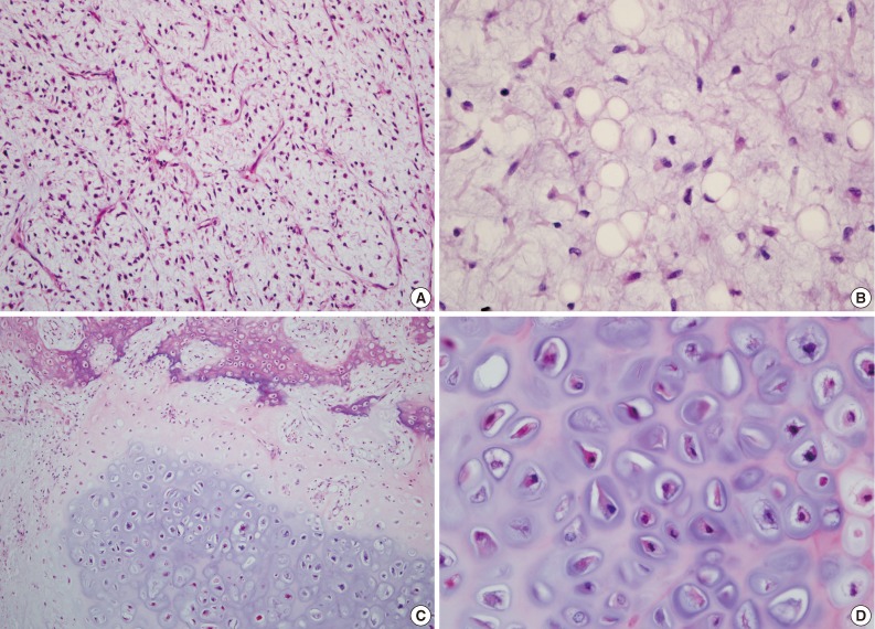
- A 45-year-old female visited the hospital with a palpable mass on the medial aspect of her thigh. The mass was observed by the patient 3 months previously and the size was perceived to be rapidly increasing. Computed tomography of the site showed an 11 cm-sized well-circumscribed mass with heterogeneous intensity, calcification and septation (Fig. 1). A diagnostic incisional biopsy was performed, and the tissue that was biopsied showed cartilage with atypical cytological features and focal necrosis. Analysis determined that myxoid stroma surrounded the cartilage, with the presence of some scattered lipoblasts within the myxoid stroma. Based on these findings, the biopsy was diagnosed as a malignant cartilage-forming tumor, and wide excision was performed on the entire mass. The specimen exhibited a well-demarcated ovoid mass with lobulation (Fig. 2), and each lobulated area was found to have different appearances that ranged from a solid gray fibrotic firm lobule, to a brown fibrotic soft lobule with a focal hemorrhage and cystic change, to a solid gray lobule with a focal glistening translucent myxoid appearance and a bluish firm resilient area with a focal calcification. Microscopic analysis revealed that the solid gray fibrotic area showed diffuse proliferation of bland fusiform cells with ovoid elongated nuclei and a moderate to abundant cytoplasm, and fine, delicate capillary vessel networks were noted within the fibrotic area (Fig. 3A). The brown fibrotic and hemorrhagic area contained coagulative necrosis of bland fusiform cells with hemorrhage, and a previous biopsy was proposed as the cause for the change in tissue. The myxoid area was characterized by a prominent basophilic myxoid matrix with scattered bland fusiform cells and lipoblasts (Fig. 3B). The lipoblasts had eccentrically located, small, elongated nuclei that were pushed to the periphery of the cell by clear, abundant intracytoplasmic vacuoles. The bluish resilient areas, which accounted for about 10% of the tumor volume, were characterized by cartilage with enchondral ossification, which showed mature bone without osteoid (Fig. 3C, D). The cartilage was moderately cellular and was composed of chondrocytes with atypical nuclei. Based on the immunohistochemistry, some of the fusiform cells and the lipoblasts were determined to be S-100 protein positive (1:300, Thermo Fisher Scientific, Waltham, MA, USA) (Fig. 4A), but were negative for smooth muscle actin (1:500, Dako, Glostrup, Denmark), desmin (1:1,000, Dako), and myogenin (1:100, Dako). Less than 5% of the cells were determined to be p53 positive (1:1,000, Dako) (Fig. 4B), and the Ki-67 (1:100, Dako) labeling index was also less than 5%. Based on these findings, a diagnosis of myxoid liposarcoma was considered. Molecular evaluation was performed on both the typical myxoid liposarcomatous and the cartilaginous areas, to confirm the proposed diagnosis. The final diagnosis was determined to be myxoid liposarcoma with cartilaginous differentiation, and the patient was scheduled for radiation therapy because the tumor was very close to the resection margin. Currently, after 6 months of follow-up, the patient has not presented with any evidence of metastasis or reoccurrence.
- Reverse transcription polymerase chain reaction investigation of mRNA expression
- Typical myxoid liposarcomatous and cartilaginous areas were marked on a paraffin block after the hematoxylin and eosin slides were reviewed. Five 10 µm sections of each area were cut from the resection specimen blocks and placed into Eppendorf tubes. The total RNA was extracted from the formalin-fixed, paraffin embedded samples using an RNeasy mini kit (Qiagen, Valencia, CA, USA) following the manufacturer's instructions. After preparation, 1 µg of each of the total RNA samples were reverse transcribed using a RevertAid First Strand cDNA Synthesis kit (Thermo Fisher Scientific), and a nested polymerase chain reaction (PCR) was performed using outer and inner primer sets that were based on a previous report.10 The outer primer set was TLS-CHOP outer forward (5'-AGCAAAGCTATAATCCCCCTCAG-3') and TLS-CHOP outer reverse (5'-GAAGGAGAAAGGCAATGACTCA-3'), and inner primer set consisted of TLS-CHOP inner forward (5'-GACAGCAGAACCAGTACAACAGCAG-3') and TLS-CHOP inner reverse (5'-GCTTTCAGGTGTGGTGATGTATGAAG-3'). The expected size of the nested PCR products for the three common types of fusion transcripts were 379 bp, 103 bp, and 412 bp for type I (exons 7-2), type II (exons 5-2), and type III (exons 8-2), respectively. The results of the nested PCR for this study exhibited type II fusion transcripts in both the typical myxoid liposarcomatous and cartilaginous areas (Fig. 5).
CASE REPORT
- Myxoid liposarcomas tend to occur in the lower extremities of young adults, and have a peak incidence in the 4th and 5th decades.3,12 Myxoid liposarcomas can be grossly identified as well-circumscribed, multinodular, intramuscular tumors with a gelatinous cut surface and occasional hemorrhage.2,12 Histologically, most of the tumors show bland fusiform cells and lipoblasts with varying degrees of cytoplasmic fat formation in the myxoid background with a prominent capillary vascular network.1,2 The fat vacuoles differ in regards to the size and number and tumor cells with a single vacuole have been described as signet ring-type lipogenic cells.1 This study observed cells with characteristics that were similar to typical myxoid liposarcoma with the exception of focal cartilaginous differentiation.
- The distinct differences between cartilaginous, leiomyomatous and osseous differentiation in myxoid liposarcoma has been described in a previous study.2 Previous studies have reported that five cases of similar myxoid liposarcomas have occurred, however there are a total of seven cases if the earlier reports by Enzinger and Winslow4 and Evans1 are counted,5,7,9 although the cartilaginous differentiation in the subtype was briefly described as metaplasia in the early reports by Enzinger and Winslow4 and Evans.1 The distribution of both cartilaginous and myxoid liposarcomatous areas has been reported in some cases to be distinct and different, but other cases have indicated that there was gradual transition between the two.1,4,5,7,9 Siebert et al.7 was the only author that described detailed histology of the cartilaginous area, and he also indicated that one of his three cases presented with cytological atypia, while the other two cases did not. This report did not discuss these specific details about the cartilaginous area, however, the image of the area indicated that there was cytological atypia of the chondrocytes.9 As Siebert et al.7 discussed, the appearance of the cartilaginous area can cause confusion with other benign or malignant soft tissue tumors. In this case, the situation could potentially be more problematic based on the biopsied material because a differential diagnosis of the myxoid chondrosarcoma from this tumor could be difficult to categorize accurately.2 But myxoid chondrosarcomas rarely show areas of mature cartilage.2 And even if the mature cartilage is noted, cytogenetical analysis of the spindle cell area and the cartilaginous area can be used to support an accurate diagnosis of the tumor.9
- Follow-up studies of the t(12;16) in myxoid liposarcoma have indicated that the translocation is a distinct characteristic feature of the tumor.10,13-16 The translocation occurrence percentage was consistently high throughout various studies and many reports showed that over 90% of the myxoid liposarcomas exhibited the cytogenetic abnormality.10,13-16 The specific translocation results in the fusion of CHOP (also known as GADD153 and DDIT3) on chromosome 12 and TLS (also referred to as FUS) on chromosome 16.14-16 To date, twelve different kinds of TLS-CHOP fusion transcripts have been detected.17 The portions of the genes that fuse can range from almost the entire CHOP gene to varying lengths of the TLS gene, which differs according to the type of fusion transcripts.17 The differences are caused by varied break points within each gene, but generally,18 three types of transcripts are commonly observed and the remaining transcripts are rare.10 Previous studies have not identified any specific correlation between the types of fusion transcript and a prognosis.11 However, this study demonstrated a correlation between the type II fusion transcript and p53 overexpression, and although the TLS-CHOP fusion is found in a high percentage of the myxoid liposarcomas, there are reports of other cytogenetic aberrations. For example, a small number of myxoid liposarcoma cases with an EWS-CHOP fusion have been reported.11 In addition, one study detected an aberration of the p53 gene in about 30% of the myxoid liposarcomas.19
- Previous studies have determined several prognostic factors of myxoid liposarcoma, which include the percentage of round cell components, tumor necrosis, and p53 overexpression.1,3,11,20 The prognostic importance of round cell components was first identified by Enzinger and Winslow4 and has been confirmed in follow-up studies.1,3,20 In addition, the overexpression of p53, determined by a 5% cut off value, was reported to be significantly associated with poor rates of survival.11
- This study is the second report of a myxoid liposarcoma with cartilaginous differentiation that was determined to have a type II TLS-CHOP fusion transcript in both the typical myxoid liposarcoma area and the cartilaginous differentiation area. Currently only a small number of cases discuss the histological aspects of the cartilaginous area, and because the chondrocytes do not present with apparent malignant or benign features, the cartilaginous area has not yet been designated into malignant or benign components. However, the study identified areas of ossification that were adjacent to cartilaginous areas. Because of the abrupt transition between the two areas and the absence of osteoid formation by the tumor cells, enchondral ossification was considered as the pathophysiological bone formation mechanism instead of osteosarcomatous differentiation. Authors of previous case reports have speculated that the heterologous component could be derived from common progenitor cells.9 However, additional follow-up studies are needed to investigate whether the cartilaginous component is created by proliferating mesenchymal cells or by metaplasia.
DISCUSSION
- 1. Evans HL. Liposarcoma: a study of 55 cases with a reassessment of its classification. Am J Surg Pathol 1979; 3: 507-523. ArticlePubMed
- 2. Weiss SW, Goldblum JR. Enzinger and Weiss's soft tissue tumors. 5th ed. Philadelphia: Mosby Elsevier, 2008.
- 3. Kilpatrick SE, Doyon J, Choong PF, Sim FH, Nascimento AG. The clinicopathologic spectrum of myxoid and round cell liposarcoma: a study of 95 cases. Cancer 1996; 77: 1450-1458. ArticlePubMed
- 4. Enzinger FM, Winslow DJ. Liposarcoma: a study of 103 cases. Virchows Arch Pathol Anat Physiol Klin Med 1962; 335: 367-388. PubMed
- 5. Dijkhuizen T, Molenaar WM, Hoekstra HJ, Wiersema J, van den Berg E. Cytogenetic analysis of a case of myxoid liposarcoma with cartilaginous differentiation. Cancer Genet Cytogenet 1996; 92: 141-143. ArticlePubMed
- 6. Shanks JH, Banerjee SS, Eyden BP. Focal rhabdomyosarcomatous differentiation in primary liposarcoma. J Clin Pathol 1996; 49: 770-772. ArticlePubMedPMC
- 7. Siebert JD, Williams RP, Pulitzer DR. Myxoid liposarcoma with cartilaginous differentiation. Mod Pathol 1996; 9: 249-252. PubMed
- 8. Govender D, Pillay P. Primary myxoid liposarcoma with rhabdomyoblastic differentiation. Arch Pathol Lab Med 1998; 122: 740-742. PubMed
- 9. Wei YC, Li CF, Eng HL, Yeh MC, Lin CN, Huang HY. Myxoid liposarcoma with cartilaginous differentiation: identification of the same type II TLS-CHOP fusion gene transcript in both lipogenic and chondroid components. Appl Immunohistochem Mol Morphol 2007; 15: 477-480. ArticlePubMed
- 10. Hisaoka M, Tsuji S, Morimitsu Y, et al. Detection of TLS/FUS-CHOP fusion transcripts in myxoid and round cell liposarcomas by nested reverse transcription-polymerase chain reaction using archival paraffin-embedded tissues. Diagn Mol Pathol 1998; 7: 96-101. ArticlePubMed
- 11. Antonescu CR, Tschernyavsky SJ, Decuseara R, et al. Prognostic impact of p53 status, TLS-CHOP fusion transcript structure, and histological grade in myxoid liposarcoma: a molecular and clinicopathologic study of 82 cases. Clin Cancer Res 2001; 7: 3977-3987. PubMed
- 12. Fletcher CD, Unni KK, Mertens F. World Health Organization calssification of tumours: pathology and genetics of tumours of soft tissue and bone. Lyon: IARC Press, 2002.
- 13. Limon J, Turc-Carel C, Dal Cin P, Rao U, Sandberg AA. Recurrent chromosome translocations in liposarcoma. Cancer Genet Cytogenet 1986; 22: 93-94. ArticlePubMed
- 14. Aman P, Ron D, Mandahl N, et al. Rearrangement of the transcription factor gene CHOP in myxoid liposarcomas with t(12;16)(q13; p11). Genes Chromosomes Cancer 1992; 5: 278-285. ArticlePubMed
- 15. Crozat A, Aman P, Mandahl N, Ron D. Fusion of CHOP to a novel RNA-binding protein in human myxoid liposarcoma. Nature 1993; 363: 640-644. ArticlePubMedPDF
- 16. Rabbitts TH, Forster A, Larson R, Nathan P. Fusion of the dominant negative transcription regulator CHOP with a novel gene FUS by translocation t(12;16) in malignant liposarcoma. Nat Genet 1993; 4: 175-180. ArticlePubMedPDF
- 17. Nishio J, Iwasaki H, Nabeshima K, Naito M. Cytogenetics and molecular genetics of myxoid soft-tissue sarcomas. Genet Res Int 2011; 2011: 497148.ArticlePubMedPMCPDF
- 18. Panagopoulos I, Mertens F, Isaksson M, Mandahl N. A novel FUS/CHOP chimera in myxoid liposarcoma. Biochem Biophys Res Commun 2000; 279: 838-845. ArticlePubMed
- 19. Dei Tos AP, Piccinin S, Doglioni C, et al. Molecular aberrations of the G1-S checkpoint in myxoid and round cell liposarcoma. Am J Pathol 1997; 151: 1531-1539. PubMedPMC
- 20. Smith TA, Easley KA, Goldblum JR. Myxoid/round cell liposarcoma of the extremities: a clinicopathologic study of 29 cases with particular attention to extent of round cell liposarcoma. Am J Surg Pathol 1996; 20: 171-180. PubMed
REFERENCES
Fig. 1Computed tomography of the left distal thigh. A well-demarcated mass (arrowheads) within muscle with heterogeneous density and calcification.
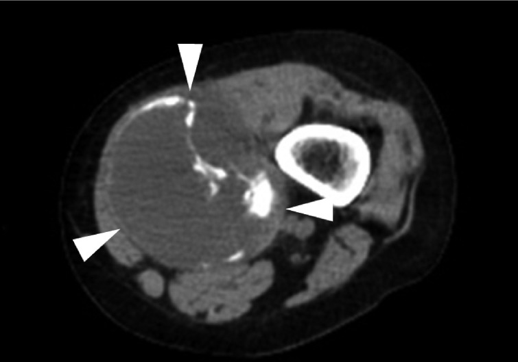

Fig. 2Gross picture of resected specimen. An 11 cm-sized, well-demarcated lobulated mass with hemorrhage and cystic change. Soft myxoid, gray firm fibrotic, and bluish resilient cartilaginous areas (arrowheads) are noted.
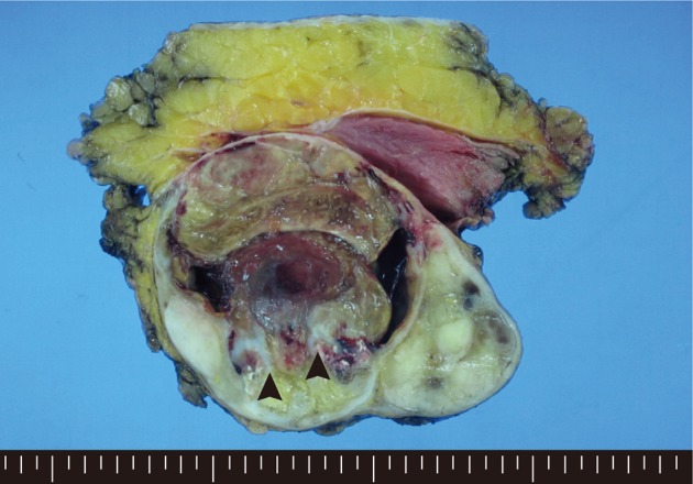

Fig. 3(A) Fusiform cells and capillary networks are observed in the fibrotic area. (B) Lipoblasts are seen in the myxoid area. (C, D) Cartilage with moderate cellularity, cytological atypia, and enchondral ossification are found in the bluish resilient area.


Fig. 4Immunohistochemical staining reveals that some of the fusiform cells and round lipoblasts are positive for S-100 protein (A). A few cells (less than 5%) are determined to be positive for p53 (B).
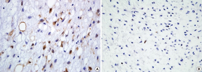

Fig. 5From the left, the designated lanes are the size marker, typical myxoid liposarcoma area and cartilaginous area. Both the typical myxoid liposarcoma and cartilaginous areas demonstrate nested polymerase chain reaction product of 103 bp, which corresponds to a type II TLS-CHOP fusion transcript.
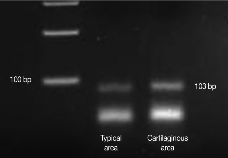

Figure & Data
References
Citations
Citations to this article as recorded by 

- Myxoid liposarcoma with nuclear pleomorphism: a clinicopathological and molecular study
Naoki Kojima, Takashi Kubo, Taisuke Mori, Kaishi Satomi, Yuko Matsushita, Shintaro Iwata, Yasushi Yatabe, Koichi Ichimura, Akira Kawai, Hitoshi Ichikawa, Akihiko Yoshida
Virchows Archiv.2024; 484(1): 71. CrossRef - The Conundrum of Dedifferentiation in a Liposarcoma at a Peculiar Location: A Case Report and Literature Review
Ana-Maria Ciongariu, Adrian-Vasile Dumitru, Cătălin Cîrstoiu, Bogdan Crețu, Maria Sajin, Dana-Antonia Țăpoi, Aminia-Diana Ciobănoiu, Adrian Bejenariu, Andrei Marin, Mariana Costache
Medicina.2023; 59(5): 967. CrossRef - Myxoid liposarcoma with cartilaginous differentiation showing DDIT3 rearrangement
Kayo Suzuki, Taketoshi Yasuda, Kenta Watanabe, Takeshi Hori, Masahiko Kanamori, Tomoatsu Kimura
Oncology Letters.2017;[Epub] CrossRef
 PubReader
 ePub Link
-
 Cite this Article
- Cite this Article
-
- Close
- Download Citation
- Close
- Figure
Myxoid Liposarcoma with Cartilaginous Differentiation: A Case Study with Cytogenetical Analysis


Fig. 1 Computed tomography of the left distal thigh. A well-demarcated mass (arrowheads) within muscle with heterogeneous density and calcification.
Fig. 2 Gross picture of resected specimen. An 11 cm-sized, well-demarcated lobulated mass with hemorrhage and cystic change. Soft myxoid, gray firm fibrotic, and bluish resilient cartilaginous areas (arrowheads) are noted.
Fig. 3 (A) Fusiform cells and capillary networks are observed in the fibrotic area. (B) Lipoblasts are seen in the myxoid area. (C, D) Cartilage with moderate cellularity, cytological atypia, and enchondral ossification are found in the bluish resilient area.
Fig. 4 Immunohistochemical staining reveals that some of the fusiform cells and round lipoblasts are positive for S-100 protein (A). A few cells (less than 5%) are determined to be positive for p53 (B).
Fig. 5 From the left, the designated lanes are the size marker, typical myxoid liposarcoma area and cartilaginous area. Both the typical myxoid liposarcoma and cartilaginous areas demonstrate nested polymerase chain reaction product of 103 bp, which corresponds to a type II TLS-CHOP fusion transcript.
Fig. 1
Fig. 2
Fig. 3
Fig. 4
Fig. 5
Myxoid Liposarcoma with Cartilaginous Differentiation: A Case Study with Cytogenetical Analysis

 E-submission


