Warning: mkdir(): Permission denied in /home/virtual/lib/view_data.php on line 81

Warning: fopen(upload/ip_log/ip_log_2024-11.txt): failed to open stream: No such file or directory in /home/virtual/lib/view_data.php on line 83

Warning: fwrite() expects parameter 1 to be resource, boolean given in /home/virtual/lib/view_data.php on line 84
Proposal of an Appropriate Decalcification Method of Bone Marrow Biopsy Specimens in the Era of Expanding Genetic Molecular Study
Skip Navigation
Skip to contents

J Pathol Transl Med : Journal of Pathology and Translational Medicine

OPEN ACCESS
SEARCH
Search

Articles

Page Path
HOME > J Pathol Transl Med > Volume 49(3); 2015 > Article
Original Article
Proposal of an Appropriate Decalcification Method of Bone Marrow Biopsy Specimens in the Era of Expanding Genetic Molecular Study
Sung-Eun Choi, Soon Won Hong, Sun Och Yoon
Journal of Pathology and Translational Medicine 2015;49(3):236-242.
DOI: https://doi.org/10.4132/jptm.2015.03.16
Published online: May 15, 2015

Department of Pathology, Yonsei University College of Medicine, Seoul, Korea

Corresponding Author Sun Och Yoon, M.D., Ph.D. Department of Pathology, Yonsei University College of Medicine, 50-1 Yonsei-ro, Seodaemun-gu, Seoul 120-752, Korea Tel: +82-2-2228-1763 Fax: +82-2-2227-7939 E-mail: revita@naver.com
• Received: January 27, 2015   • Revised: February 24, 2015   • Accepted: March 16, 2015

© 2015 The Korean Society of Pathologists/The Korean Society for Cytopathology

This is an Open Access article distributed under the terms of the Creative Commons Attribution Non-Commercial License (http://creativecommons.org/licenses/by-nc/3.0/) which permits unrestricted noncommercial use, distribution, and reproduction in any medium, provided the original work is properly cited.

prev next
  • 13,617 Views
  • 291 Download
  • 45 Web of Science
  • 47 Crossref
  • 53 Scopus
  • Background:
    The conventional method for decalcification of bone specimens uses hydrochloric acid (HCl) and is notorious for damaging cellular RNA, DNA, and proteins, thus complicating molecular and immunohistochemical analyses. A method that can effectively decalcify while preserving genetic material is necessary.
  • Methods:
    Pairs of bilateral bone marrow biopsies sampled from 53 patients were decalcified according to protocols of two comparison groups: EDTA versus HCl and RDO GOLD (RDO) versus HCl. Pairs of right and left bone marrow biopsy samples harvested from 28 cases were allocated into the EDTA versus HCl comparison group, and 25 cases to the RDO versus HCl comparison group. The decalcification protocols were compared with regards to histomorphology, immunohistochemistry, and molecular analysis. For molecular analysis, we randomly selected 5 cases from the EDTA versus HCl and RDO versus HCl groups.
  • Results:
    The decalcification time for appropriate histomorphologic analysis was the longest in the EDTA method and the shortest in the RDO method. EDTA was superior to RDO or HCl in DNA yield and integrity, assessed via DNA extraction, polymerase chain reaction, and silver in situ hybridization using DNA probes. The EDTA method maintained intact nuclear protein staining on immunohistochemistry, while the HCl method produced poor quality images. Staining after the RDO method had equivocal results. RNA in situ hybridization using kappa and lambda RNA probes measured RNA integrity; the EDTA and RDO method had the best quality, followed by HCl.
  • Conclusions:
    The EDTA protocol would be the best in preserving genetic material. RDO may be an acceptable alternative when rapid decalcification is necessary.
Sampling bone tissue is usually performed for the diagnosis of hematologic malignancy, metastatic tumor, or primary bone tumor. The processing of bone specimens usually follows decalcification and microtome sectioning in pathology laboratories. Inorganic acids such as nitric acid or HCl are used in decalcification, and limit diagnostic options by damaging DNA and RNA. As a result, gene testing is usually not plausible with these decalcified bone specimens, even though certain cancers need further genetic studies for diagnostic and therapeutic purposes. Therefore, there is a growing need for new decalcification agents that adequately preserve DNA and RNA [1-3].
A variety of molecular testing techniques are necessary to diagnose hematologic malignancies. Fluorescence in situ hybridization, gene rearrangement studies of immunoglobulin and T cell receptor genes, and in situ hybridization for kappa and lambda light chains and Epstein-Barr virus–encoded small RNAs are commonly used molecular tools in diagnosis of hematologic malignancies. However, further molecular study from bone marrow biopsy specimens is often impossible due to DNA or RNA damage by decalcification. Furthermore, immunohistochemistry may be required for differentiating and subtyping hematolymphoid lesions in conjunction with histomorphologic features of paratrabecular, interstitial, intrasinusoidal, or intravascular aggregates within bone marrow structures. HCl degrades both protein quality and quantity, resulting in poor immunohistochemical staining that cannot be used for accurate diagnosis.
With consideration of these limitations, we evaluated modified bone marrow decalcification protocols and compared them to the HCl method.
This was a prospective study. To eliminate bias due to variables among cases, cases were enrolled when pairs of bilaterally biopsied bone marrow specimens were available. Among bone marrow specimens sampled from the right and left iliac crests between January 2013 and July 2014 at Gangnam Severance Hospital, 53 cases were finally included. For the 53 selected cases, 28 right and left bone marrow samples were allocated to the EDTA (Sigma-Aldrich, St. Louis, MO, USA) protocol and HCl (Calci-Clear Rapid, National Diagnostics, Atlanta, GA, USA) protocol, respectively. Samples from the 25 remaining cases were assigned to the RDO GOLD (RDO) group (Apex Engineering Products Corporation, Aurora, IL, USA) protocol and HCl protocol. Concentration, processing time, and temperature are summarized in Table 1.
To test DNA quality, five cases were randomly selected from each of the three groups (EDTA, HCl, and RDO). DNA was extracted, and the quantity and quality were confirmed using NanoDrop ND-2000 spectrophotometer (Thermo Fisher Scientific Inc., Waltham, MA, USA). The BRAF PNA clamping method (Panagene, Daejeon, Korea) and comparison with Ct values of internal controls were used to evaluate the efficacy of polymerase chain reaction (PCR) DNA amplification. To evaluate the efficacy of DNA in situ hybridization, HER2 dual color silver in situ hybridization (Ventana, Tucson, AZ, USA) was applied to 10 pairs of bone marrow samples. RNA in situ hybridization and immunohistochemical studies were performed prospectively. According to the potential differential diagnoses for suspicious lesions observed within bone marrow, appropriate RNA probes or protein antibodies were applied. For immunohistochemistry, the following primary antibodies were used and the details are summarized in Table 2: cyclin D1, Ki67, Bcl2, Bcl6, TdT, CD138, CD20, CD79a, CD3, CD5, CD23, CD10, CD30, and myeloperoxidase. To state the process, after deparaffinization and rehydration, the sections were incubated in BenchMark XT automated slide stainer (Ventana) for 16 minutes at 37°C and then counterstained with hematoxylin reagent. Two pathologists reviewed the hematoxylin and eosin (H&E), immunohistochemistry, and in situ hybridization slides. The quality of immunohistochemistry and RNA in situ hybridization were assessed using a 3-tiered grading scale: good, equivocal, or poor. HER2 silver in situ hybridization was assessed by detecting two signals of HER2 and CEP17 per nucleus from the normal hematopoietic cells of bone marrow.
The differences in variables were analyzed using the Mann-Whitney U test. All statistical analyses were carried out by SPSS ver. 20.0 for Windows (IBM Co., Armonk, NY, USA).
Isolated DNA quality
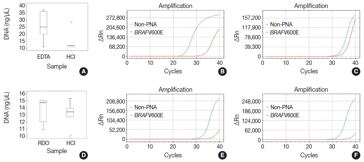
DNA quantity, purity, and Ct values of internal controls after real-time PCR in the EDTA versus HCl group and RDO versus HCl group are depicted in Table 3 and Fig. 1. Although differences were not statistically significant, the DNA yield of the EDTA protocol was about 2 times higher than the HCl protocol. In addition, the Ct value of the former protocol was significantly lower than that of the latter (p<.001) with the estimated difference being about 7. Furthermore, the Ct values of EDTA processed samples were lower than 30, demonstrating that the amount of intact DNA feasible for PCR with the EDTA protocol is better preserved by a factor of 2 [7] than the HCl protocol.
There were no significant differences between DNA yield and Ct values of RDO and HCl methods. The yield of extracted DNA after RDO decalcification was similar to that of HCl. The Ct values of both protocols were above 33, indicating that the amount of intact DNA feasible for PCR was very small.
Morphological comparison of DNA, RNA, and protein expression
The morphological comparison and quality assessment of H&E stain, HER2/CEP17 dual color silver in situ hybridization, kappa/lambda in situ hybridization, and immunohistochemistry studies were analyzed. The rates of high quality staining for each study were compared between the three protocols. The results are summarized in Table 4.
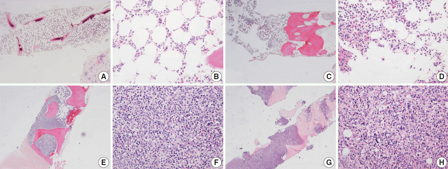
There was no difficulty in microtome dissection of 4 µm or less in thickness in any of the three methods. The morphological quality of H&E slide was similar in all three protocols, showing well-preserved histological features of the bone marrow (Fig. 2).
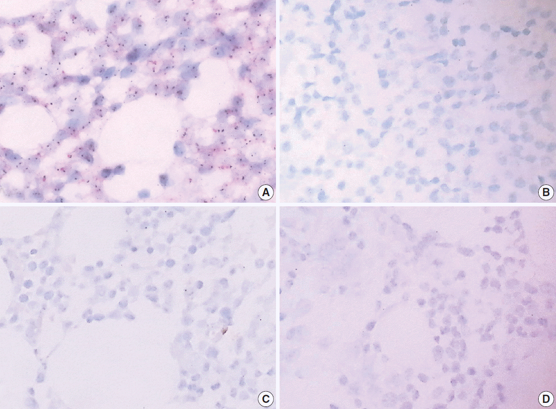
All five cases in the RDO versus HCl group had severe DNA breakdown on HER2/CEP17 dual color silver in situ hybridization, revealing no HER2 or CEP17 nuclear signal in bone marrow hematopoietic cells. However, in the EDTA versus HCl group, all 5 cases in the EDTA protocol showed two HER2 and CEP17 nuclear signals from almost all of the hematopoietic cells, whereas nearly no nuclear signal was detected in samples from the HCl protocol (Fig. 3).
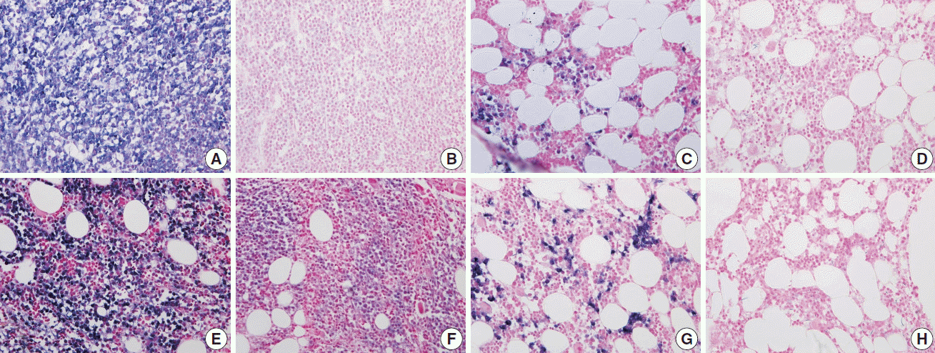
Bone marrow specimens from the EDTA and RDO protocols that underwent kappa/lambda RNA in situ hybridization showed well preserved RNA signal in the nuclei of plasma cells, while those in the HCl group had severe breakdown of RNA signals (Fig. 4).
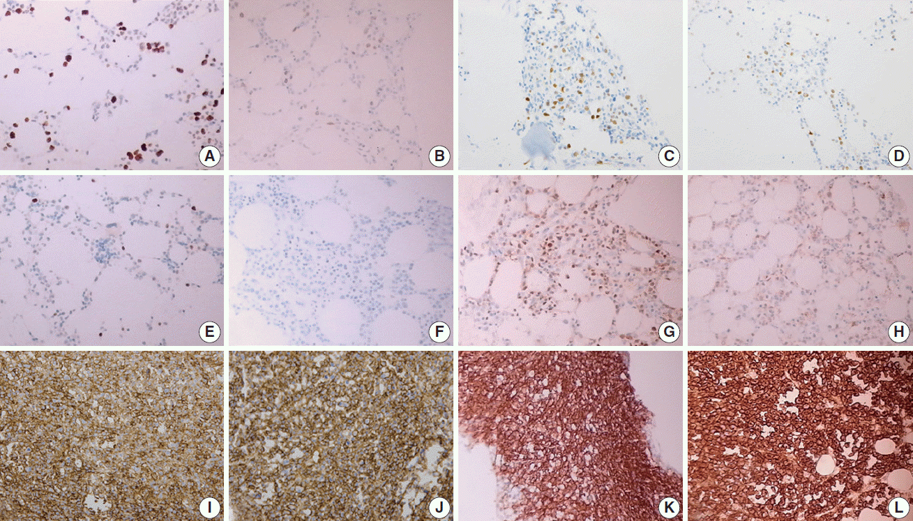
Nuclear proteins such as Ki67, cyclin D1, and TdT were relatively better preserved on immunohistochemistry with both EDTA and RDO protocols, while samples from the HCl protocol showed breakdown and lower quality of nuclear protein staining (Fig. 5AH). Immunohistochemistry targeting the cytoplasmic membrane or cytoplasmic CD markers was well preserved in all three protocols (Fig. 5IL).
There have been several studies comparing several types of decalcification protocols to date. Some retrospective studies compared the morphology of in situ hybridization or immunohistochemistry by using stored tissues containing bone. Other studies extracted nucleic acid from stored tissues containing bone and compared the quantity, purity, and Ct value of the DNA and/or RNA after real-time PCR [1-4]. Few studies have used specimens sampled with diagnostic purpose in clinical practice. We investigated the quality of nucleic acid and protein in decalcified bone marrow tissue employing several types of genetic tools, immunohistochemistry, and morphological assessments. In addition, we compared the effect of decalcification protocol while limiting bias variance, which may be caused by sampling from different patients and cell degeneration from long-term storage. We prospectively investigated pairs of bone marrow biopsy specimens from the same patients sampled for diagnostic purposes in a clinical setting. To our knowledge, this is one of the first studies utilizing clinical samples to assess decalcification protocols.
We compared the conventional protocol using HCl, the well-known alternative protocol using EDTA, and the new protocol using RDO.
All three methods had equally good performance with respect to microtome dissection and preservation of cytomorphologic and histomorphologic features. The EDTA protocol was superior in preserving nucleic acid (DNA and RNA), allowing for the feasibility of genetic studies, such as real time PCR and in situ hybridization. The EDTA protocol would be an appropriate option for genetic studies in bone-contained tissues. The present study also confirmed that the HCl protocols are inappropriate for genetic studies due to the severe damage of genetic material. The quality of nucleic acids in the RDO group was equivocal, but this method also didn’t seem to be suitable for genetic studies. In immunohistochemistry targeting nuclear protein, both EDTA and RDO were relatively superior to the HCl protocol. All three methods showed intact staining in immunohistochemistry targeting cytoplasmic membranes or cytoplasm. EDTA protocols seem to be the most appropriate in that comprehensive immunohistochemical stains can be applied to the specimens processed by this protocol. The feasibility of immunohistochemistry using specimens processed with the HCl protocol would be limited in many cases, especially when immunostaining for nuclear proteins is necessary. Immunohistochemical markers can be more widely applied in samples processed with the RDO protocol than with the HCl protocol, but diagnostic options are still more limited than the EDTA protocol.
In this study, the EDTA protocol was the most feasible method for several types of genetic studies and immunohistochemistry. Such advantages of EDTA decalcification protocol are already widely known [1,5-7]. The present study confirms again the superiority of the EDTA protocol in a wide range of ancillary tests for pathologic diagnosis. The potential of the RDO protocol is higher than the conventional HCl protocol; however, it is not superior to the EDTA protocol and thus, it cannot be considered an alternative to EDTA protocols, particularly when genetic studies are needed.
When choosing decalcification agents, cost-effectiveness and turn-around time from biopsy to final pathologic diagnosis are important issues. HCl costs less and requires less time than using EDTA. Small bone tissues like bone marrow take about three to twenty four hours for decalcification in EDTA protocol, but most of the other bone specimens need more time when using the same protocol. RDO protocols can shorten decalcification time, but this agent is also expensive.
In many cases, bone biopsies are needed for diagnosis of hematologic cancer, metastatic tumors, and primary bone sarcoma; these types of cancers usually need further gene-based diagnosis. Bone marrow biopsy is widely performed in patients with hematologic malignancies and pediatric sarcomas or blastomas. Therefore, preserving genetic materials is critical in handling bone marrow tissues, and cost and turn-around time may be less important in such cases. Preserving intact nucleic acid, as well as intact proteins, is critical to providing not only accurate pathologic diagnosis but also diverse therapeutic options to the patients. Considering this, in this study the EDTA protocol was the most appropriate method for handling bone marrow specimens. RDO may also be useful in that it requires less decalcification time and it enables more ancillary tests than HCl, but its usefulness is limited by less potential for genetic studies in processed samples than EDTA.
In this era of expanding genetic molecular study, better tissue handling methods are needed. The present study suggests an appropriate approach in handling bone marrow tissues, and this approach should be expanded to other types of tissue specimens.
This study was supported by a faculty research grant of Yonsei University College of Medicine for 2014 (6-2014-0133).
Fig. 1.
The quality, quantity, and feasibility of real time PCR study is compared between EDTA, RDO, and HCl protocols. The first row demonstrates EDTA versus HCl, and the second row RDO versus HCl (A, D, DNA yield; B, PCR result of EDTA; E, PCR result of RDO; C, F, PCR results of HCl). D, PCR result of RDO. PCR, polymerase chain reaction; EDTA, ethylenediaminetetraacetic acid disodium salt dehydrate; RDO, RDO GOLD; HCl, hydrochloric acid; PNA, peptide nucleic acid.
jptm-49-3-236f1.gif
Fig. 2.
In EDTA versus HCl comparison of a pair of bone marrow sampled from the same patient (A–D, EDTA versus HCl group; A, B, EDTA protocol; C, D, HCl protocol), and in RDO versus HCl comparison of a pair of bone marrow sampled from the same patient (E–H, RDO versus HCl group; E, F, RDO protocol; G, H, HCl protocol), all the three methods of EDTA, RDO, and HCl protocols demonstrate intact and well-preserved histological features of bone marrow. EDTA, ethylenediaminetetraacetic acid disodium salt dehydrate; HCl, hydrochloric acid; RDO, RDO GOLD.
jptm-49-3-236f2.gif
Fig. 3.
In HER2 dual color silver in situ hybridization study, almost every nucleus in cases of EDTA protocol demonstrates two intact signals of HER2 and CEP17, while cases of RDO or HCl protocol barely demonstrate HER2 or CEP17 signals from the nucleus due to the severe breakdown of DNA (A, B, comparison of EDTA versus HCl in a pair of bone marrow from the same patients; C, D, comparison of RDO versus HCl in a pair of bone marrow from the same patients). EDTA, ethylenediaminetetraacetic acid disodium salt dehydrate; RDO, RDO GOLD; HCl, hydrochloric acid.
jptm-49-3-236f3.gif
Fig. 4.
The quality of RNA is compared in kappa light chain (A–D) and lambda light chain (E–H) RNA in situ hybridization using a pair of bone marrow specimens from the same patient. In a case of kappa light chain-restricted plasma cell myeloma, EDTA protocol (A) reveals intact quality while HCl (B) protocol shows poor quality in kappa light chain RNA in situ hybridization. In a case of lambda light chain-restricted plasma cell myeloma, EDTA protocol (E) reveals intact quality while HCl (F) protocol shows poor quality. In a case of polyclonal plasma cell infiltration within the bone marrow, the EDTA and RDO protocol (C and G, respectively) reveal intact quality while HCl (D, H) protocol show poor quality in kappa and lambda light chain RNA in situ hybridization, respectively. EDTA, ethylenediaminetetraacetic acid disodium salt dehydrate; HCl, hydrochloric acid; RDO, RDO GOLD.
jptm-49-3-236f4.gif
Fig. 5.
In immunohistochemistry of Ki67 (A–D), EDTA (A) shows intact quality while HCl (B) shows poor quality in a pair of bone marrow sample from the same patient. A similar result is noted in comparison of RDO (C) versus HCl (D). Nuclear staining of cyclin D1 is intact in EDTA (E), while it is not in HCl (F) of a paired bone marrow from the same patient. Nuclear staining of TdT is intact in RDO (G), while it is poor in HCl (H) of a paired bone marrow from the same patient. Cytoplasmic membrane staining of CD138 (I–L) reveals intact quality in all three protocols: EDTA (I), RDO (K), and HCl (J, L). EDTA, ethylenediaminetetraacetic acid disodium salt dehydrate; HCl, hydrochloric acid; RDO, RDO GOLD.
jptm-49-3-236f5.gif
Table 1.
The decalcification protocols of three methods
Solution Processing time (hr) Processing temperature
HCl (100%) 3 Room temperature
EDTA (12.5%) 3 or 24a Room temperature
RDO (100%) 0.5–1 Room temperature

HCl, hydrochloric acid; EDTA, ethylenediaminetetraacetic acid disodium salt dehydrate; RDO, RDO GOLD.

aThe processing time of EDTA method was mostly 3 hours. It was 24 hours for few cases that contained more cortical bone due to the oblique direction when inserting biopsy-needle.

Table 2.
Primary antibodies used
Product name Dilution Clonality Clone Company
Cyclin D1 (SP4) 1:50 Monoclonal SP4 LabVisiona
Ki67 1:1,000 Monoclonal MIB-1 DAKOb
Bcl2 Bond-III 1:50 Monoclonal bcl2/100/D5 Novocastrac
Bcl6 Prediluent Monoclonal LN22 Novocastra
TdT 1:100 Polyclonal - Cell Marqued
CD138 Prediluent Monoclonal ML15 DAKO
CD20 1:400 Monoclonal L26 Novocastra
CD79a (B cell) 1:100 Monoclonal JCB117 DAKO
CD3 1:200 Monoclonal SP7 LabVision
CD5 1:100 Monoclonal 4C7 Novocastra
CD23 1:100 Monoclonal SP23 LabVision
CD10 1:75 Monoclonal 56C6 Novocastra
CD30 1:50 Monoclonal Ber-H2 DAKO
Myeloperoxidase 1:2,000 Polyclonal - DAKO

aLab vision, Waltham, MA;

bDAKO, Carpinteria, CA;

cNovocastra, Buffalo Grave, IL;

dCell Marque, Rocklin, CA.

Table 3.
Quantity and quality of DNA according to decalcification protocols
DNA yield (median, range) p-value Ct value (median, range) p-value
EDTA vs HCl EDTA 25 (11.0–37.0) .168 25.0 (24.6–27.2) <.001
HCl 12 (11.0–28.0) 32.7 (28.9–33.3)
RDO vs HCl RDO 14.7 (10.9–15.0) .753 33.6 (33.0–34.1) .754
HCl 13.4 (10.0–15.3) 33.5 (33.2–35.2)

Mann-Whitney U test was used to compare the median of each variables.

HCl, hydrochloric acid; EDTA, ethylenediaminetetraacetic acid disodium salt dehydrate; RDO, RDO GOLD.

Table 4.
Comparison of the immunohistochemical results
Item EDTA vs HCl
RDO vs HCl
EDTA HCl RDO HCl
HER2/CEP17 SISH 5/5 (100)a 0/5 (0) 0/5 (0) 0/5 (0)
Kappa ISH 7/7 (100) 4/7 (57.1) 2/3 (66.7) 2/3 (66.7)
Lambda ISH 5/5 (100) 1/5 (20) 2/2 (100) 0/2 (0)
CyclinD1 9/9 (100) 2/9 (22.2) 3/3 (100) 1/3 (33.3)
Ki67 9/9 (100) 5/9 (55.6) 10/10 (100) 6/10 (45.5)
Bcl2 1/1 (100) 1/1 (100) 4/4 (100) 4/4 (100)
Bcl6 No data No data 1/1 (100) 1/1 (100)
TdT No data No data 1/1 (100) 0/1 (0)
CD138 7/7 (100) 7/7 (100) 3/3 (100) 3/3 (100)
CD20 9/9 (100) 9/9 (100) 9/9 (100) 9/9 (100)
CD79a (B cell) No data No data 2/2 (100) 2/2 (100)
CD3 5/5 (100) 5/5 (100) 4/4 (100) 4/4 (100)
CD5 No data No data 3/3 (100) 3/3 (100)
CD23 No data No data 1/1 (100) 1/1 (100)
CD10 1/1 (100) 1/1 (100) 1/1 (100) 1/1 (100)
CD30 1/1 (100) 1/1 (100) No data No data
Myeloperoxidase 1/1 (100) 1/1 (100) 2/2 (100) 2/2 (100)

HCl, hydrochloric acid; EDTA, ethylenediaminetetraacetic acid disodium salt dehydrate; RDO, RDO GOLD; SISH, silver in situ hybridization; ISH, in situ hybridization.

aCase number showing intact stain result among overall case number stained with each item (%). Positive expression in the indicated tumor cells or internal controls was considered as intact stain. For example, positive expression of cyclin D1 in mantle cell lymphoma cells or endothelial cells was interpreted as intact stain result.

  • 1. Singh VM, Salunga RC, Huang VJ, et al. Analysis of the effect of various decalcification agents on the quantity and quality of nucleic acid (DNA and RNA) recovered from bone biopsies. Ann Diagn Pathol 2013; 17: 322-6. ArticlePubMed
  • 2. Reineke T, Jenni B, Abdou MT, et al. Ultrasonic decalcification offers new perspectives for rapid FISH, DNA, and RT-PCR analysis in bone marrow trephines. Am J Surg Pathol 2006; 30: 892-6. ArticlePubMed
  • 3. Brown RS, Edwards J, Bartlett JW, Jones C, Dogan A. Routine acid decalcification of bone marrow samples can preserve DNA for FISH and CGH studies in metastatic prostate cancer. J Histochem Cytochem 2002; 50: 113-5. ArticlePubMed
  • 4. Alers JC, Krijtenburg PJ, Vissers KJ, van Dekken H. Effect of bone decalcification procedures on DNA in situ hybridization and comparative genomic hybridization. EDTA is highly preferable to a routinely used acid decalcifier. J Histochem Cytochem 1999; 47: 703-10. ArticlePubMed
  • 5. Adegboyega PA, Gokhale S. Effect of decalcification on the immunohistochemical expression of ABH blood group isoantigens. Appl Immunohistochem Mol Morphol 2003; 11: 194-7. ArticlePubMed
  • 6. Castania VA, Silveira JW, Issy AC, et al. Advantages of a combined method of decalcification compared to EDTA. Microsc Res Tech 2015; 78: 111-8. ArticlePubMed
  • 7. Wickham CL, Sarsfield P, Joyner MV, Jones DB, Ellard S, Wilkins B. Formic acid decalcification of bone marrow trephines degrades DNA: alternative use of EDTA allows the amplification and sequencing of relatively long PCR products. Mol Pathol 2000; 53: 336.ArticlePubMedPMC

Figure & Data

References

    Citations

    Citations to this article as recorded by  
    • Optimization of Formic Acid-Formalin-Based Decalcification Protocol for Rat Calvarial Bone Histology
      S. Amitha Banu, Khan Sharun, Merlin Mamachan, Athira Subash, Vadapalli Deekshita, Kirtika Sharma, Karikalan Mathesh, Obli Rajendran Vinodh kumar, Swapan Kumar Maiti, Abhijit M. Pawde, Laith Abualigah, Kuldeep Dhama, Amarpal
      Journal of Experimental Biology and Agricultural Sciences.2024; 12(2): 218.     CrossRef
    • Effects of fixation and demineralization on histomorphology and DNA amplification of canine bone marrow
      Gabriella M. L. Diamantino, Janet Beeler-Marfisi, Robert A. Foster, William Sears, Alice Defarges, William Vernau, Dorothee Bienzle
      Veterinary Pathology.2024; 61(6): 943.     CrossRef
    • In situ metabolomic analysis of osteonecrosis of the femoral head (ONFH) using MALDI MSI
      Chen Li, Jikun Liu, Yiqi Sheng, Yinghao Wang, Lan Jia, Yinguang Zhang, Jiantao Li, Shuangshuang Di, Honggang Nie, Yehua Han
      Analytical and Bioanalytical Chemistry.2024; 416(23): 5155.     CrossRef
    • A set of pretreatment reagents including improved formula fixation and decalcification facilitating immunohistochemistry and DNA analyses of formalin-fixed paraffin-embedded bone marrow trephine biopsy
      Ting Sun, Liming Xu, Hongtian Yao, Jing Zhao, Zhen Chen, Zexin Chen, Bo Wang, Wei Ding
      Acta Histochemica.2024; 126(8): 152188.     CrossRef
    • Germline and somatic testing for homologous repair deficiency in patients with prostate cancer (part 1 of 2)
      Andrew J. Armstrong, Amy Taylor, Michael C. Haffner, Wassim Abida, Alan H. Bryce, Lawrence I. Karsh, Scott T. Tagawa, Przemyslaw Twardowski, Anthony V. Serritella, Joshua M. Lang
      Prostate Cancer and Prostatic Diseases.2024;[Epub]     CrossRef
    • To Freeze or Not to Freeze? Recommendations for Intraoperative Examination and Gross Prosection of Thyroid Glands
      Fouad R. Zakka, Nicole A. Cipriani
      Surgical Pathology Clinics.2023; 16(1): 15.     CrossRef
    • Effect of Surface Decalcification With Hydrochloric Acid on the Determination of Estrogen Receptor, Progesterone Receptor, Ki67, and Human Epidermal Growth Factor Receptor 2 Expressions in Invasive Breast Carcinoma Based on Immunohistochemistry and Fluore
      Wu Ping, Rao Xin, Zhang Li, Chen Yupeng, Song Fangling, Ren Caihong, Hu Shun, Zhang Sheng
      Applied Immunohistochemistry & Molecular Morphology.2023; 31(4): 232.     CrossRef
    • Diagnostic value of MDM2 RNA in situ hybridization for low-grade osteosarcoma: Consistency comparison of RNA in situ hybridization, fluorescence in situ hybridization, and immunohistochemistry
      Chen Chen, Xin He, Min Chen, Tianhai Du, Weiji Qin, Wenyi Jing, Hongying Zhang
      Virchows Archiv.2023; 482(6): 999.     CrossRef
    • Bone marrow fibrosis is associated with non‐response to CD19 CAR T‐cell therapy in B‐acute lymphoblastic leukemia
      Joshua Anil, Ahab Alnemri, Andrew Lytle, Brian Lockhart, Ashley E. Anil, Michael Baumgartner, Kirubel Gebre, Jared McFerran, Stephan A. Grupp, Susan R. Rheingold, Vinodh Pillai
      American Journal of Hematology.2023; 98(12): 1888.     CrossRef
    • Epithelioid haemangioendothelioma of the mandible – A case report and review of the literature
      Ali Rizvi, Tim K. Blackburn, Guy N. J. Betts
      Oral Surgery.2022; 15(3): 387.     CrossRef
    • Evaluation of EDTA and nitric acid solutions for decalcification of joints in AG/WT, BALB/c, C57, DBA1/J mice, and in Wistar rats
      Eduarda Correa Freitas, Suelen Pizzolatto Dalmolin, Mateus Müller da Silva, Francine Hehn de Oliveira, Emily Ferreira Salles Pilar
      Biotechnic & Histochemistry.2022; 97(5): 372.     CrossRef
    • Coupling Lipid Labeling and Click Chemistry Enables Isolation of Extracellular Vesicles for Noninvasive Detection of Oncogenic Gene Alterations
      Na Sun, Benjamin V. Tran, Zishan Peng, Jing Wang, Ceng Zhang, Peng Yang, Tiffany X. Zhang, Josephine Widjaja, Ryan Y. Zhang, Wenxi Xia, Alexandra Keir, Jia‐Wei She, Hsiao‐hua Yu, Jing‐Jong Shyue, Hongguang Zhu, Vatche G. Agopian, Renjun Pei, James S. Toml
      Advanced Science.2022;[Epub]     CrossRef
    • The Expressions of CD30 and CD123 of Mastocytosis in Taiwan
      Ching-Fen Yang, Chih-Yi Hsu
      Applied Immunohistochemistry & Molecular Morphology.2022; 30(4): 278.     CrossRef
    • Unusual Patterns of HER2 Expression in Breast Cancer: Insights and Perspectives
      Dora Grassini, Eliano Cascardi, Ivana Sarotto, Laura Annaratone, Anna Sapino, Enrico Berrino, Caterina Marchiò
      Pathobiology.2022; 89(5): 278.     CrossRef
    • Expert opinion on NSCLC small specimen biomarker testing — Part 1: Tissue collection and management
      Frédérique Penault-Llorca, Keith M. Kerr, Pilar Garrido, Erik Thunnissen, Elisabeth Dequeker, Nicola Normanno, Simon J. Patton, Jenni Fairley, Joshua Kapp, Daniëlle de Ridder, Aleš Ryška, Holger Moch
      Virchows Archiv.2022; 481(3): 335.     CrossRef
    • Comparison of bone demineralisation procedures for DNA recovery from burned remains
      Meghan Mckinnon, Denice Higgins
      Forensic Science International: Genetics.2021; 51: 102448.     CrossRef
    • A review of the current understanding of burned bone as a source of DNA for human identification
      Meghan Mckinnon, Maciej Henneberg, Denice Higgins
      Science & Justice.2021; 61(4): 332.     CrossRef
    • Time is bone — Quantitative comparison of decalcification solvents in human femur samples using dual-X-ray-absorptiometry and computed tomography
      Joshua Gawlitza, Jakob Steinhäuser, Arno Bücker, Gabriela Krasteva-Christ, Thomas Tschernig
      Annals of Anatomy - Anatomischer Anzeiger.2021; 235: 151696.     CrossRef
    • Molecular biomarker testing for non–small cell lung cancer: consensus statement of the Korean Cardiopulmonary Pathology Study Group
      Sunhee Chang, Hyo Sup Shim, Tae Jung Kim, Yoon-La Choi, Wan Seop Kim, Dong Hoon Shin, Lucia Kim, Heae Surng Park, Geon Kook Lee, Chang Hun Lee
      Journal of Pathology and Translational Medicine.2021; 55(3): 181.     CrossRef
    • Effect of EDTA decalcification on estrogen receptor and progesterone receptor immunohistochemistry and HER2/neu fluorescence in situ hybridization in breast carcinoma
      Erik Washburn, Xiaoyu Tang, Carla Caruso, Michelle Walls, Bing Han
      Human Pathology.2021; 117: 108.     CrossRef
    • Performances of single tube nested polymerase chain reaction and GeneXpert ultra on Formalin fixed paraffin embedded tissues in the diagnosis of tuberculous spondylodiscitis
      Emna Romdhane, Soumaya Rammeh, Chelli Mouna Bouaziz, Hend Riahi, Meriam Rekaya Ben, Meriam Ksentini, Yosra Chebbi, Wafa Achour, Asma Ferjani, Ben Boubaker Ilhem Boutiba, Leila Slim-Saidi, Mohamed Fethi Ladeb
      Clinical Rheumatology.2021; 40(10): 4317.     CrossRef
    • Molecular Characterization of Prostate Cancers in the Precision Medicine Era
      Emilio Francesco Giunta, Laura Annaratone, Enrico Bollito, Francesco Porpiglia, Matteo Cereda, Giuseppe Luigi Banna, Alessandra Mosca, Caterina Marchiò, Pasquale Rescigno
      Cancers.2021; 13(19): 4771.     CrossRef
    • Increased NF-κB Activity in Osteoprogenitor-Lineage Cells Impairs the Balance of Bone Versus Fat in the Marrow of Skeletally Mature Mice
      Tzuhua Lin, Jukka Pajarinen, Yusuke Kohno, Akira Nabeshima, Laura Lu, Karthik Nathan, Zhenyu Yao, Joy Y. Wu, Stuart Goodman
      Regenerative Engineering and Translational Medicine.2020; 6(1): 69.     CrossRef
    • Percutaneous CT-guided biopsy of lytic bone lesions in patients clinically suspected of lung cancer: Diagnostic performances for pathological diagnosis and molecular testing
      Anne-Claire Toffart, Stéphane Asfari, Anne Mc Leer, Emilie Reymond, Adrien Jankowski, Denis Moro-Sibilot, Olivier Stephanov, Julien Ghelfi, Sylvie Lantuejoul, Gilbert R. Ferretti
      Lung Cancer.2020; 140: 93.     CrossRef
    • Effect of decalcification protocols on immunohistochemistry and molecular analyses of bone samples
      Elodie Miquelestorena-Standley, Marie-Lise Jourdan, Christine Collin, Corinne Bouvier, Frédérique Larousserie, Sébastien Aubert, Anne Gomez-Brouchet, Jean-Marc Guinebretière, Matthias Tallegas, Bénédicte Brulin, Louis-Romée Le Nail, Anne Tallet, François
      Modern Pathology.2020; 33(8): 1505.     CrossRef
    • Identifying Opportunities and Challenges for Patients With Sarcoma as a Result of Comprehensive Genomic Profiling of Sarcoma Specimens
      Margaret A. Hay, Eric A. Severson, Vincent A. Miller, David A. Liebner, Jo-Anne Vergilio, Sherri Z. Millis, James L. Chen
      JCO Precision Oncology.2020; (4): 176.     CrossRef
    • Comparison of Methods for the Histological Evaluation of Odontocete Spiral Ganglion Cells
      Tania Ramírez, Simona Sacchini, Yania Paz, Rubén S. Rosales, Nakita Câmara, Marisa Andrada, Manuel Arbelo, Antonio Fernández
      Animals.2020; 10(4): 683.     CrossRef
    • Molecular Pathology of Primary Non-small Cell Lung Cancer
      David Ilan Suster, Mari Mino-Kenudson
      Archives of Medical Research.2020; 51(8): 784.     CrossRef
    • Comparison of ethylenediaminetetraacetic acid and rapid decalcificier solution for studying human temporal bones by immunofluorescence
      Sumana Ghosh, Mark B. Lewis, Bradley J. Walters
      Laryngoscope Investigative Otolaryngology.2020; 5(5): 919.     CrossRef
    • A novel cryo-embedding method for in-depth analysis of craniofacial mini pig bone specimens
      Pavla Ticha, Igor Pilawski, Xue Yuan, Jie Pan, Ustun S. Tulu, Benjamin R. Coyac, Waldemar Hoffmann, Jill A. Helms
      Scientific Reports.2020;[Epub]     CrossRef
    • Accelerating precision medicine in metastatic prostate cancer
      Joaquin Mateo, Rana McKay, Wassim Abida, Rahul Aggarwal, Joshi Alumkal, Ajjai Alva, Felix Feng, Xin Gao, Julie Graff, Maha Hussain, Fatima Karzai, Bruce Montgomery, William Oh, Vaibhav Patel, Dana Rathkopf, Matthew Rettig, Nikolaus Schultz, Matthew Smith,
      Nature Cancer.2020; 1(11): 1041.     CrossRef
    • Tissue Morphology and Antigenicity in Mouse and Rat Tibia: Comparing 12 Different Decalcification Conditions
      Kristofor Bogoevski, Anna Woloszyk, Keith Blackwood, Maria A. Woodruff, Vaida Glatt
      Journal of Histochemistry & Cytochemistry.2019; 67(8): 545.     CrossRef
    • Cellular and collagen reference values of gingival and periodontal ligament tissues in rats: a pilot study
      Antoine Alves, Nina Attik, Carine Wirth, Yves Bayon, Alexis Piat, Brigitte Grosgogeat, Kerstin Gritsch
      Histochemistry and Cell Biology.2019; 152(2): 145.     CrossRef
    • Implementing Precision Medicine Programs and Clinical Trials in the Community-Based Oncology Practice: Barriers and Best Practices
      Jennifer L. Ersek, Lora J. Black, Michael A. Thompson, Edward S. Kim
      American Society of Clinical Oncology Educational Book.2018; (38): 188.     CrossRef
    • Integration of next-generation sequencing in clinical diagnostic molecular pathology laboratories for analysis of solid tumours; an expert opinion on behalf of IQN Path ASBL
      Zandra C Deans, Jose Luis Costa, Ian Cree, Els Dequeker, Anders Edsjö, Shirley Henderson, Michael Hummel, Marjolijn JL Ligtenberg, Marco Loddo, Jose Carlos Machado, Antonio Marchetti, Katherine Marquis, Joanne Mason, Nicola Normanno, Etienne Rouleau, Ed S
      Virchows Archiv.2017; 470(1): 5.     CrossRef
    • Protocolo para el estudio de muestras y estandarización del informe patológico de tumores óseos
      Isidro Machado, José Juan Pozo, David Marcilla, Julia Cruz, Juan C. Tardío, Aurora Astudillo, Sílvia Bagué
      Revista Española de Patología.2017; 50(1): 34.     CrossRef
    • Extremely Well-Differentiated Papillary Thyroid Carcinoma Resembling Adenomatous Hyperplasia Can Metastasize to the Skull: A Case Report
      Ju Yeon Pyo, Jisup Kim, Sung-eun Choi, Eunah Shin, Seok-Woo Yang, Cheong Soo Park, Seok-Mo Kim, SoonWon Hong
      Yonsei Medical Journal.2017; 58(1): 255.     CrossRef
    • Treatment of steroid-induced osteonecrosis of the femoral head using porous Se@SiO2 nanocomposites to suppress reactive oxygen species
      Guoying Deng, Kerun Niu, Feng Zhou, Buxiao Li, Yingjie Kang, Xijian Liu, Junqing Hu, Bo Li, Qiugen Wang, Chengqing Yi, Qian Wang
      Scientific Reports.2017;[Epub]     CrossRef
    • Precision Medicine Starts With Preanalytics: Real-Time Assessment of Tissue Fixation Quality by Ultrasound Time-of-Flight Analysis
      Melissa L. Lerch, Daniel R. Bauer, David Chafin, Abbey Theiss, Michael Otter, Geoffrey S. Baird
      Applied Immunohistochemistry & Molecular Morphology.2017; 25(3): 160.     CrossRef
    • Good Laboratory Standards for Clinical Next-Generation Sequencing Cancer Panel Tests
      Jihun Kim, Woong-Yang Park, Nayoung K. D. Kim, Se Jin Jang, Sung-Min Chun, Chang-Ohk Sung, Jene Choi, Young-Hyeh Ko, Yoon-La Choi, Hyo Sup Shim, Jae-Kyung Won
      Journal of Pathology and Translational Medicine.2017; 51(3): 191.     CrossRef
    • An international survey about nail histology processing techniques
      Christina Wlodek, Pauline Lecerf, Josette Andre, Beth S. Ruben, David de Berker
      Journal of Cutaneous Pathology.2017; 44(9): 749.     CrossRef
    • pSTAT5 and ERK exhibit different expression in myeloproliferative neoplasms
      Ewa Wiśniewska-Chudy, Łukasz Szylberg, Grzegorz Dworacki, Ewa Mizera-Nyczak, Andrzej Marszałek
      Oncology Reports.2017; 37(4): 2295.     CrossRef
    • How we do: optimizing bone marrow biopsy logistics for sign-out within 2 days
      I. de Laak–de Vries, A. G. Siebers, L. Burgers, C. Diepenbroek, M. Link, P. Groenen, J. H. J. M. van Krieken, K. M. Hebeda
      Journal of Hematopathology.2016; 9(2): 67.     CrossRef
    • Do More With Less: Tips and Techniques for Maximizing Small Biopsy and Cytology Specimens for Molecular and Ancillary Testing: The University of Colorado Experience
      Dara L. Aisner, Mathew D. Rumery, Daniel T. Merrick, Kimi L. Kondo, Hala Nijmeh, Derek J. Linderman, Robert C. Doebele, Natalie Thomas, Patrick C. Chesnut, Marileila Varella-Garcia, Wilbur A. Franklin, D. Ross Camidge
      Archives of Pathology & Laboratory Medicine.2016; 140(11): 1206.     CrossRef
    • Analysis of the Effects of Bone Marrow Biopsy Decalcification Methods on Histopathological Examination
      Ji Young Park, Kyung Hee Han
      The Korean Journal of Clinical Laboratory Science.2016; 48(4): 371.     CrossRef
    • Distinguishing between Microbial Habitats Unravels Ecological Complexity in Coral Microbiomes
      Amy Apprill, Laura G. Weber, Alyson E. Santoro, Nicole S. Webster
      mSystems.2016;[Epub]     CrossRef
    • Optimal Fixation and Decalcification Methods for Bone Marrow Biopsy
      Myung-Sub Choi, Hyunsup Lee, Hyuk-Chul Kwon, Moon-Hwan Bae, Young-Hye Ko, Hee-Jin Kim, Beom-Se Lee, Bon-Kyung Koo
      Korean Journal of Clinical Laboratory Science.2015; 47(4): 243.     CrossRef

    • PubReader PubReader
    • ePub LinkePub Link
    • Cite this Article
      Cite this Article
      export Copy Download
      Close
      Download Citation
      Download a citation file in RIS format that can be imported by all major citation management software, including EndNote, ProCite, RefWorks, and Reference Manager.

      Format:
      • RIS — For EndNote, ProCite, RefWorks, and most other reference management software
      • BibTeX — For JabRef, BibDesk, and other BibTeX-specific software
      Include:
      • Citation for the content below
      Proposal of an Appropriate Decalcification Method of Bone Marrow Biopsy Specimens in the Era of Expanding Genetic Molecular Study
      J Pathol Transl Med. 2015;49(3):236-242.   Published online May 15, 2015
      Close
    • XML DownloadXML Download
    Figure
    • 0
    • 1
    • 2
    • 3
    • 4
    Proposal of an Appropriate Decalcification Method of Bone Marrow Biopsy Specimens in the Era of Expanding Genetic Molecular Study
    Image Image Image Image Image
    Fig. 1. The quality, quantity, and feasibility of real time PCR study is compared between EDTA, RDO, and HCl protocols. The first row demonstrates EDTA versus HCl, and the second row RDO versus HCl (A, D, DNA yield; B, PCR result of EDTA; E, PCR result of RDO; C, F, PCR results of HCl). D, PCR result of RDO. PCR, polymerase chain reaction; EDTA, ethylenediaminetetraacetic acid disodium salt dehydrate; RDO, RDO GOLD; HCl, hydrochloric acid; PNA, peptide nucleic acid.
    Fig. 2. In EDTA versus HCl comparison of a pair of bone marrow sampled from the same patient (A–D, EDTA versus HCl group; A, B, EDTA protocol; C, D, HCl protocol), and in RDO versus HCl comparison of a pair of bone marrow sampled from the same patient (E–H, RDO versus HCl group; E, F, RDO protocol; G, H, HCl protocol), all the three methods of EDTA, RDO, and HCl protocols demonstrate intact and well-preserved histological features of bone marrow. EDTA, ethylenediaminetetraacetic acid disodium salt dehydrate; HCl, hydrochloric acid; RDO, RDO GOLD.
    Fig. 3. In HER2 dual color silver in situ hybridization study, almost every nucleus in cases of EDTA protocol demonstrates two intact signals of HER2 and CEP17, while cases of RDO or HCl protocol barely demonstrate HER2 or CEP17 signals from the nucleus due to the severe breakdown of DNA (A, B, comparison of EDTA versus HCl in a pair of bone marrow from the same patients; C, D, comparison of RDO versus HCl in a pair of bone marrow from the same patients). EDTA, ethylenediaminetetraacetic acid disodium salt dehydrate; RDO, RDO GOLD; HCl, hydrochloric acid.
    Fig. 4. The quality of RNA is compared in kappa light chain (A–D) and lambda light chain (E–H) RNA in situ hybridization using a pair of bone marrow specimens from the same patient. In a case of kappa light chain-restricted plasma cell myeloma, EDTA protocol (A) reveals intact quality while HCl (B) protocol shows poor quality in kappa light chain RNA in situ hybridization. In a case of lambda light chain-restricted plasma cell myeloma, EDTA protocol (E) reveals intact quality while HCl (F) protocol shows poor quality. In a case of polyclonal plasma cell infiltration within the bone marrow, the EDTA and RDO protocol (C and G, respectively) reveal intact quality while HCl (D, H) protocol show poor quality in kappa and lambda light chain RNA in situ hybridization, respectively. EDTA, ethylenediaminetetraacetic acid disodium salt dehydrate; HCl, hydrochloric acid; RDO, RDO GOLD.
    Fig. 5. In immunohistochemistry of Ki67 (A–D), EDTA (A) shows intact quality while HCl (B) shows poor quality in a pair of bone marrow sample from the same patient. A similar result is noted in comparison of RDO (C) versus HCl (D). Nuclear staining of cyclin D1 is intact in EDTA (E), while it is not in HCl (F) of a paired bone marrow from the same patient. Nuclear staining of TdT is intact in RDO (G), while it is poor in HCl (H) of a paired bone marrow from the same patient. Cytoplasmic membrane staining of CD138 (I–L) reveals intact quality in all three protocols: EDTA (I), RDO (K), and HCl (J, L). EDTA, ethylenediaminetetraacetic acid disodium salt dehydrate; HCl, hydrochloric acid; RDO, RDO GOLD.
    Proposal of an Appropriate Decalcification Method of Bone Marrow Biopsy Specimens in the Era of Expanding Genetic Molecular Study
    Solution Processing time (hr) Processing temperature
    HCl (100%) 3 Room temperature
    EDTA (12.5%) 3 or 24a Room temperature
    RDO (100%) 0.5–1 Room temperature
    Product name Dilution Clonality Clone Company
    Cyclin D1 (SP4) 1:50 Monoclonal SP4 LabVisiona
    Ki67 1:1,000 Monoclonal MIB-1 DAKOb
    Bcl2 Bond-III 1:50 Monoclonal bcl2/100/D5 Novocastrac
    Bcl6 Prediluent Monoclonal LN22 Novocastra
    TdT 1:100 Polyclonal - Cell Marqued
    CD138 Prediluent Monoclonal ML15 DAKO
    CD20 1:400 Monoclonal L26 Novocastra
    CD79a (B cell) 1:100 Monoclonal JCB117 DAKO
    CD3 1:200 Monoclonal SP7 LabVision
    CD5 1:100 Monoclonal 4C7 Novocastra
    CD23 1:100 Monoclonal SP23 LabVision
    CD10 1:75 Monoclonal 56C6 Novocastra
    CD30 1:50 Monoclonal Ber-H2 DAKO
    Myeloperoxidase 1:2,000 Polyclonal - DAKO
    DNA yield (median, range) p-value Ct value (median, range) p-value
    EDTA vs HCl EDTA 25 (11.0–37.0) .168 25.0 (24.6–27.2) <.001
    HCl 12 (11.0–28.0) 32.7 (28.9–33.3)
    RDO vs HCl RDO 14.7 (10.9–15.0) .753 33.6 (33.0–34.1) .754
    HCl 13.4 (10.0–15.3) 33.5 (33.2–35.2)
    Item EDTA vs HCl
    RDO vs HCl
    EDTA HCl RDO HCl
    HER2/CEP17 SISH 5/5 (100)a 0/5 (0) 0/5 (0) 0/5 (0)
    Kappa ISH 7/7 (100) 4/7 (57.1) 2/3 (66.7) 2/3 (66.7)
    Lambda ISH 5/5 (100) 1/5 (20) 2/2 (100) 0/2 (0)
    CyclinD1 9/9 (100) 2/9 (22.2) 3/3 (100) 1/3 (33.3)
    Ki67 9/9 (100) 5/9 (55.6) 10/10 (100) 6/10 (45.5)
    Bcl2 1/1 (100) 1/1 (100) 4/4 (100) 4/4 (100)
    Bcl6 No data No data 1/1 (100) 1/1 (100)
    TdT No data No data 1/1 (100) 0/1 (0)
    CD138 7/7 (100) 7/7 (100) 3/3 (100) 3/3 (100)
    CD20 9/9 (100) 9/9 (100) 9/9 (100) 9/9 (100)
    CD79a (B cell) No data No data 2/2 (100) 2/2 (100)
    CD3 5/5 (100) 5/5 (100) 4/4 (100) 4/4 (100)
    CD5 No data No data 3/3 (100) 3/3 (100)
    CD23 No data No data 1/1 (100) 1/1 (100)
    CD10 1/1 (100) 1/1 (100) 1/1 (100) 1/1 (100)
    CD30 1/1 (100) 1/1 (100) No data No data
    Myeloperoxidase 1/1 (100) 1/1 (100) 2/2 (100) 2/2 (100)
    Table 1. The decalcification protocols of three methods

    HCl, hydrochloric acid; EDTA, ethylenediaminetetraacetic acid disodium salt dehydrate; RDO, RDO GOLD.

    The processing time of EDTA method was mostly 3 hours. It was 24 hours for few cases that contained more cortical bone due to the oblique direction when inserting biopsy-needle.

    Table 2. Primary antibodies used

    Lab vision, Waltham, MA;

    DAKO, Carpinteria, CA;

    Novocastra, Buffalo Grave, IL;

    Cell Marque, Rocklin, CA.

    Table 3. Quantity and quality of DNA according to decalcification protocols

    Mann-Whitney U test was used to compare the median of each variables.

    HCl, hydrochloric acid; EDTA, ethylenediaminetetraacetic acid disodium salt dehydrate; RDO, RDO GOLD.

    Table 4. Comparison of the immunohistochemical results

    HCl, hydrochloric acid; EDTA, ethylenediaminetetraacetic acid disodium salt dehydrate; RDO, RDO GOLD; SISH, silver in situ hybridization; ISH, in situ hybridization.

    Case number showing intact stain result among overall case number stained with each item (%). Positive expression in the indicated tumor cells or internal controls was considered as intact stain. For example, positive expression of cyclin D1 in mantle cell lymphoma cells or endothelial cells was interpreted as intact stain result.


    J Pathol Transl Med : Journal of Pathology and Translational Medicine
    TOP