 E-submission
Articles
- Page Path
- HOME > J Pathol Transl Med > Volume 54(2); 2020 > Article
-
Original Article
Colorectal epithelial neoplasm associated with gut-associated lymphoid tissue -
Yo Han Jeon
 , Ji Hyun Ahn, Hee Kyung Chang
 -
Journal of Pathology and Translational Medicine 2020;54(2):135-145.
DOI: https://doi.org/10.4132/jptm.2019.11.06
Published online: January 29, 2020
Department of Pathology, Kosin University College of Medicine, Busan, Korea
- Corresponding Author: Hee Kyung Chang, MD, Department of Pathology, Kosin University College of Medicine, 262 Gamcheon-ro, Seo-gu, Busan 49267, Korea Tel: 82-51-990-6323, Fax: 82-51-241-7420, E-mail: changhkg@ns.kosinmed.or.kr
© The Korean Society of Pathologists/The Korean Society for Cytopathology
This is an Open Access article distributed under the terms of the Creative Commons Attribution Non-Commercial License (http://creativecommons.org/licenses/by-nc/4.0) which permits unrestricted non-commercial use, distribution, and reproduction in any medium, provided the original work is properly cited.


Figure & Data
References
Citations
- Redefining GALT-associated carcinoma: a distinct subtype of colorectal adenocarcinoma
Jennifer Fallas, Marianna Arvanitaki, Sophie Lecomte, Jean-Yves Bonnet, Sarah De Clercq, Audrey Verrellen, Nicky D’Haene, María Gómez Galdón, Laurine Verset
Virchows Archiv.2026; 488(3): 695. CrossRef - Family adenomatous polyposis come across dome type adenocarcinoma: a case report and literature review
Ying-Ying Chang, Xiao-Long Zhang, Yao-Hui Wang, Ting-Sheng Ling
Diagnostic Pathology.2025;[Epub] CrossRef - Radiation-induced injury and the gut microbiota: insights from a microbial perspective
Qiaoli Wang, Guoqiang Xu, Ouying Yan, Shang Wang, Xin Wang
Therapeutic Advances in Gastroenterology.2025;[Epub] CrossRef
 PubReader
 ePub Link
-
 Cite this Article
- Cite this Article
-
- Close
- Download Citation
- Close
- Figure
-
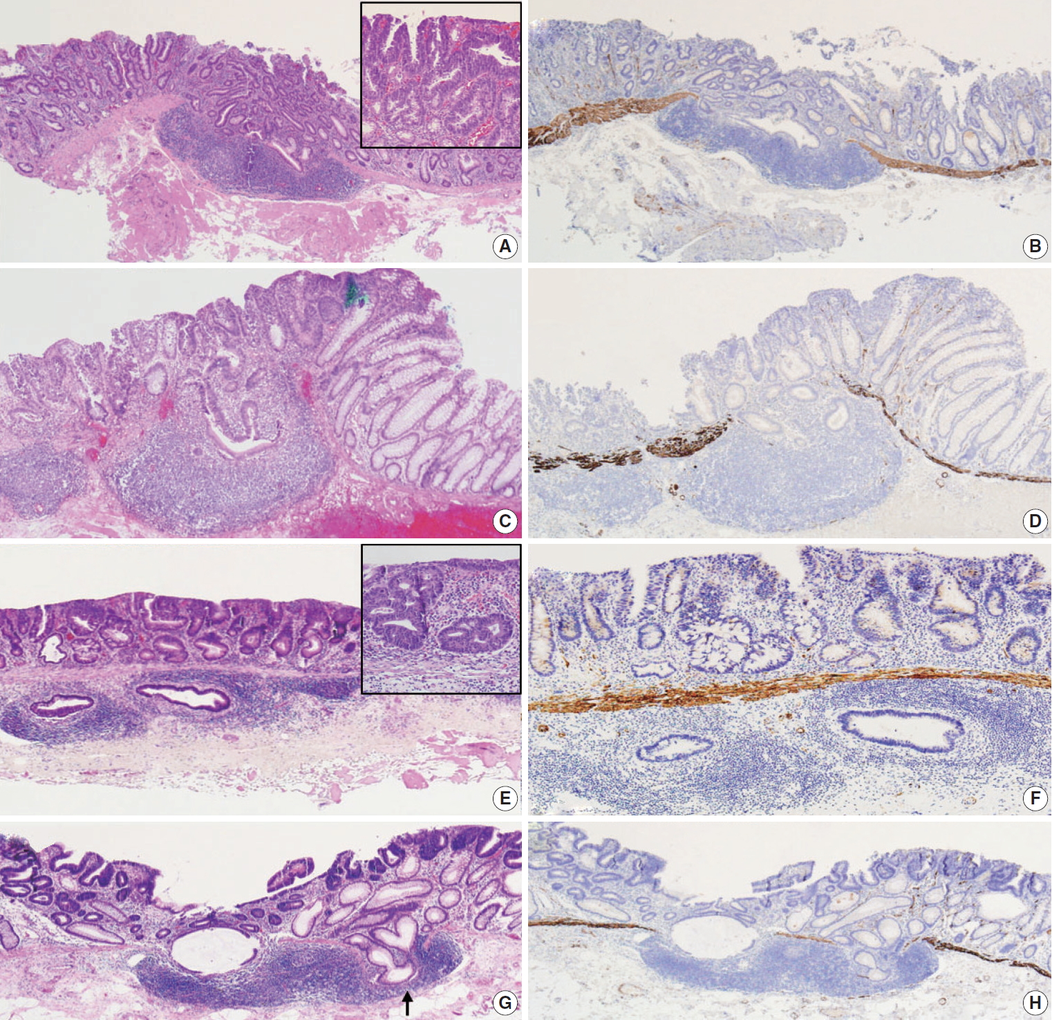
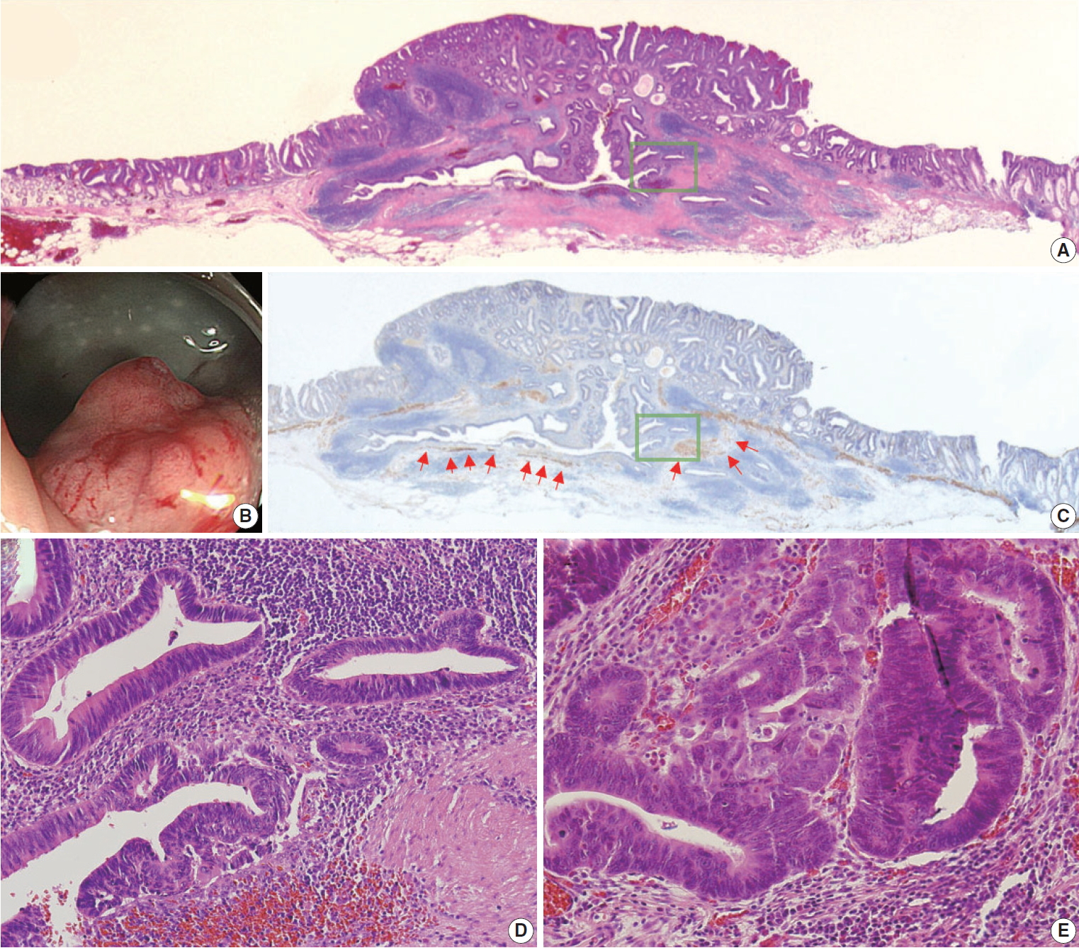


Fig. 1.
Fig. 2.
| Case 1 | Case 2 | Case 3 | Case 4 | Case 5 | Case 6 | Case 7 | Case 8 | Case 9 | Case 10 | Case 11 | |
|---|---|---|---|---|---|---|---|---|---|---|---|
| Age (yr) | 56 | 58 | 66 | 53 | 70 | 71 | 55 | 73 | 73 | 59 | 54 |
| Sex | F | M | F | M | M | M | M | F | M | M | M |
| Location | Rectum | Ascending | Sigmoid | Ascending | Transverse | Ascending | Rectum | Transverse | Transverse | HF | Transverse |
| Endoscopic appearance |
0-IIa + Is | 0-IIa | 0-Is | 0-IIa | 0-IIa | 0-IIa | 0-IIc | 0-IIa | 0-IIa + Is | 0-IIa | 0-IIa |
| Pathologic diagnosis | TA w/HGD | TA w/HGD | TA w/HGD | TA w/HGD | TA w/HGD | TA w/HGD | TA w/HGD | TA w/HGD | TA w/HGD | TA w/LGD | TA w/LGD |
| Dysplasia in the SM GALT | LGD | LGD | HGD | LGD | LGD | LGD | LGD | HGD | HGD | LGD | LGD |
| Continuity of SM glands with surface adenomatous component | + | – | + | + | + | + | – | + | + | + | + |
| Focal defect of MM adjacent to the SM GALT | + | – | + | + | + | + | – | + | + | + | – |
| Hemosiderin deposit in the SM | – | – | – | – | – | – | – | – | – | – | – |
| Rounded contour of involved SM GALT | + | + | – | + | + | + | + | + | + | + | + |
| Cystic dilatation of SM glands | + | – | + | – | – | – | – | – | – | – | + |
| Admixture of SM glands with normal colonic epithelium | – | – | – | – | – | – | – | – | – | – | + |
| Desmoplasia | – | – | – | – | – | – | – | – | – | – | – |
| Single or small clusters of tumor cells in the SM GALT | – | – | – | – | – | – | – | – | – | – | – |
| Lymphovascular invasion | – | – | – | – | – | – | – | – | – | – | – |
| Oncocytic cytoplasm of the SM glands | – | – | – | – | – | – | – | – | – | – | – |
| Goblet cells in the SM glands | + | + | – | – | + | – | + | + | + | – | + |
| Histologic size of the entire tumor (cm) | 1.7 | 1.4 | 1.5 | 2.2 | 1.2 | 2.1 | 1.5 | 1.8 | 1.2 | 2.7 | 1.8 |
| The largest diameter of isolated SM GALT (cm) | 0.14 | 0.13 | 0.33 | 0.17 | 0.14 | 0.21 | 0.29 | 0.12 | 0.23 | 0.14 | 0.19 |
| Total (n = 11) | HGD in the SM (n = 3) | LGD in the SM (n = 8) | p-value | |
|---|---|---|---|---|
| Age (yr) | 59 (53–73) | 73 (66–73) | 57 (53–71) | |
| Sex | .138 | |||
| Male | 8 (72.7) | 1 (33.3) | 7 (87.5) | |
| Female | 3 (27.3) | 2 (66.7) | 1 (12.5) | |
| Endoscopic appearance |
.001 | |||
| Protruding | 1 (9.1) | 1 (33.3) | 0 | |
| Non-protruding | 10 (90.9) | 2 (66.7) | 8 (100) | |
| Location | .138 | |||
| Right-sided | 8 (72.7) | 2 (66.7) | 6 (75.0) | |
| Left-sided | 3 (27.3) | 1 (33.3) | 2 (25.0) | |
| Pathologic diagnosis of entire ESD specimen | .491 | |||
| HGD | 8 (72.7) | 3 (100) | 5 (62.5) | |
| LGD | 3 (27.3) | 0 | 3 (37.5) | |
| Continuity of SM glands with surface adenomatous component | > .99 | |||
| Continued | 9 (81.8) | 3 (100.0) | 6 (75.0) | |
| Discontinued | 2 (18.2) | 0 (0.0) | 2 (25.0) | |
| Focal defect of MM adjacent to SM GALT | > .99 | |||
| Continued | 2 (18.2) | 0 | 2 (25.0) | |
| Discontinued | 9 (81.8) | 3 (100) | 6 (75.0) | |
| Hemosiderin deposit in the SM | – | |||
| Present | 0 | 0 | 0 | |
| Absent | 11 (100) | 3 (100) | 8 (100) | |
| Contour of involved SM GALT | .273 | |||
| Rounded | 10 (90.9) | 2 (66.7) | 8 (100) | |
| Irregular | 1 (9.1) | 1 (33.3) | 0 | |
| Cystic dilatation of SM glands | > .99 | |||
| Present | 3 (27.3) | 1 (33.3) | 2 (25.0) | |
| Absent | 8 (72.7) | 2 (66.7) | 6 (75.0) | |
| Admixture of SM glands with normal colonic epithelium | > .99 | |||
| Present | 1 (9.1) | 0 | 1 (12.5) | |
| Absent | 10 (90.9) | 3 (100) | 7 (87.5) | |
| Desmoplasia | – | |||
| Present | 0 | 0 | 0 | |
| Absent | 11 (100) | 3 (100) | 8 (100) | |
| Single or small clusters of tumor cells in the SM GALT | – | |||
| Present | 0 | 0 | 0 | |
| Absent | 11 (100) | 3 (100) | 8 (100) | |
| Lymphovascular invasion | – | |||
| Present | 0 | 0 | 0 | |
| Absent | 11 (100) | 3 (100) | 8 (100) | |
| Oncocytic cytoplasm of the SM glands | – | |||
| Present | 0 | 0 | 0 | |
| Absent | 11 (100) | 3 (100) | 8 (100) | |
| Goblet cells in the SM glands | – | |||
| Present | 7 (63.6) | 2 (66.7) | 5 (62.5) | |
| Absent | 4 (36.4) | 1 (33.3) | 3 (37.5) | |
| Pathologic size of entire lesion (cm) | 1.7 (1.2–2.7) | 1.5 (1.2–1.8) | 1.75 (1.2–2.7) | |
| The largest diameter of isolated SM GALT (cm) | 0.17 (0.12 –0.33) | 0.23 (0.12– 0.33) | 0.155 (0.13–0.29) |
Values are presented as median (range) or number (%). GALT, gut-associated lymphoid tissue; HGD, high-grade dysplasia; SM, submucosa or submucosal; LGD, low-grade dysplasia; ESD, endoscopic submucosal dissection. Endoscopic appearance was classified according to the Paris classification. GALT, gut-associated lymphoid tissue; F, female; M, male; TA, tubular adenoma; w/, with; HGD, high-grade dysplasia; LGD, low-grade dysplasia; SM, submucosa or submucosal; 0-Is, protruding and sessile type; 0-IIa, flat elevated type; 0-IIc, slightly depressed type; +, present; –, absent; ±, inconspicuous. Endoscopic appearance was classified according to the Paris classification. 0-IIa+Is corresponds to “nodular mixed type of the granular laterally spreading tumor.” 0-IIa corresponds to either “homogeneous type of granular laterally spreading tumor” or “flat elevated type of non-granular laterally spreading tumor.”

