 E-submission
Articles
- Page Path
- HOME > J Pathol Transl Med > Volume 54(3); 2020 > Article
-
Case Study
Gastric IgG4-related disease presenting as a mass lesion and masquerading as a gastrointestinal stromal tumor -
Banumathi Ramakrishna1
 , Rohan Yewale2, Kavita Vijayakumar1, Patta Radhakrishna3, Balakrishnan Siddartha Ramakrishna2
 -
Journal of Pathology and Translational Medicine 2020;54(3):258-262.
DOI: https://doi.org/10.4132/jptm.2020.02.10
Published online: March 4, 2020
1Department of Pathology, SRM Institutes for Medical Science, Vadapalani, India
2Department of Medical Gastroenterology, SRM Institutes for Medical Science, Vadapalani, India
3Department of Surgical Gastroenterology, SRM Institutes for Medical Science, Vadapalani, India
- Corresponding Author: Banumathi Ramakrishna, MBBS, MD, Department of Pathology, SRM Institutes for Medical Science, 1 Jawaharlal Nehru Road, Vadapalani, Chennai 600026, India Tel: +91-44-20002001, Fax: +91-44-49211455, E-mail: banu_ramakrishna@hotmail.com
© 2020 The Korean Society of Pathologists/The Korean Society for Cytopathology
This is an Open Access article distributed under the terms of the Creative Commons Attribution Non-Commercial License (http://creativecommons.org/licenses/by-nc/4.0) which permits unrestricted non-commercial use, distribution, and reproduction in any medium, provided the original work is properly cited.
Abstract
- IgG4-related disease of the stomach is a rare disorder, and only a few cases have been reported. We present two cases that were identified over a 2-month period in our center. Two male patients aged 52 and 48 years presented with mass lesion in the stomach, which were clinically thought to be gastrointestinal stromal tumor, and they underwent excision of the lesion. Microscopic examination revealed marked fibrosis, which was storiform in one case, associated with diffuse lymphoplasmacytic infiltration and an increase in IgG4-positive plasma cells on immunohistochemistry. Serum IgG4 level was markedly elevated. Although rare, IgG4-related disease should be considered in the differential diagnosis of gastric submucosal mass lesions.
- Case 1
- A 42-year-old male patient was admitted to our hospital with a diagnosis of acute pyelonephritis. Computed tomography (CT) of the kidney, ureter, and bladder revealed an incidental mass lesion in the posterior wall of the stomach. Upper gastrointestinal endoscopy revealed submucosal lesions in the esophagus and stomach that were clinically suspected to be GIST. The patient underwent endoscopic ultrasound-guided fine-needle aspiration biopsy of the esophageal lesion and endoscopic mucosal biopsy of the stomach, both of which were inconclusive. The patient also underwent wedge resection of the gastric lesion. Histopathological examination revealed a wedge of the gastric wall with a globular submucosal lesion measuring 6.5×6.0×4.0 cm. The cut surface had a greyish white appearance with foci of calcification. Microscopically, there was marked fibrous expansion of the submucosa with collagenization extending through the muscularis propria to the subserosa, diffuse infiltrates of predominantly plasma cells arranged in cords and clusters admixed with lymphocytes and eosinophils, and several lymphoid follicles with reactive germinal centers (Fig. 1A, B). Small foci of calcification were also present. The muscle coat was disorganized and had muscular hypertrophy in foci. There was no storiform fibrosis or evidence of obliterative or non-obliterative phlebitis. Based on these characteristics, a possible diagnosis of IgG4-RD was suggested.
- IgG4 immunohistochemistry (IHC) and serum IgG4 level were assessed. The IgG4 IHC showed 35–40 immunoreactive plasma cells per high power field (hpf), and serum IgG4 level was elevated (4.36 g/L). These tests confirmed the diagnosis of IgG4-RD. Subsequent serum levels after 3 and 4 months were 2.71g/L and 2.53 g/L, respectively. The patient was treated with steroids and azathioprine. He experienced postsurgery complications that required revision of the gastric anastomosis. During follow-up, his prognosis while receiving medical treatment has been good.
- Case 2
- The second patient was a 58-year-old male found to have erosive gastritis and a submucosal swelling in the body of the stomach in December 2017, while undergoing upper gastrointestinal endoscopy for investigation of dyspepsia. CT examination showed a well-defined, 3×2.9 cm, round, homogeneous, enhancing soft tissue lesion in the distal body of the stomach along the lesser curvature, which was suspected to be GIST or leiomyoma. He was advised to undergo follow-up and elective surgery. His gastric symptoms worsened over the next 10 months, and he underwent excision of the gastric submucosal lesion in October 2018. Histopathological examination revealed a wellcircumscribed globular mass measuring 4×3.3×3.5 cm, and whorling was seen on the cut surface. Microscopically, the lesion was composed of extensively fibrotic and sclerotic stroma with a storiform pattern of fibrosis in foci (Fig. 1C). Discrete, cords and clusters of plasma cells admixed with lymphocytes, a few eosinophils and a few scattered lymphoid aggregates and follicles were present. Perivascular aggregates of plasma cells were also present. There was no evidence of obliterative or nonobliterative phlebitis. Bundles of smooth muscle were identified at the periphery on one aspect. The possibility of IgG4-RD of the stomach was suggested, and IgG4 IHC and serum estimation were recommended. The IgG4 IHC revealed 20–30 immuno-reactive plasma cells/hpf (Fig. 1D). Serum IgG4 was elevated at 3.11g/L, well above the 1.35 g/L cut-off for diagnosis of IgG4-RD. The patient did not return for follow-up.
- Ethics statement
- The authors certify that they obtained patient consent for publication, and the study was approved by the Institutional Review Board of Sri Ramaswamy Memorial Institutes for Medical Science, Chennai, India (IEC NO: SIMS IEC/other/18/2019).
CASE REPORT
- IgG4-RD is an immune-mediated fibroinflammatory lesion, first described in patients with sclerosing cholangitis associated with autoimmune pancreatitis type 1 [5]. Later, it was identified in other organs including the liver, bile ducts, salivary glands, retroperitoneum, lymph nodes, and lungs. It is characterized by diffuse or partial enlargement of the organ and histologically as dense lymphoplasmacytic infiltration with an increase in IgG4 plasma cells on immunostaining, a storiform pattern of fibrosis and obliterative or non-obliterative phlebitis, and an increase in serum IgG4 level.
- IgG4-RD of the gastrointestinal tract is very rare and can present as diffuse wall thickening or as polyp or mass-like lesion [2,3]. Even though obliterative phlebitis was not present in these two cases, the presence of dense fibrosis, which was storiform in one case, and dense lymphoplasmacytic infiltration with lymphoid aggregates and follicles, presenting as a submucosal masslike lesion, suggested the possibility of IgG4-RD, which was confirmed by IHC and elevated serum IgG4 level. The presence of at least two histological features is required for confident diagnosis of IgG4-RD; in most cases, dense lymphoplasmacytic infiltrate and diffuse/storiform fibrosis are seen. Additional clinical, serological (serum level >135 mg/dL or 1.35 g/L), or radiological evidence is required to confirm IgG4-RD [1].
- The cut-off point for the presence of IgG4 plasma cells in tissues varies and can range from >30 plasma cells/hpf to >50/hpf, which is highly specific [1,5]. In biopsy specimens, more than 10 IgG4 plasma cells/hpf were reported in one study [16]. However, the cut-off points vary depending on organ system. Some studies have suggested that IgG4+/IgG+ plasma cell ratio >0.4 is a marker of IgG4-RD in the presence of classic histopathological features and with a compatible clinical features [1,17].
- IgG4-RD can involve multiple organs or any sites in the body synchronously or metachronously [18]. Patients can present with non-specific symptoms and swelling or mass-like lesion. Patients with biliary or pancreatic lesion can present with jaundice, weight loss, and vague abdominal pain. The disease can be an incidental finding during radiological examination and can be mistaken for malignancy, as there are no specific radiological features characteristic of this disease [18,19]. Most cases of gastric IgG4-RD have been reported in middle-aged patients, and both men and women are affected [3,6]. Both patients in this report were middle-aged men.
- IgG4-RD of the stomach was first described in 2004 by Shinji et al. [20], presenting as a gastric ulcer. Because it is difficult to diagnose clinically, especially in isolated cases, most of the reported patients have undergone surgery. Because this disease involves a submucosal lesion in the stomach, these cases are often misdiagnosed as GIST [3,7,18,19] and are difficult to diagnose on endoscopic forceps biopsy, similar to our cases. Gastric lesions that have been mistaken for GIST or gastric cancer have been reported in the literature (Table 1) [3,6-15]. IgG4-RD of the stomach that involved the regional lymph nodes has also been reported [6].
- Although steroids are the first therapeutic option for treating IgG4-RD, it is difficult to diagnose gastric IgG4-RD without histopathological examination. Almost all cases have been reported after surgical resection. Therefore, this disease should be considered in the differential diagnosis of gastric submucosal mass lesion.
- To conclude, we present two cases of IgG4-RD of the stomach that presented as a mass lesion and were clinically suspected to be GIST. The diagnosis was made only after histopathological examination of resection specimens. This highlights the importance of considering this disease in differential diagnosis to avoid surgical resection.
DISCUSSION
Author contributions
Conceptualization: BR.
Data curation: BR, RY.
Investigation: BR, KV, PR, RY, BSR.
Visualization: BR.
Writing—original draft: BR.
Writing—review and editing: BR, RY, BSR.
Conflicts of Interest
The authors declare that they have no potential conflicts of interest.
Funding
No funding to declare.

| Case No. | Age (yr) | Sex | Endoscopic finding/Clinical diagnosis | Location | Size (mm) | Histopathology/IHC | Serum IgG4 levels | Treatment | Study |
|---|---|---|---|---|---|---|---|---|---|
| 1 | 48 | F | Mass/GIST/NET | Mid body | 36 × 22 | SF, LP, OP, IgG4 + 210/hpf, IgG4/IgG ratio about 85% | NA | WR | Woo et al. [3] |
| 2 | 62 | F | Mass/gastic cancer | Antrum | 80 × 30 | SF, LP, OP, IgG4 + ve lymphoplasmacytes > 50/hpf | Elevated | DG | Bulanov et al. [6] |
| 3 | 59 | F | Mass/GIST | NA | 33 × 14 | Abundant LP, SF, lymphoid follicles, IgG4 > 50/hpf | Normal | WR | Kim et al. [7] |
| 4 | 56 | F | Mass/GIST | NA | 21 × 15 | Abundant LP, SF, calcification, IgG4 > 50/hpf | Normal | WR | Kim et al. [7] |
| 5 | 60 | F | Nodule/NA | Fundus | 10 × 15 | Fibrosis, dense LP, IgG4 > 80/hpf | Normal | WR | Chetty et al. [8] |
| 6 | 45 | M | Multiple nodules/NA | Antrum | Up to 22 | LP, many eosinophils, IgG4/IgG ratio 0.84 | NA | DG | Chetty et al. [8] |
| 7 | 56 | M | Nodule/NA | Body | 8 | SF, LP, IgG4-40-102/hpf IgG4/IgG ratio 80%–90% | NA | ESR | Na et al. [9] |
| 8 | 58 | M | Nodule/AIP | Fundus and body | 14 | Dense LP, extensive IgG and IgG4 + staining | Normal | Steroid | Baez et al. [10] |
| 9 | 55 | F | Nodule/GIST | Body | 20 | Dense hyalinization, LP, IgG4/IgG ratio 41% | Normal | ESR | Zhang et al. [11] |
| 10 | 75 | F | Polyp/GIST | Body | 56 × 50 | Fibrosis, LP, many eosinophils, IgG4–39/hpf | Normal | WR | Rollins et al. [12] |
| 11 | 44 | M | Mass/GIST | Body | 20 × 18 | Fibrosis, LP, IgG4 + ve lymphoplasmacytes | Normal | ESR | Otsuka et al. [13] |
| 12 | 27 | F | Mass/GIST/NET | Fundus | 40 | Dense fibrosis, LP, IgG4/IgG ratio 25.3% | Normal | WR | Cheong et al. [14] |
| 13 | 29 | F | Mass/GIST | Body | 20 × 15 | Fibrosis, LP, IgG4 + ve plasma cells 150/hpf | NA | WR | Skorus et al. [15] |
| 14 | 43 | M | Mass/GIST | Antrum | 70 × 50 | Dense LP, IgG4 + plasma cells 35–40/hpf | Elevated | WR + Steroids | Present case 1 |
| 15 | 58 | M | Mass/GIST | Distal body | 45 × 40 | LP, SP, IgG4 + ve plasma cells 20–30/hpf | Elevated | WR | Present case 2 |
IHC, immunohistochemistry; GIST, gastrointestinal stromal tumor; NET, neuroendocrine tumor; SF, storiform fibrosis; LP, lymphoplasmacytic infiltrate; OP, obliterative phlebitis; hpf, high power field; NA, not available; WR, wedge resection; DG, distal gastrectomy; ESR, endoscopic submucosal resection; AIP, autoimmune pancreatitis.
- 1. Deshpande V, Zen Y, Chan JK, et al. Consensus statement on the pathology of IgG4-related disease. Mod Pathol 2012; 25: 1181-92. PubMedPDF
- 2. Koizumi S, Kamisawa T, Kuruma S, et al. Immunoglobulin G4-related gastrointestinal diseases, are they immunoglobulin G4-related diseases? World J Gastroenterol 2013; 19: 5769-74. ArticlePubMedPMC
- 3. Woo CG, Yook JH, Kim AY, Kim J. IgG4-related disease presented as a mural mass in the stomach. J Pathol Transl Med 2016; 50: 67-70. ArticlePubMedPDF
- 4. Stone JH, Zen Y, Deshpande V. IgG4-related disease. N Engl J Med 2012; 366: 539-51. ArticlePubMed
- 5. Kamisawa T, Funata N, Hayashi Y, et al. A new clinicopathological entity of IgG4-related autoimmune disease. J Gastroenterol 2003; 38: 982-4. ArticlePubMedPDF
- 6. Bulanov D, Arabadzhieva E, Bonev S, et al. A rare case of IgG4-related disease: a gastric mass, associated with regional lymphadenopathy. BMC Surg 2016; 16: 37.ArticlePubMedPMCPDF
- 7. Kim DH, Kim J, Park DH, et al. Immunoglobulin G4-related inflammatory pseudotumor of the stomach. Gastrointest Endosc 2012; 76: 451-2. ArticlePubMed
- 8. Chetty R, Serra S, Gauchotte G, Markl B, Agaimy A. Sclerosing nodular lesions of the gastrointestinal tract containing large numbers of IgG4 plasma cells. Pathology 2011; 43: 31-5. ArticlePubMed
- 9. Na KY, Sung JY, Jang JY, et al. Gastric nodular lesion caused by IgG4-related disease. Pathol Int 2012; 62: 716-8. ArticlePubMed
- 10. Baez JC, Hamilton MJ, Bellizzi A, Mortelé KJ. Gastric involvement in autoimmune pancreatitis: MDCT and histopathologic features. JOP 2010; 11: 610-3. PubMed
- 11. Zhang H, Jin Z, Ding S. Gastric calcifying fibrous tumor: a case of suspected immunoglobulin G4-related gastric disease. Saudi J Gastroenterol 2015; 21: 423-6. ArticlePubMedPMC
- 12. Rollins KE, Mehta SP, O'Donovan M, Safranek PM. Gastric IgG4-related autoimmune fibrosclerosing pseudotumour: a novel location. ISRN Gastroenterol 2011; 2011: 873087.ArticlePubMedPDF
- 13. Otsuka R, Kano M, Hayashi H, et al. Probable IgG4-related sclerosing disease presenting as a gastric submucosal tumor with an intense tracer uptake on PET/CT: a case report. Surg Case Rep 2016; 2: 33.ArticlePubMedPMCPDF
- 14. Cheong HR, Lee BE, Song GA, Kim GH, An SG, Lim W. Immunoglobulin G4-related inflammatory pseudotumor presenting as a solitary mass in the stomach. Clin Endosc 2016; 49: 197-201. ArticlePubMedPMCPDF
- 15. Skorus U, Kenig J, Mastalerz K. IgG4-related disease manifesting as an isolated gastric lesion- a literature review. Pol Przegl Chir 2018; 90: 41-5.
- 16. Chari ST. Diagnosis of autoimmune pancreatitis using its five cardinal features: introducing the Mayo Clinic’s HISORt criteria. J Gastroenterol 2007; 42 Suppl 18: 39-41. ArticlePubMedPDF
- 17. Miyabe K, Zen Y, Cornell LD, et al. Gastrointestinal and extra-intestinal manifestations of IgG4-related disease. Gastroenterology 2018; 155: 990-1003. ArticlePubMed
- 18. Seo HS, Jung YJ, Park CH, Song KY, Jung ES. IgG4-related disease in the stomach which was confused with gastrointestinal stromal tumor (GIST): two case reports and review of the literature. J Gastric Cancer 2018; 18: 99-107. ArticlePubMedPMCPDF
- 19. Inoue D, Yoneda N, Yoshida K, et al. Imaging and pathological features of gastric lesion of immunoglobulin G4-related disease: A case report and review of the recent literature. Mod Rheumatol 2019; 29: 377-82. ArticlePubMedPDF
- 20. Shinji A, Sano K, Hamano H, et al. Autoimmune pancreatitis is closely associated with gastric ulcer presenting with abundant IgG4-bearing plasma cell infiltration. Gastrointest Endosc 2004; 59: 506-11. ArticlePubMed
REFERENCES
Figure & Data
References
Citations
- Isolated IgG4-related disease of terminal ileum: Report of a rare case and review of literature
Subham Bhowmik, Hemanga K. Bhattacharjee, Joyner Abraham, Raju Sharma, Prasenjit Das
Journal of Cancer Research and Therapeutics.2025; 21(1): 200. CrossRef - Great Mimics in Oncology: A Retrospective Study from a Tertiary Care Centre of Eastern India
Suvendu Maji, Jayesh kumar Jha, Vikram Chaturvedi
Indian Journal of Surgical Oncology.2025; 16(1): 64. CrossRef - Systemic diseases affecting the GI tract: A review of clinical and histopathologic manifestations
Maryam K. Pezhouh, Dora Lam-Himlin, Atif Zaheer, Lysandra Voltaggio
Annals of Diagnostic Pathology.2024; 73: 152351. CrossRef - Utilizing Immunoglobulin G4 Immunohistochemistry for Risk Stratification in Patients with Papillary Thyroid Carcinoma Associated with Hashimoto Thyroiditis
Faridul Haq, Gyeongsin Park, Sora Jeon, Mitsuyoshi Hirokawa, Chan Kwon Jung
Endocrinology and Metabolism.2024; 39(3): 468. CrossRef - Compromiso gástrico por enfermedad relacionada con IgG4
Gilberto Jaramillo Trujillo, Oscar Fernando Ruiz, Melissa González Pabón, Maria Andrea Jaramillo Trujillo
Revista Repertorio de Medicina y Cirugía.2024; 33(3): 319. CrossRef - CGB5, INHBA and TRAJ19 Hold Prognostic Potential as Immune Genes for Patients with Gastric Cancer
Bei Ji, Lili Qiao, Wei Zhai
Digestive Diseases and Sciences.2023; 68(3): 791. CrossRef - IgG4-related diseases of the digestive tract
J.-Matthias Löhr, Miroslav Vujasinovic, Jonas Rosendahl, John H. Stone, Ulrich Beuers
Nature Reviews Gastroenterology & Hepatology.2022; 19(3): 185. CrossRef - Clinicopathological characteristics of gastric IgG4‐related disease: Systematic scoping review
Haruki Sawada, Torrey Czech, Krixie Silangcruz, Landon Kozai, Adham Obeidat, Eric Andrew Wien, Midori Filiz Nishimura, Asami Nishikori, Yasuharu Sato, Yoshito Nishimura
Journal of Gastroenterology and Hepatology.2022; 37(10): 1865. CrossRef - Utility of gastric biopsy in diagnosing IgG4‐related gastrointestinal disease
Kaori Uchino, Kenji Notohara, Takeshi Uehara, Yasuhiro Kuraishi, Junya Itakura, Akihiro Matsukawa
Pathology International.2021; 71(2): 124. CrossRef
 PubReader
 ePub Link
-
 Cite this Article
- Cite this Article
-
- Close
- Download Citation
- Close
- Figure
-
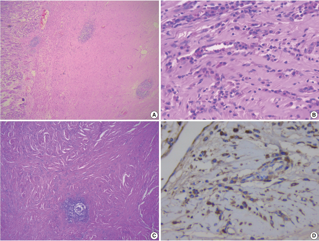

Fig. 1.
| Case No. | Age (yr) | Sex | Endoscopic finding/Clinical diagnosis | Location | Size (mm) | Histopathology/IHC | Serum IgG4 levels | Treatment | Study |
|---|---|---|---|---|---|---|---|---|---|
| 1 | 48 | F | Mass/GIST/NET | Mid body | 36 × 22 | SF, LP, OP, IgG4 + 210/hpf, IgG4/IgG ratio about 85% | NA | WR | Woo et al. [3] |
| 2 | 62 | F | Mass/gastic cancer | Antrum | 80 × 30 | SF, LP, OP, IgG4 + ve lymphoplasmacytes > 50/hpf | Elevated | DG | Bulanov et al. [6] |
| 3 | 59 | F | Mass/GIST | NA | 33 × 14 | Abundant LP, SF, lymphoid follicles, IgG4 > 50/hpf | Normal | WR | Kim et al. [7] |
| 4 | 56 | F | Mass/GIST | NA | 21 × 15 | Abundant LP, SF, calcification, IgG4 > 50/hpf | Normal | WR | Kim et al. [7] |
| 5 | 60 | F | Nodule/NA | Fundus | 10 × 15 | Fibrosis, dense LP, IgG4 > 80/hpf | Normal | WR | Chetty et al. [8] |
| 6 | 45 | M | Multiple nodules/NA | Antrum | Up to 22 | LP, many eosinophils, IgG4/IgG ratio 0.84 | NA | DG | Chetty et al. [8] |
| 7 | 56 | M | Nodule/NA | Body | 8 | SF, LP, IgG4-40-102/hpf IgG4/IgG ratio 80%–90% | NA | ESR | Na et al. [9] |
| 8 | 58 | M | Nodule/AIP | Fundus and body | 14 | Dense LP, extensive IgG and IgG4 + staining | Normal | Steroid | Baez et al. [10] |
| 9 | 55 | F | Nodule/GIST | Body | 20 | Dense hyalinization, LP, IgG4/IgG ratio 41% | Normal | ESR | Zhang et al. [11] |
| 10 | 75 | F | Polyp/GIST | Body | 56 × 50 | Fibrosis, LP, many eosinophils, IgG4–39/hpf | Normal | WR | Rollins et al. [12] |
| 11 | 44 | M | Mass/GIST | Body | 20 × 18 | Fibrosis, LP, IgG4 + ve lymphoplasmacytes | Normal | ESR | Otsuka et al. [13] |
| 12 | 27 | F | Mass/GIST/NET | Fundus | 40 | Dense fibrosis, LP, IgG4/IgG ratio 25.3% | Normal | WR | Cheong et al. [14] |
| 13 | 29 | F | Mass/GIST | Body | 20 × 15 | Fibrosis, LP, IgG4 + ve plasma cells 150/hpf | NA | WR | Skorus et al. [15] |
| 14 | 43 | M | Mass/GIST | Antrum | 70 × 50 | Dense LP, IgG4 + plasma cells 35–40/hpf | Elevated | WR + Steroids | Present case 1 |
| 15 | 58 | M | Mass/GIST | Distal body | 45 × 40 | LP, SP, IgG4 + ve plasma cells 20–30/hpf | Elevated | WR | Present case 2 |
IHC, immunohistochemistry; GIST, gastrointestinal stromal tumor; NET, neuroendocrine tumor; SF, storiform fibrosis; LP, lymphoplasmacytic infiltrate; OP, obliterative phlebitis; hpf, high power field; NA, not available; WR, wedge resection; DG, distal gastrectomy; ESR, endoscopic submucosal resection; AIP, autoimmune pancreatitis.

