Articles
- Page Path
- HOME > J Pathol Transl Med > Volume 56(5); 2022 > Article
-
Original Article
Landscape of EGFR mutations in lung adenocarcinoma: a single institute experience with comparison of PANAMutyper testing and targeted next-generation sequencing -
Jeonghyo Lee1,2
 , Yeon Bi Han1,2
 , Hyun Jung Kwon1,2
 , Song Kook Lee1
 , Hyojin Kim1,2
 , Jin-Haeng Chung1,2,3
-
Journal of Pathology and Translational Medicine 2022;56(5):249-259.
DOI: https://doi.org/10.4132/jptm.2022.06.11
Published online: September 13, 2022
1Department of Pathology and Translational Medicine, Seoul National University Bundang Hospital, Seongnam, Korea
2Department of Pathology, Seoul National University College of Medicine, Seoul, Korea
3Artificial Intelligence Institute, Seoul National University, Seoul, Korea
-
Corresponding Author: Jin-Haeng Chung, MD, PhD, Department of Pathology and Translational Medicine, Seoul National University Bundang Hospital, 82 Gumi-ro 173 beon-gil, Bundang-gu, Seongnam 13620, Korea, Tel: +82-31-787-7713, Fax: +82-31-787-4012, E-mail: chungjh@snu.ac.kr
Corresponding Author: Yeon Bi Han, MD, PhD, Department of Pathology and Translational Medicine, Seoul National University Bundang Hospital, 82 Gumi-ro 173 beon-gil, Bundang-gu, Seongnam 13620, Korea, Tel: +82-31-787-7710, Fax: +82-31-787-4012, E-mail: yeonbimoya@gmail.com
© 2022 The Korean Society of Pathologists/The Korean Society for Cytopathology
This is an Open Access article distributed under the terms of the Creative Commons Attribution Non-Commercial License (https://creativecommons.org/licenses/by-nc/4.0) which permits unrestricted non-commercial use, distribution, and reproduction in any medium, provided the original work is properly cited.
Figure & Data
References
Citations

- Comparison of tissue-based and plasma-based testing for EGFR mutation in non–small cell lung cancer patients
Yoon Kyung Kang, Dong Hoon Shin, Joon Young Park, Chung Su Hwang, Hyun Jung Lee, Jung Hee Lee, Jee Yeon Kim, JooYoung Na
Journal of Pathology and Translational Medicine.2025; 59(1): 60. CrossRef - Localization of epidermal growth factor receptor-mutations using PNA:DNA probes in clinical specimens from patients with non-small cell lung cancer
Haruo Miyata, Hajime Shigeto, Tomoatsu Ikeya, Tadashi Ashizawa, Akira Iizuka, Yasufumi Kikuchi, Chie Maeda, Akari Kanematsu, Kazue Yamashita, Kenichi Urakami, Yuji Shimoda, Takeshi Nagashima, Keiichi Ohshima, Yasuhisa Ohde, Mitsuhiro Isaka, Takashi Sugino
Scientific Reports.2025;[Epub] CrossRef - Molecular characteristics and responses to EGFR tyrosine kinase inhibitors in non-small cell lung cancer patients with EGFR exon 19 insertions
Yang Li, Yunfeng Ni, Feng Lv, Yan Shi, Yedan Chen, Xiaoying Wu, Jiaohui Pang, Long Huang, Yang Shao, Tao Wang, Jie Min, Yang Song
BMC Medicine.2025;[Epub] CrossRef - Detection of EGFR exon 20 insertion mutations in non-small cell lung cancer: implications for consistent nomenclature in precision medicine
Jieun Park, Boram Lee, Ji-Young Song, Minjung Sung, Mi Jeong Kwon, Chae Rin Kim, Sangjin Lee, Young Kee Shin, Yoon-La Choi
Pathology.2024; 56(5): 653. CrossRef - Histo-pillar strip for optimal histogel block construction and biomarker analysis in 3D-lung cancer patient-derived organoids
Sang-Yun Lee, Eunyoung Lee, Ji-O Ryu, Kyuhwan Kim, Yongki Hwang, Bosung Ku, Seok Whan Moon, Mi Hyoung Moon, Kyung Soo Kim, Kwanyong Hyun, Jeong Uk Lim, Chan Kwon Park, Sung Won Kim, Chang Dong Yeo, Dong Woo Lee, Seung Joon Kim
Biofabrication.2024; 16(4): 045017. CrossRef
 PubReader
 ePub Link
-
 Cite this Article
- Cite this Article
-
- Close
- Download Citation
- Close
- Figure


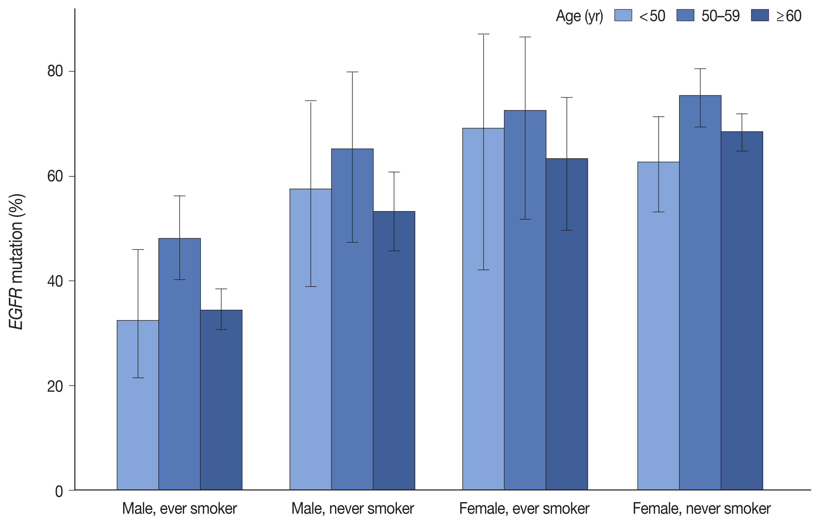
Fig. 1
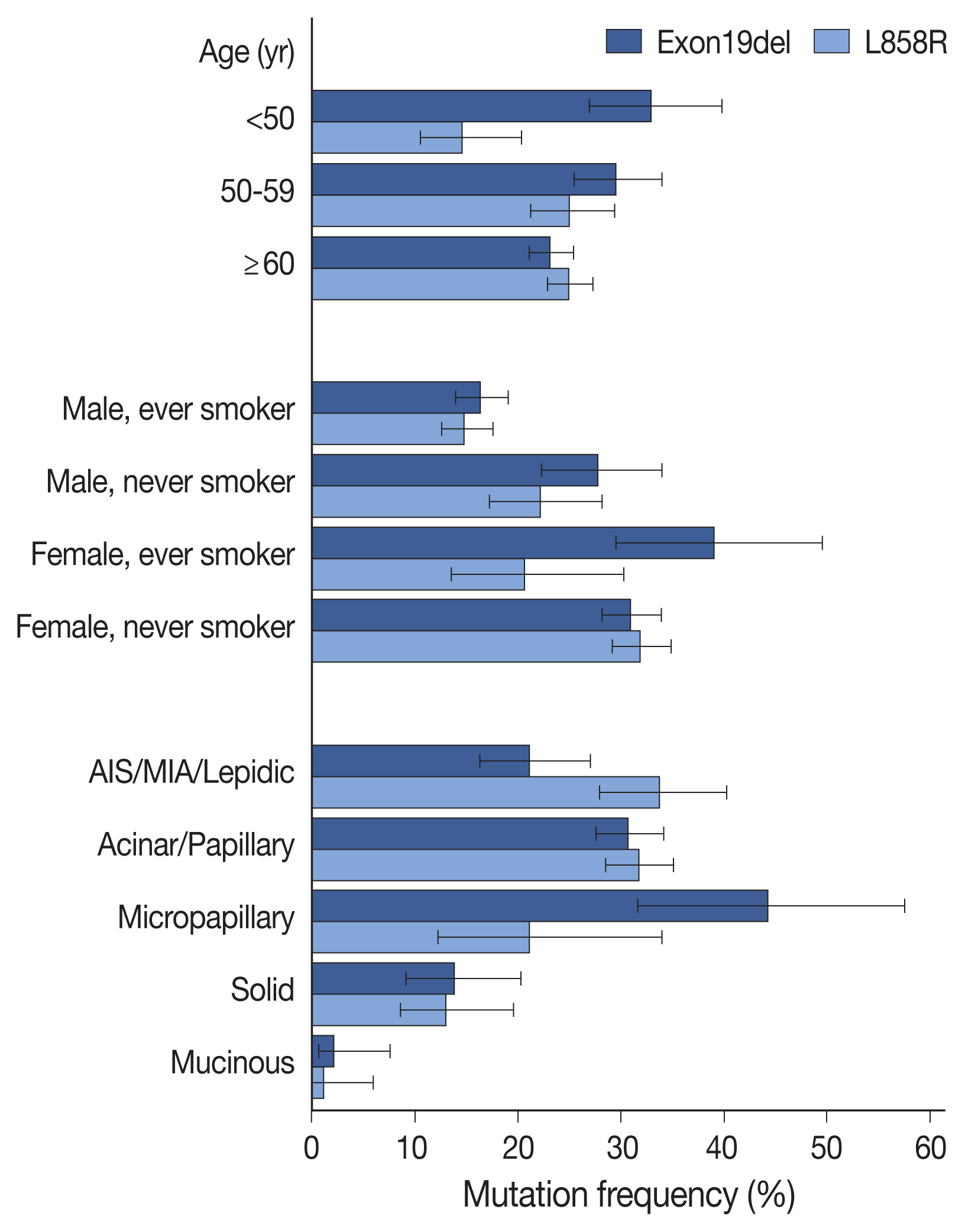
Fig. 2
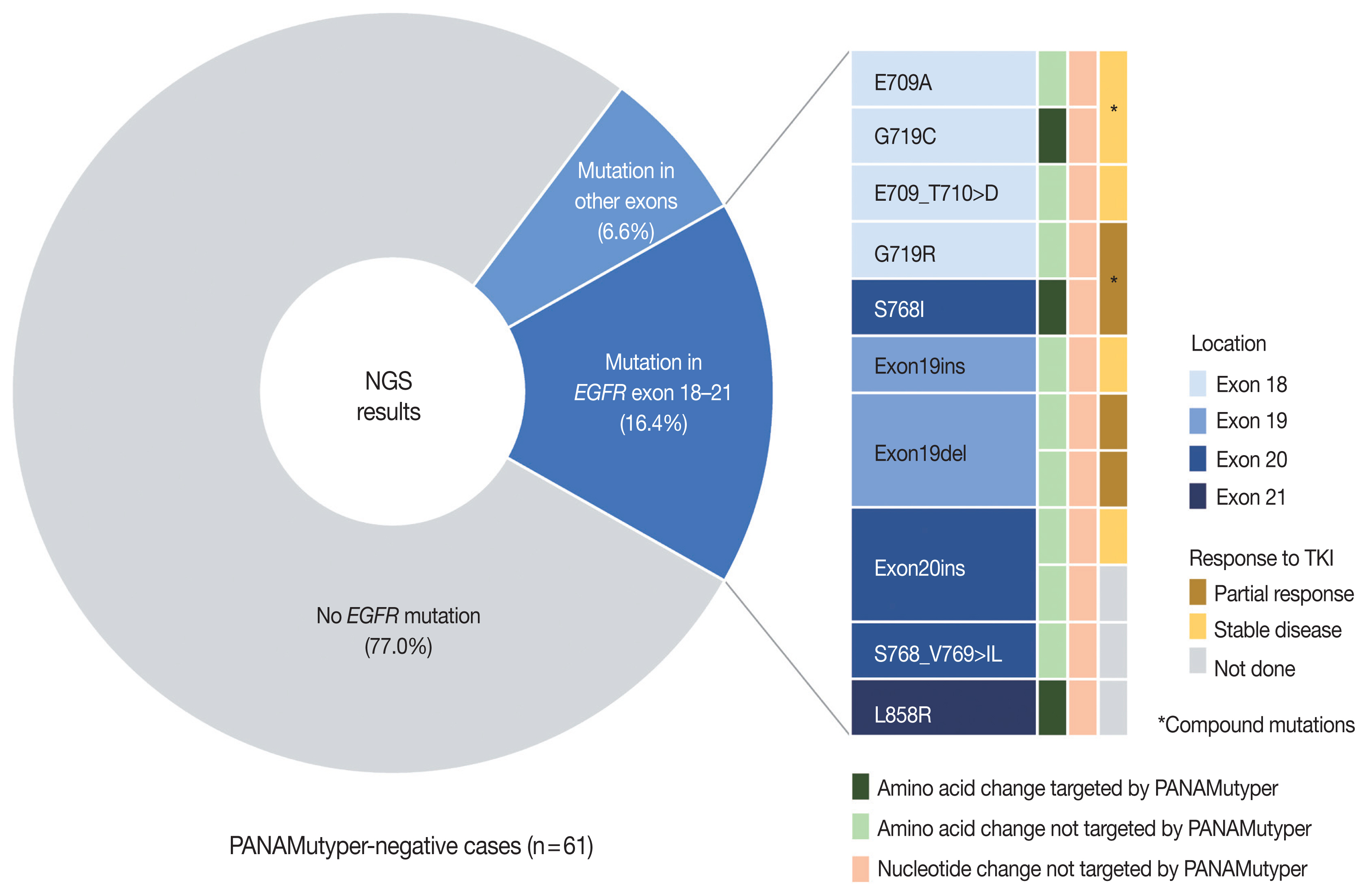
Fig. 3
| Characteristic | Examined No. | EGFR mutation | p-value |
|---|---|---|---|
| Total | 2,088 (100) | 1,162 (55.7) | |
| Age (yr) | |||
| < 40 | 36 (1.7) | 17 (47.2) | .001 |
| 40–49 | 161 (7.7) | 90 (55.9) | |
| 50–59 | 427 (20.5) | 277 (64.9) | |
| 60–69 | 673 (32.2) | 370 (55.0) | |
| 70–79 | 638 (30.6) | 334 (52.4) | |
| ≥ 80 | 153 (7.3) | 74 (48.4) | |
| Sex | |||
| Male | 1,005 (48.1) | 412 (41.0) | < .001 |
| Female | 1,083 (51.9) | 750 (69.3) | |
| Smoking status | |||
| Never | 1,205 (57.7) | 808 (67.1) | < .001 |
| Ever | 873 (41.8) | 349 (40.0) | |
| Sex and smoking status | |||
| Male, never smoker | 216 (10.3) | 120 (55.6) | < .001 |
| Male, ever smoker | 786 (37.6) | 291 (37.0) | |
| Female, never smoker | 989 (47.4) | 688 (69.6) | |
| Female, ever smoker | 87 (4.2) | 58 (66.7) | |
| Specimen type | |||
| Resection | 1,300 (62.3) | 773 (59.5) | < .001 |
| Primary lesion | 1,261 (60.4) | 746 (59.2) | |
| Metastatic lesion | 39 (1.9) | 27 (69.2) | |
| Biopsy | 776 (37.2) | 385 (49.6) | |
| Primary lesion | 463 (22.2) | 241 (52.1) | |
| PCNB | 345 (16.5) | 179 (51.9) | |
| Bronchoscopic biopsy | 117 (5.6) | 62 (53.0) | |
| Thoracoscopic biopsy | 1 (0) | 0 | |
| Metastatic lesion | 313 (15.0) | 144 (46.0) | |
| PCNB (other than LN) | 52 (2.5) | 29 (55.8) | |
| PCNB (LN) | 75 (3.6) | 32 (42.7) | |
| EBUS-TBNA biopsy (mediastinal LN) | 154 (7.4) | 64 (41.6) | |
| Thoracoscopic biopsy | 29 (1.4) | 18 (62.1) | |
| Other biopsies | 3 (0.1) | 1 (33.3) | |
| Cytology | 12 (0.6) | 4 (33.3) | |
| EBUS-TBNA | 4 (0.2) | 0 | |
| Pleural fluid | 8 (0.4) | 4 (50.0) | |
| Histologic subtype | |||
| Adenocarcinoma in situ | 16 (1.3) | 5 (31.3) | < .001 |
| Minimally invasive adenocarcinoma | 145 (11.5) | 92 (63.4) | |
| Lepidic adenocarcinoma | 55 (4.4) | 36 (65.5) | |
| Acinar adenocarcinoma | 404 (32.0) | 279 (69.1) | |
| Papillary adenocarcinoma | 348 (27.6) | 244 (70.1) | |
| Micropapillary adenocarcinoma | 52 (4.1) | 37 (71.2) | |
| Solid adenocarcinoma | 145 (11.5) | 48 (33.1) | |
| Invasive mucinous adenocarcinoma |
91 (7.2) | 5 (5.5) | |
| Colloid adenocarcinoma | 2 (0.2) | 0 | |
| Enteric-type adenocarcinoma | 3 (0.2) | 0 | |
| Total occurrence | Positive rate (%) | Relative proportion (%) | Form of mutation, n (%) | ||
|---|---|---|---|---|---|
|
| |||||
| Singlet | Compound | ||||
| G719X | 49 | 2.3 | 4.2 | 31 (63.3) | 18 (36.7) |
| Exon19del | 530 | 25.4 | 45.7 | 521 (98.3) | 9 (1.7) |
| T790M |
12 | 0.6 | 1.0 | 3 (25.0) | 9 (75.0) |
| S768I | 12 | 0.6 | 1.0 | 3 (25.0) | 9 (75.0) |
| Exon20ins | 70 | 3.4 | 6.0 | 64 (91.4) | 6 (8.6) |
| L858R | 502 | 24.0 | 43.3 | 487 (97.0) | 15 (3.0) |
| L861Q | 19 | 0.9 | 1.6 | 15 (78.9) | 4 (21.1) |
| Case | Sex | Age (yr) | PANA results | NGS results | PANA-NGS samples | Sample type (PANA vs. NGS) | TKI treatment | Best response (TTD, mo) | |||
|---|---|---|---|---|---|---|---|---|---|---|---|
|
| |||||||||||
| Exon | Nucleotide change | Amino acid change | VAF (%) | ||||||||
| N01 | M | 55 | Not identified | 18 18 |
c.2126A > C c.2154_2155delGGinsTT |
p.E709A p.G719C |
32.1 31.0 |
Same | Lung, PCNB | Afatinib | SD (32) |
| N02 | M | 55 | Not identified | 18 | c.2127_2129delAAC | p.E709_T710delinsD | 68.5 | Different | Lung, lobectomy (different block) | Afatinib | SD (15) |
| N03 | M | 54 | Not identified | 18 20 |
c.2155G > C c.2303_2304delGCinsTT |
p.G719R p.S768I |
27.5 33.7 |
Same | Lung, PCNB | Erlotinib Afatinib Osimertinib |
PR (11) SD (7) PD (ND, 2) |
| N04 | M | 68 | Not identified | 19 | c.2214_2231dupTAAAATTCCCGTCGCTAT | p.I744_K745insKIPVAI | 33.7 | Different | Lung, lobectomy (different block) | Erlotinib | SD (24) |
| N05 | F | 34 | Not identified | 19 | c.2239_2253delTTAAGAGAAGCAACAinsAAC | p.L747_T751delinsN | 47.8 | Different | Lung, lobectomy (different block) | Erlotinib | PR (NR, 24+) |
| N06 | F | 50 | Not identified | 19 26 |
c.2253_2276delATCTCCGAAAGCCAACAAGGAAATinsTTCCGC c.3143C > T |
p.P753_I759delinsA p.A1048V |
10.8 50.0 |
Same | LN, PCNB | Erlotinib | PR (4) |
| N07 | F | 59 | Not identified | 20 | c.2284-5_2290dupTCCAGGAAGCCT | p.A763_Y764insFQEA | 20.2 | Same | LN, PCNB | Amivantamab | SD (NR, 9+) |
| N08 | M | 67 | Not identified | 20 26 |
c.2303_2305delGCGinsTCT c.3143C > T |
p.S768_V769 > IL p.A1048V |
51.7 60.7 |
Same | Lung, PCNB | Erlotinib |
PR (22) |
| N09 | F | 71 | Not identified | 20 | c.2311_2319dupAACCCCCAC | p.N771_H773dup | 13.0 | Same | Lung, PCNB | Not done | - |
| N10 | M | 54 | Not identified | 21 | c.2573_2574delTGinsGC | p.L858R | 16.5 | Different | Peritoneum, excision (different lesion) | Not done | - |
| N11 | F | 75 | Not identified | 4 | c.500T > C | p.I167T | 3.8 | Same | Lung, PCNB | Not done | - |
| N12 | M | 68 | Not identified | 11 | c.1282G > A | p.G428S | 2.3 | Same | Lung, PCNB | Not done | - |
| N13 | M | 55 | Not identified | 26 | c.3143C > T | p.A1048V | 44.0 | Different | LN, EBUS-TBNA Bx (different node) | Not done | - |
| N14 | F | 49 | Not identified | 26 | c.3143C > T | p.A1048V | 49.9 | Same | Lung, PCNB | Not done | - |
| N15 | F | 58 | G719X | 18 18 |
c.2125G > A c.2156G > C |
p.E709K p.G719A |
42.2 41.0 |
Same | LN, PCNB | Erlotinib Afatinib |
SD (6) N/A (ND, 4) |
| N16 | F | 36 | Exon19del | 18 19 |
c.2170G > A c.2237_2255delAATTAAGAGAAGCAACATCinsT |
p.G724S p.E746_S752delinsV |
9.8 32.7 |
Same | Pleura, VATS biopsy | Gefitinib Erlotinib |
SD (12.5) SD (9) |
| N17 | M | 66 | Exon19del | 17 | c.1996C > T | p.L666F | 45.9 | Same | Lung, PCNB | Erlotinib | SD (ND, 2) |
| N18 | F | 50 | Exon19del | Not identified | Different | LN, EBUS-TBNA Bx vs. pleural fluid | Erlotinib Erlotinib |
PD (1) PD (1) | |||
Values are presented as number (%). EGFR, epidermal growth factor receptor; PCNB, percutaneous needle biopsy; LN, lymph node; EBUS-TBNA, endobronchial ultrasound-transbronchial needle aspiration. Smoking status missing from 10 patients; For primary resection specimens only; Including 11 mixed invasive mucinous and non-mucinous adenocarcinomas (2/11).
EGFR, epidermal growth factor receptor. Excluding acquired T790M mutations.
EGFR, epidermal growth factor receptor; NGS, next-generation sequencing; PANA, PANAMutyper; VAF, variant allele frequency; TKI, tyrosine kinase inhibitor; TTD, time-to-treatment discontinuation; M, male; F, female; PCNB, percutaneous needle biopsy; SD, stable disease; PR, partial response; PD, progressive disease; ND, not detected; NR, not reached; LN, lymph node; EBUS-TBNA, endobronchial ultrasound-transbronchial needle aspiration; Bx, biopsy; N/A, not available; VATS, video-assisted thoracoscopic surgery. Treatment done before NGS test.

 E-submission


