Articles
- Page Path
- HOME > J Pathol Transl Med > Volume 56(6); 2022 > Article
-
Original Article
Diagnostic distribution and pitfalls of glandular abnormalities in cervical cytology: a 25-year single-center study -
Jung-A Sung1
 , Ilias P. Nikas2
 , Haeryoung Kim1
 , Han Suk Ryu1,3
 , Cheol Lee,1
-
Journal of Pathology and Translational Medicine 2022;56(6):354-360.
DOI: https://doi.org/10.4132/jptm.2022.09.05
Published online: November 9, 2022
1Department of Pathology, Seoul National University College of Medicine, Seoul, Korea
2School of Medicine, European University Cyprus, Nicosia, Cyprus
3Center for Medical Innovation, Biomedical Research Institute, Seoul National University Hospital, Seoul, Korea
- Corresponding Author: Cheol Lee, MD, PhD, Department of Pathology, Seoul National University College of Medicine, 101 Daehak-ro, Jongno-gu, Seoul 03080, Korea Tel: +82-2-2072-4919, Fax: +82-2-743-5530, E-mail: fe98134@snu.ac.kr
© 2022 The Korean Society of Pathologists/The Korean Society for Cytopathology
This is an Open Access article distributed under the terms of the Creative Commons Attribution Non-Commercial License (http://creativecommons.org/licenses/by-nc/4.0) which permits unrestricted non-commercial use, distribution, and reproduction in any medium, provided the original work is properly cited.
Figure & Data
References
Citations

- Expertise in Gynecological Pathology Impacts Diagnosis of Atypical Glandular Cell Category in Cervical Cytology
Havva Gökce Terzioglu, Alessa Aragao, Julieta E. Barroeta
Journal of Lower Genital Tract Disease.2025; 29(4): 297. CrossRef - Analysis of atypical glandular cells in ThinPrep Pap smear and follow-up histopathology
Tengfei Wang, Yinan Hua, Lina Liu, Bing Leng
Baylor University Medical Center Proceedings.2024; 37(3): 403. CrossRef
 PubReader
 ePub Link
-
 Cite this Article
- Cite this Article
-
- Close
- Download Citation
- Close
- Figure


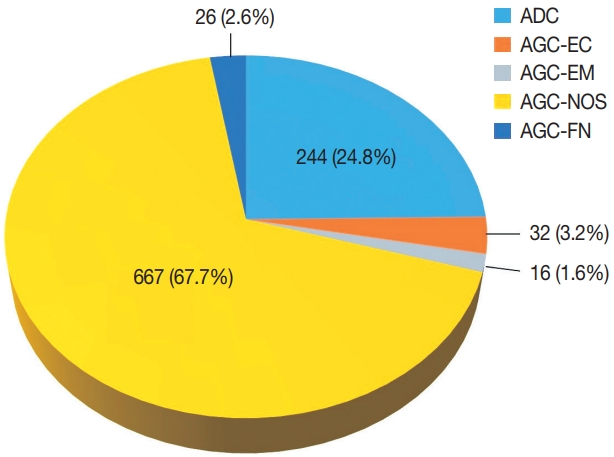
Fig. 1.
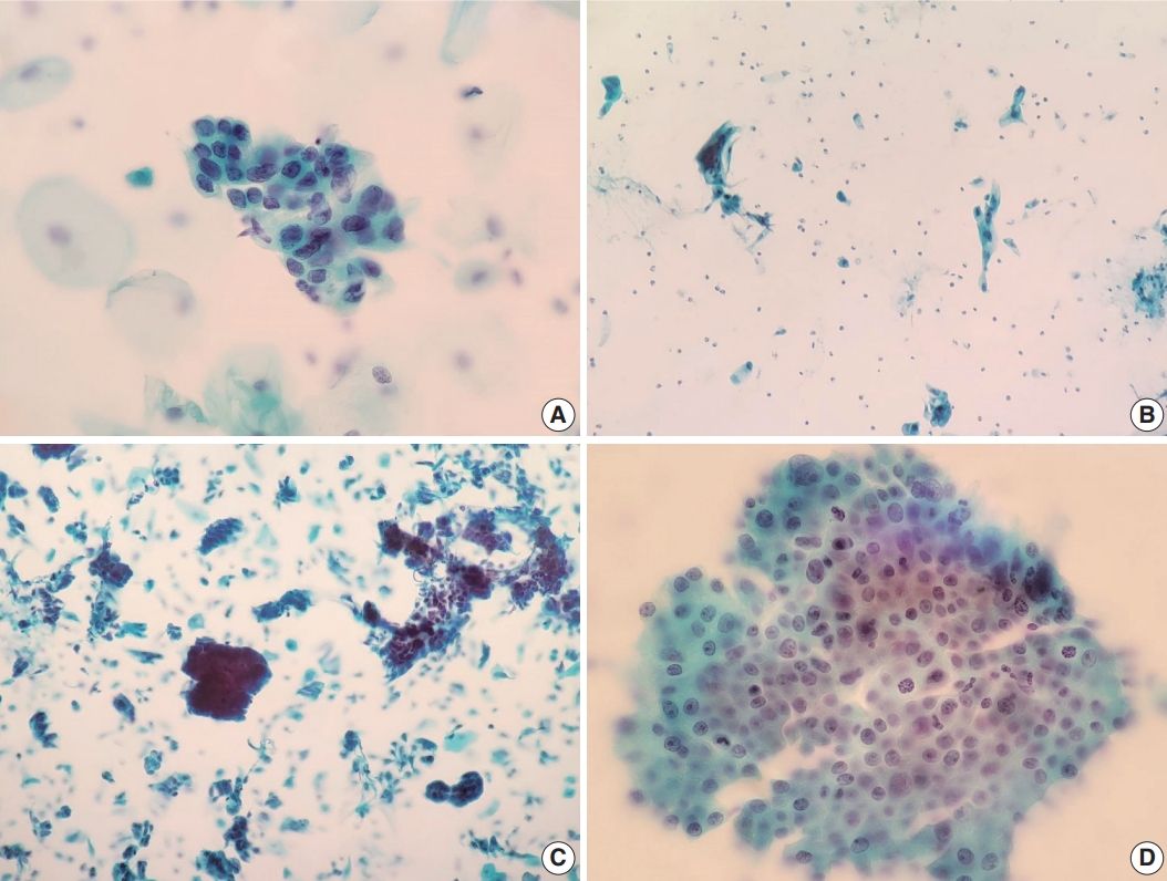
Fig. 2.
| Histopathologic result | Pap test result |
||||
|---|---|---|---|---|---|
| AGC-NOS | AGC-EC | AGC-EM | AGC-FN | ADC | |
| Benign | 197 | 9 | 3 | 4 | 9 |
| Endometrial lesion | |||||
| Endometrioid adenocarcinoma | 68 | 1 | 5 | 6 | 34 |
| Serous adenocarcinoma | 7 | - | - | - | 15 |
| Clear cell carcinoma | 4 | - | - | - | 3 |
| Adenosquamous carcinoma | 2 | ||||
| Carcinosarcoma | 5 | - | - | - | 3 |
| EM other malignant | 3 | - | - | - | - |
| EM hyperplasia | 5 | - | 1 | - | - |
| Cervical squamous lesion | |||||
| LSIL | 21 | - | - | - | 2 |
| HSIL | 22 | 1 | - | - | 4 |
| Squamous cell carcinoma | 4 | - | - | - | - |
| Cervical glandular lesion | |||||
| Adenocarcinoma in situ | 13 | - | - | - | 3 |
| Adenocarcinoma | 33 | - | - | 7 | 83 |
| Adenosquamous carcinoma | - | - | - | 1 | 6 |
| Cervical other malignant | 2 | - | - | - | 3 |
| Ovarian lesion | |||||
| Serous adenocarcinoma | 9 | - | - | - | 12 |
| Mucinous adenocarcinoma | - | - | - | - | 1 |
| Clear cell carcinoma | - | - | - | - | 2 |
| Endometrioid adenocarcinoma | 1 | - | - | - | - |
| Ovary other malignant | 1 | 1 | - | - | 1 |
| Vaginal lesion | |||||
| Poorly differentiated carcinoma | 1 | - | - | - | 1 |
| Metastatic tumor | 5 | - | - | 2 | 22 |
| Other | 2 | - | - | 1 | - |
| TIFD, unknown | 6 | - | - | - | - |
| Total | 409 | 12 | 9 | 21 | 206 |
| Cytomorphological feature | Histologic diagnosis |
p-value |
|
|---|---|---|---|
| Benign (n = 51) | ADC/AIS (n = 23) | ||
| Architectural features | |||
| Overlapping | .058 | ||
| Present | 30 | 18 | |
| Absent | 21 | 5 | |
| Single-cell pattern | < .001 | ||
| Present | 1 | 8 | |
| Absent | 50 | 15 | |
| Three-dimensional clusters | < .001 | ||
| Present | 0 | 8 | |
| Absent | 51 | 15 | |
| Nuclear features | |||
| Increased N/C ratio | .434 | ||
| Present | 50 | 22 | |
| Absent | 1 | 1 | |
| Hyperchromasia | .233 | ||
| Present | 39 | 18 | |
| Absent | 12 | 5 | |
| Membrane irregularity | .012 | ||
| Present | 18 | 15 | |
| Absent | 33 | 8 | |
| Prominent nucleoli | .124 | ||
| Present | 16 | 10 | |
| Absent | 35 | 13 | |
| Mitoses | .009 | ||
| 0/HPF | 49 | 17 | |
| > 1/HPF | 2 | 6 | |
Pap, Papanicolaou; AGC-NOS, atypical glandular cells not otherwise specified; AGC-EC, atypical endocervical cells; AGC-EM, atypical endometrial cells; AGC-FN, atypical glandular cells favor neoplastic; ADC, adenocarcinoma; EM, endometrial; LSIL, low grade squamous intraepithelial lesion; HSIL, high grade squamous intraepithelial lesion; TIFD, tissue insufficient for diagnosis.
Pap, Papanicolaou; ADC, adenocarcinoma; AIS, adenocarcinoma in situ; N/C, nuclear-to-cytoplasm; HPF, high-power field. The Fisher exact test was used. p-value of < .05 was considered statistically significant.

 E-submission


