
 E-submission
Articles
- Page Path
- HOME > J Pathol Transl Med > Volume 59(3); 2025 > Article
-
Original Article
Diagnostic yield of fine needle aspiration with simultaneous core needle biopsy for thyroid nodules -
Mohammad Ali Hasannia1,2
 , Ramin Pourghorban3,4, Hoda Asefi4, Amir Aria5, Elham Nazar6, Hojat Ebrahiminik7, Alireza Mohamadian1,2
 -
Journal of Pathology and Translational Medicine 2025;59(3):180-187.
DOI: https://doi.org/10.4132/jptm.2025.03.04
Published online: March 28, 2025
1Department of Radiology, School of Medicine, Tehran University of Medical Sciences, Tehran, Iran
2Advanced Diagnostic and Interventional Radiology Research Center (ADIR), Tehran University of Medical Sciences, Tehran, Iran
3Department of Medical Imaging, Nepean Hospital, Kingswood, New South Wales, Australia
4Department of Radiology, Sina Hospital, Tehran University of Medical Sciences, Tehran, Iran
5Department of Internal Medicine, Alzahra Hospital, Isfahan University of Medical Sciences, Isfahan, Iran
6Department of Pathology, Sina Hospital, Tehran University of Medical Sciences, Tehran, Iran
7Department of Interventional Radiology and Radiation Sciences Research Center, Aja University of Medical Sciences, AJA Campus, Tehran, Iran
- Corresponding Author Alireza Mohamadian, MD, MPH Department of Radiology, School of Medicine, Tehran University of Medical Sciences, Sina Hospital, Imam Khomeini St., Tehran 11367-46911, Iran Tel: +98-937-323-3713, Fax: +98-21-8889-8532, E-mail: alirezamohamadian.md@gmail.com
© The Korean Society of Pathologists/The Korean Society for Cytopathology
This is an Open Access article distributed under the terms of the Creative Commons Attribution Non-Commercial License (http://creativecommons.org/licenses/by-nc/4.0) which permits unrestricted non-commercial use, distribution, and reproduction in any medium, provided the original work is properly cited.


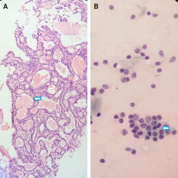
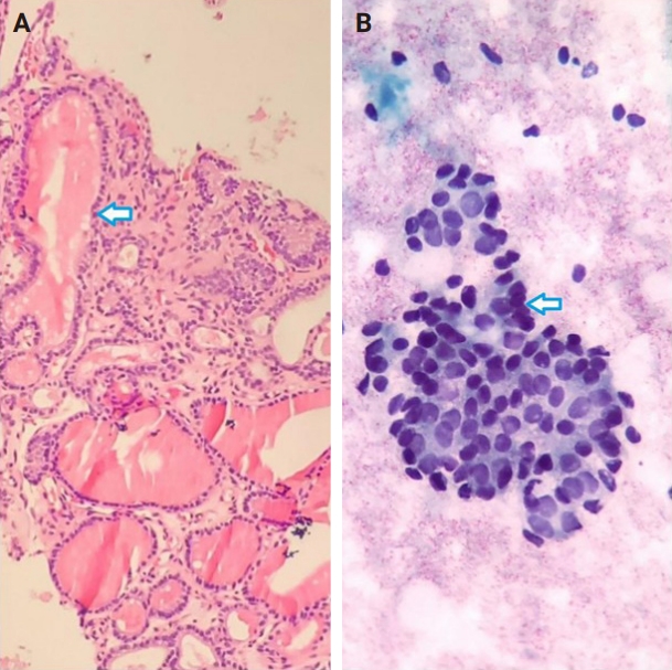
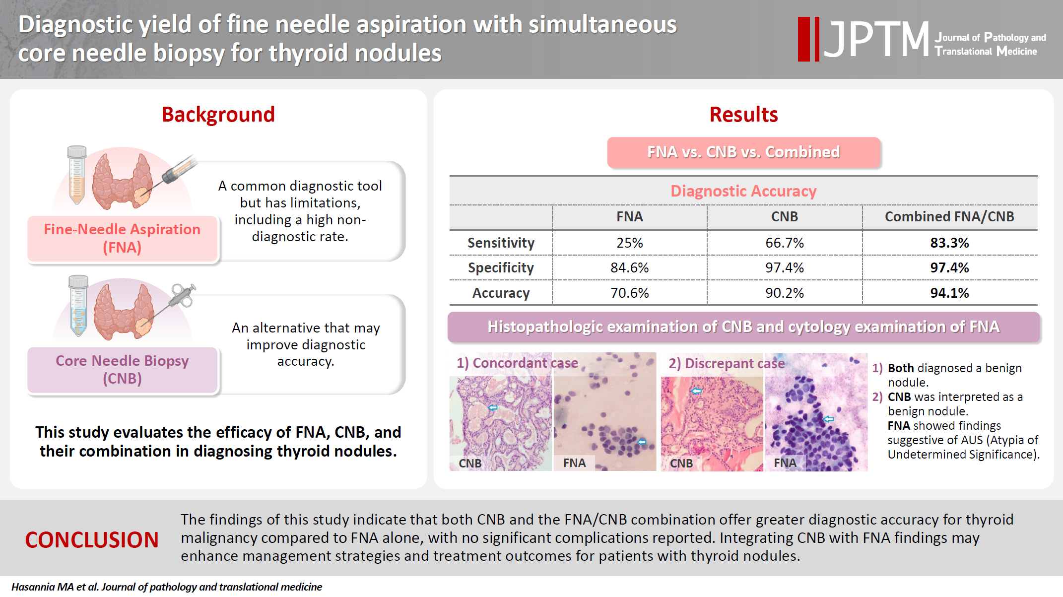
Figure & Data
References
Citations

- Clinicopathological Correlation of Core-Needle Biopsy in Thyroid Nodule Assessment: Insights from a Tertiary Care Center
Neha Nigam, Rishabh Sahai, Anil Singh, Anjali Mishra, Sabaretnam Mayilvaganan, Ojas Gupta, Prabhakar Mishra, Nirbhay Kumar, Amit Rastogi
Journal of Cytology.2026; 43(4): 235. CrossRef
 PubReader
 ePub Link
-
 Cite this Article
- Cite this Article
-
- Close
- Download Citation
- Close
 XML Download
- Figure
-
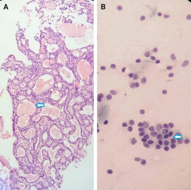
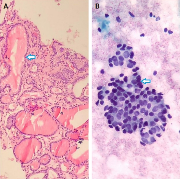

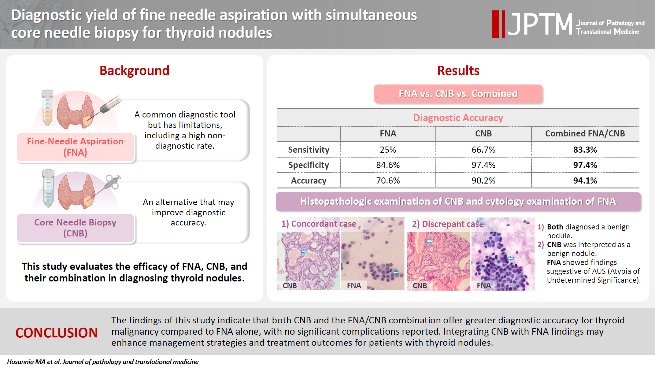
Fig. 1.
Fig. 2.
Graphical abstract
| Variable | Value |
|---|---|
| Age (y/o) | 54.23 ± 8.63 |
| Size of nodule (mm) | 27.0 ± 12.3 |
| Distance from skin (mm) | 12.2 ± 4.7 |
| TI-RADS | |
| 3 | 19 (33.9) |
| 4 | 29 (51.8) |
| 5 | 8 (14.3) |
| Calcification type | |
| None | 30 (53.6) |
| Large comet-tail artifact | 3 (5.4) |
| Macrocalcification | 10 (17.9) |
| Peripheral/rim calcification | 1 (1.8) |
| Punctate echogenic foci | 12 (21.4) |
| Category | FNA | CNB | FNA/CNB |
|---|---|---|---|
| I | 6 (11.8) | 0 | 0 |
| II | 34 (66.7) | 39 (76.5) | 38 (74.5) |
| III | 4 (7.8) | 2 (3.9) | 3 (5.9) |
| IV | 1 (1.9) | 2 (3.9) | 0 |
| V | 3 (5.9) | 0 | 0 |
| VI | 3 (5.9) | 8 (15.7) | 10 (19.6) |
| Category | FNA |
CNB |
FNA/CNB |
|||
|---|---|---|---|---|---|---|
| Benign (n = 39) | Malignant (n = 12) | Benign (n = 39) | Malignant (n = 12) | Benign (n = 39) | Malignant (n = 12) | |
| I | 6 (15.4) | 0 | 0 | 0 | 0 | 0 |
| II | 33 (84.6) | 1 (8.3) | 38 (97.4) | 1 (8.3) | 38 (97.4) | 0 |
| III | 0 | 4 (33.3) | 1 (2.6) | 1 (8.3) | 1 (2.6) | 2 (16.7) |
| IV | 0 | 1 (8.3) | 0 | 2 (16.7) | 0 | 0 |
| V | 0 | 3 (25) | 0 | 0 | 0 | 0 |
| VI | 0 | 3 (25) | 0 | 8 (66.7) | 0 | 10 (83.3) |
| Diagnostic values | FNA | CNB | FNA/CNB |
|---|---|---|---|
| Sensitivity (%) | 25 | 66.7 | 83.3 |
| Specificity (%) | 84.6 | 97.4 | 97.4 |
| Positive predictive value (%) | 100 | 100 | 100 |
| Negative predictive value (%) | 97.1 | 97.4 | 100 |
| Accuracy | 70.6 | 90.2 | 94.1 |
Values are presented as mean ± SD or number (%). SD, standard deviation; TI-RADS, Thyroid Imaging Reporting and Data System.
Values are presented as number (%). CNB, core needle biopsy; FNA, fine needle aspiration.
Values are presented as number (%). FNA, fine needle aspiration; CNB, core needle biopsy.
FNA, fine needle aspiration; CNB, core needle biopsy.


