 E-submission
Articles
- Page Path
- HOME > J Pathol Transl Med > Volume 46(6); 2012 > Article
-
Original Article
Fine-Needle Aspiration Cytology of Pleomorphic Carcinomas of the Lung - Hee Seung Choi, Hyesil Seol, Il Yeong Heo, Chang Won Jung, Soo Youn Cho, Sunhoo Park, Jae Soo Koh, Seung-Sook Lee
-
Korean Journal of Pathology 2012;46(6):576-582.
DOI: https://doi.org/10.4132/KoreanJPathol.2012.46.6.576
Published online: December 26, 2012
Department of Pathology, Korea Cancer Center Hospital, Korea Institute of Radiological and Medical Sciences, Seoul, Korea.
- Corresponding Author: Hyesil Seol, M.D. Department of Pathology, Korea Cancer Center Hospital, 75 Nowon-ro, Nowon-gu, Seoul 139-706, Korea. Tel: +82-2-970-2545, Fax: +82-2-970-2430, Hyesilseol@gmail.com
© 2012 The Korean Society of Pathologists/The Korean Society for Cytopathology
This is an Open Access article distributed under the terms of the Creative Commons Attribution Non-Commercial License (http://creativecommons.org/licenses/by-nc/3.0/) which permits unrestricted non-commercial use, distribution, and reproduction in any medium, provided the original work is properly cited.


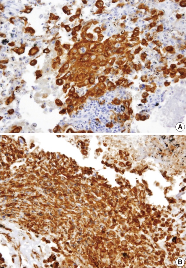
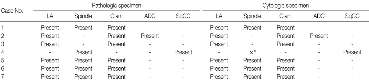

Figure & Data
References
Citations
- Sarcomatoid carcinoma in cytology: Report of a rare entity presenting in pleural and pericardial fluid preparations
Atreyee Basu, Andre L. Moreira, Anthony Simms, Tamar C. Brandler
Diagnostic Cytopathology.2019; 47(8): 813. CrossRef - Cytological Evaluation of Pleomorphic Carcinoma of the Lung
Kevin Kuan, Samer N. Khader, Siba El Hussein
Diagnostic Cytopathology.2019; 47(9): 961. CrossRef - Combined small cell carcinoma with giant cell carcinoma component of the lung: A case successfully diagnosed by computed tomography‑guided fine‑needle aspiration cytology
Yusuke Ebisu, Mitsuaki Ishida, Tomohito Saito, Tomohiro Murakawa, Yoshiko Uemura, Koji Tsuta
Oncology Letters.2017;[Epub] CrossRef - Pulmonary Pleomorphic Carcinoma Detected as a Result of Pneumothorax and the Subsequent Occurrence of Multiple Cystic Metastases
Hideaki Yamakawa, Masahiro Yoshida, Masami Yabe, Yuri Baba, Emiri Baba, Hiroaki Katagi, Takeo Ishikawa, Masamichi Takagi, Takeo Nakada, Tadashi Akiba, Kazuyoshi Kuwano
Case Reports in Medicine.2014; 2014: 1. CrossRef - Pulmonary pleomorphic carcinoma with multiple metastases to the right posterior knee complicated by paraneoplastic hypercalcemia
PENG-FEI LI, CHENG-HSIANG LO, SHAN-HAN YANG, PING-YING CHUNG, CHING-LIANG HO
Oncology Letters.2014; 7(2): 452. CrossRef
 PubReader
 Cite this Article
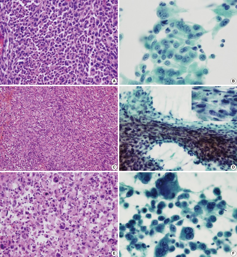
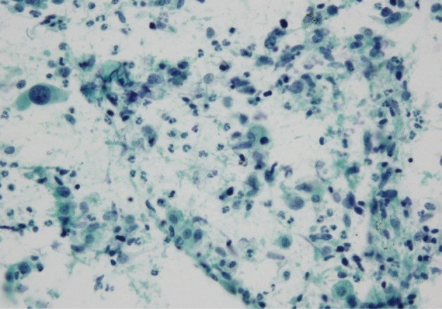


Fig. 1
Fig. 2
Fig. 3


NSCCM, male; RUL, right upper lobe; Chemo, chemotherapy; LUL, left upper lobe; NSCC, non-small cell carcinoma; ADC, adenocarcinoma; LLL, left lower lobe; F, female; SqCC, squamous cell carcinoma; rad, radiotherapy; RLL, right lower lobe. aThis size is assessed using computed tomography. Metastasectomy is performed in case 5.
LA, large cell carcinoma; ADC, adenocarcinoma; SqCC, squamous cell carcinoma. aThe spindle cell component is not observed in the cytological specimens from case 4, contrary to that observed in the corresponding case 4 pathological specimen.
±, positive in the variable minority of tumor cells; +, positive in the majority of tumor cells; -, negative in the tumor cells.

