Articles
- Page Path
- HOME > J Pathol Transl Med > Volume 47(2); 2013 > Article
-
Case Study
ThinPrep Cytological Findings of Desmoplastic Small Round Cell Tumor with Extensive Glandular Differentiation: A Case Study - Hyun-Jung Kim, Byeong Seok Sohn1, Ji-Eun Kwon2, Jeong Yeon Kim, Kyeongmee Park
-
Korean Journal of Pathology 2013;47(2):182-187.
DOI: https://doi.org/10.4132/KoreanJPathol.2013.47.2.182
Published online: April 24, 2013
Department of Pathology, Sanggye Paik Hospital, Inje University College of Medicine, Seoul, Korea.
1Department of Internal Medicine, Sanggye Paik Hospital, Inje University College of Medicine, Seoul, Korea.
2Department of Pathology, Ajou University Hospital, Ajou University School of Medicine, Suwon, Korea.
- Corresponding Author: Hyun-Jung Kim, M.D. Department of Pathology, Sanggye Paik Hospital, Inje University,1342 Dongil-ro, Nowon-gu, Seoul 139-707, Korea. Tel: +82-2-950-1261, Fax: +82-2-951-6964, hjkim@paik.ac.kr
• Received: July 20, 2012 • Revised: November 28, 2012 • Accepted: December 17, 2012
© 2013 The Korean Society of Pathologists/The Korean Society for Cytopathology
This is an Open Access article distributed under the terms of the Creative Commons Attribution Non-Commercial License (http://creativecommons.org/licenses/by-nc/3.0/) which permits unrestricted non-commercial use, distribution, and reproduction in any medium, provided the original work is properly cited.
Figure & Data
References
Citations
Citations to this article as recorded by 

- A Rare and Aggressive Abdominopelvic Tumor: A Case of Desmoplastic Small Round Cell Tumor in a Young Male
Yağmur Sena Tosun, Aytan Babazade, Emine Sena Sözen, Betül Erişmiş, Enes Seyda Şahiner
Caucasian Medical Journal.2025; 3(1): 4. CrossRef - Desmoplastic Small Round Cell Tumor Involving Serous Fluid: Cytologic Features and Diagnostic Pitfalls: A Series of 8 Cases
Nibras L Fakhri, Qiong Gan
American Journal of Clinical Pathology.2023; 160(4): 417. CrossRef - A Review of Effusion Cytomorphology of Small Round Cell Tumors
Lucy M. Han, Christopher J. VandenBussche, Mads Abildtrup, Ashish Chandra, Poonam Vohra
Acta Cytologica.2022; 66(4): 336. CrossRef - Intra-abdominal desmoplastic small blue round cell tumor: A case report
Tareq Hamed Al Taei, Hasan Al Fardan, Sarah Ali Al Mail
Radiology Case Reports.2022; 17(12): 4502. CrossRef - Desmoplastic Small Round Cell Tumor of the Kidney: Report of a Case, Literature Review, and Comprehensive Discussion of the Distinctive Morphologic, Immunohistochemical, and Molecular Features in the Differential Diagnosis of Small Round Cell Tumors Affec
Carlos A. Galliani, Michele Bisceglia, Antonio Del Giudice, Giuseppe Cretì
Advances in Anatomic Pathology.2020; 27(6): 408. CrossRef - Intra-abdominal desmoplastic small round cell tumors: CT and FDG-PET/CT findings with histopathological association
JINGJING CHEN, ZENGJIE WU, BINBIN SUN, DACHENG LI, ZHENGUANG WANG, FANGJUN LIU, HUI HUA
Oncology Letters.2016; 11(5): 3298. CrossRef - Desmoplastic small round cell tumor with sphere‐like clusters mimicking adenocarcinoma
Yukinori Hattori, Akihiko Yoshida, Naoshi Sasaki, Yasuo Shibuki, Kenji Tamura, Koji Tsuta
Diagnostic Cytopathology.2015; 43(3): 214. CrossRef - Tumor intraabdominal desmoplásico de células pequeñas y redondas
Andrés Alejandro Briseño-Hernández, Deissy Roxana Quezada-López, Lilia Edith Corona-Cobián, Agar Castañeda-Chávez, Alfonso Tonatiuh Duarte-Ojeda, Michel Dassaejv Macías-Amezcua
Cirugía y Cirujanos.2015; 83(3): 243. CrossRef - Intra-abdominal desmoplastic small round cell tumour
Andrés Alejandro Briseño-Hernández, Deissy Roxana Quezada-López, Lilia Edith Corona-Cobián, Agar Castañeda-Chávez, Alfonso Tonatiuh Duarte-Ojeda, Michel Dassaejv Macías-Amezcua
Cirugía y Cirujanos (English Edition).2015; 83(3): 243. CrossRef - Diagnostic Pitfalls of Differentiating Desmoplastic Small Round Cell Tumor (DSRCT) From Wilms Tumor (WT)
Michael A. Arnold, Lynn Schoenfield, Berkeley N. Limketkai, Christina A. Arnold
American Journal of Surgical Pathology.2014; 38(9): 1220. CrossRef
 PubReader
 ePub Link
-
 Cite this Article
- Cite this Article
-
- Close
- Download Citation
- Close
- Figure
ThinPrep Cytological Findings of Desmoplastic Small Round Cell Tumor with Extensive Glandular Differentiation: A Case Study


Fig. 1 (A) The abdomino-pelvic computer-tomography image discloses a 5.5 cm-sized mass at the right lower quadrant abdominal cavity (red arrow) and seeding nodules and mesenteric lymphadenopathy (gathered in blue inverted triangle). (B) The colonoscopic findings are compatible with extrinsic masses. The mucosa is intact. (C) The laparoscopic findings disclose multiple disseminated, variable sized whitish firm masses.
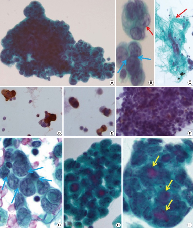
Fig. 2 (A) The clusters of tumor cells are arranged in finger-like budding with common outer cell borders. (B) The tumor cells show intra-cytoplasmic vacuoles, convoluted nuclei (red arrow), and relatively prominent nucleoli, with a few foci of nuclear molding (blue arrows). (C) Thin-Prep cytology of peritoneal washing shows a few fragments of fibrillary stroma (red arrow). (D-F) Immunocytochemistry reveals a positive reaction to vimentin (D), pancytokeratin (E), and weak positive reaction to desmin (F). (G) The conventional smear of pleural effusion fluid shows clusters of angulated nuclei and distinct nuclear moldings (blue arrows). (H, I) The ThinPrep cytology of pleural effusion shows rounded clusters with a three-dimensional structures (H), with extracellular mucinous content (I, yellow arrows), and more glandular differentiation.
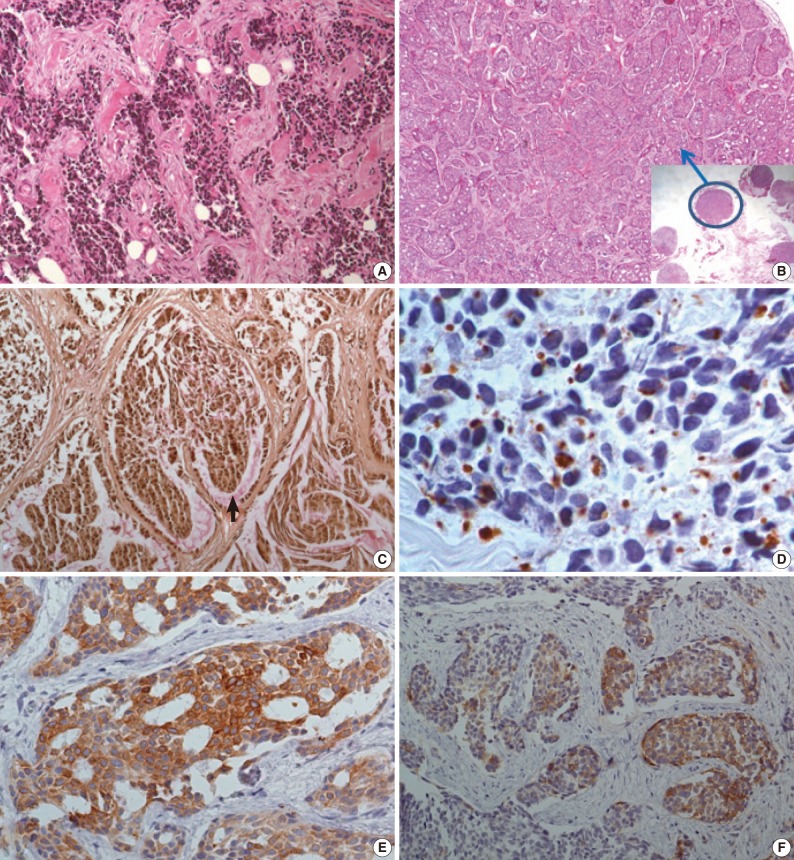
Fig. 3 (A) The initial biopsy discloses typical findings of desmoplastic small round cell tumor; desmoplastic stroma, and variable sized nests of small round cells. (B) In the second biopsy specimen, there are multiple nodules, some of them (blue circle) show extensive glandular differentiation. (C) Mucicarmine staining is positive for intra-glandular content. (D-F) The immunohistochemical staining is also divergent for each nodule. The dot-like desmin reactivity is well demonstrated in a typical desmoplastic small round cell tumor appearing nodule (D), but pancytokeratin reactivity is strong in a glandular differentiated nodule (E), and the neuronal differentiation (CD56) is variable (F).
Fig. 1
Fig. 2
Fig. 3
ThinPrep Cytological Findings of Desmoplastic Small Round Cell Tumor with Extensive Glandular Differentiation: A Case Study

 E-submission


