Articles
- Page Path
- HOME > J Pathol Transl Med > Volume 47(5); 2013 > Article
-
Brief Case Report
Neuroendocrine Dysplasia Combined in a Tubular Adenoma of Rectum: A Case Report - So-Young Lee, Dae-Yong Hwang1, Tae Sook Hwang, Wan-Seop Kim, So Dug Lim, Wook Youn Kim, Se-Hun Kim, Hye Seung Han
-
Korean Journal of Pathology 2013;47(5):495-498.
DOI: https://doi.org/10.4132/KoreanJPathol.2013.47.5.495
Published online: October 25, 2013
Department of Pathology, Konkuk University Medical Center, Konkuk University School of Medicine, Seoul, Korea.
1Department of General Surgery, Konkuk University Medical Center, Konkuk University School of Medicine, Seoul, Korea.
- Corresponding Author: Hye Seung Han, M.D. Department of Pathology, Konkuk University Medical Center, Konkuk University School of Medicine, 120-1 Neungdong-ro, Gwangjin-gu, Seoul 143-729, Korea. Tel: +82-2-2030-5644, Fax: +82-2-2030-5629, aphsh@kuh.ac.kr
• Received: February 1, 2013 • Revised: March 14, 2013 • Accepted: March 18, 2013
© 2013 The Korean Society of Pathologists/The Korean Society for Cytopathology
This is an Open Access article distributed under the terms of the Creative Commons Attribution Non-Commercial License (http://creativecommons.org/licenses/by-nc/3.0/) which permits unrestricted non-commercial use, distribution, and reproduction in any medium, provided the original work is properly cited.
- A 56-year-old female had a chief complaint of frequent loose stools and visited the local clinic in September 2012. The patient did not have any prior gastrointestinal problems. She had a previous surgical history for stress urinary incontinence in 2006, and had received re-operation for small bowel perforation that occurred in the sling operation. She was referred to the Kunkuk University Medical Center, Seoul, Korea for further evaluation. A colonoscopic examination was performed and revealed a large, laterally spreading circumferential mass at the rectum. The mass was greater than 5 cm in size. Due to the clinical suspicion of malignancy, a low anterior resection with extensive lymph node dissection was performed.
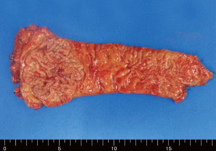
- Upon gross examination, a well-circumscribed, slightly elevated circumferential mass was identified in the rectum, measuring 6.0×5.0 cm (Fig. 1). Grossly, the mass was extending to the submucosa, exhibiting a fibrotic appearance with blurring of the muscularis mucosae. The lesion was entirely embedded and fully evaluated.
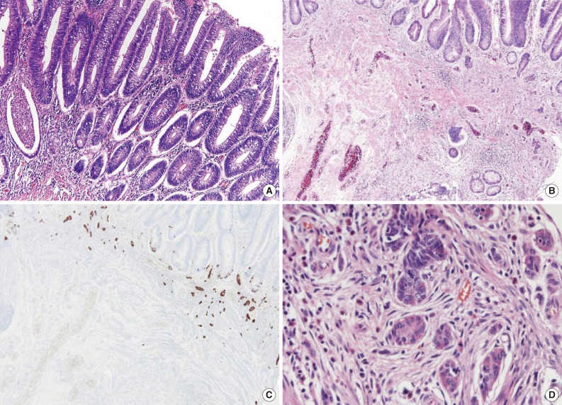
- Histologically, the elevated lesion was a typical tubular adenoma with low grade dysplasia, showing nuclear stratification and elongation (Fig. 2A). Beneath the adenomatous polyp, multiple small cellular nests were observed at the base of the adenomatous polyp (Fig. 2B, C). The cellular nests were clustered forming micronodules, and some of them fused together. At higher magnification, the cellular nests were composed of monotonous cells with eosinophilic, finely granular cytoplasm and stippled chromatin with central nuclei, reminiscent of neuroendocrine cells (Fig. 2D). Prominent nucleoli or mitosis were absent or inconspicuous. These cell nests were infiltrating the lamina propria and muscularis mucosae, and some of the cell nests seemed to intermingle with the adenomatous polyp. A desmoplastic reaction was not observed. The endocrine cell clusters were scattered multifocally along the polyp, measuring 3 cm in total diameter. The largest individual neuroendocrine cell nest measured up to 220 µm in its greatest dimension. Twenty-three regional lymph nodes were dissected, but metastatic tumor cells were not identified.
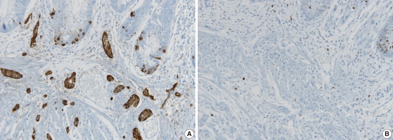
- For confirmative diagnosis, immunohistochemical staining of the chromogranin, synaptophysin, p53, and Ki-67 labeling index were performed. The cells were diffuse, strongly positive in chromogranin (Fig. 3A), and weakly positive in synaptophysin. The chromogranin staining was not only positive in the neuroendocrine cell nests, but also focally positive in the basal cells of the adenomatous polyp. The p53 staining and Ki-67 labeling index were both negative in the neuroendocrine cell nests (Fig. 3B). The patient recovered well without any complications, and follow-up colonoscopic examination 3 months after surgery found no evidence of recurrence.
CASE REPORT
- Neuroendocrine dysplasia is an extremely rare disease condition in the large intestine. It differs from neuroendocrine hyperplasia and microcarcinoid mainly in size. Other than the size criteria, neuroendocrine dysplasia has the following four characteristic morphologic features: 1) enlarged micronodules (comprising of nodules that measure larger than 150 µm), 2) fused micronodules, 3) microinfiltration of the lamina propria, and 4) nodules with surrounding newly formed stroma.3
- Although not common, composite endocrine-glandular neoplasms in the large intestine have been previously reported by several groups.5,6 Colorectal composite endocrine-glandular neoplasms encompass both benign and malignant forms, but malignant forms are much more common.5 Among them, the composite adenoma-carcinoid tumor is a rare benign counterpart that is characterized by the coexistence of the benign glandular component and benign neuroendocrine tumor (carcinoid tumor). Large intestinal microcarcinoids have been reported by some other groups, the majority of which were reported as multifocal microscopic lesions, and seem to have an association with chronic colitides, most with ulcerative colitis.7
- The present case shows the extraordinary features of combined neuroendocrine dysplasia in a tubular adenoma of the rectum. The overlying adenomatous polyp was large, measuring 6 cm in diameter, and had a fibrotic cut surface giving the clinical impression for malignancy. In the end, however, the mass turned out to be low grade dysplasia without any focus of high grade dysplasia. The fibrotic portion of the mass was, indeed, incidentally determined to be neuroendocrine dysplasia. In some portion, the neuroendocrine cell nests seemed to intermingle with the adenomatous polyp, simulating that they were neuroendocrine cells originating from the polyp. The present case differs from the neuroendocrine tumor with abundant stroma in the rectum or appendix, in the point that the neuroendocrine cell nests do not form a discrete mass, but rather are isolated small cell clusters which infiltrate lamina propria or muscularis mucosae without an adjacent stromal reaction. In a previous report by Sigel and Goldblum1 which reported on a series of 14 neuroendocrine tumors in patients with inflammatory bowel disease, they suggested that neuroendocrine differentiation may evolve from multipotent dysplastic cells on the basis of the fact that epithelial dysplasia was observed in the adjacent mucosa in more than one-third of cases. Van den Ingh et al.8 reviewed 218 endoscopically resected colorectal adenomas and investigated for the occurrence of neuroendocrine cells. According to that report, argyrophil cells were observed in 59% of the adenomas, and in 8% of the adenomas, the cells were numerous and intricately blended with the other cell types that could be regarded as an intrinsic part of the tumour.8 The morphologic and immunohistochemical features in present case raises the possibility that pluripotent epithelium located at the base of crypts differentiated into various neoplastic conditions. However, the previous report does not indicate the precise size or location of the neuroendocrine cell nests. Thus, it is unable to classify the neuroendocrine cell nests into certain categories.8
- The association between inflammatory bowel disease and neuroendocrine tumors has been reported by some other groups.1,7 The patient, however, had no history of inflammatory bowel disease or other gastrointestinal problems but has a history of a sling operation due to stress urinary incontinence in 2006. Oien et al.9 reported on two cases of neuroendocrine cell hyperplasia in colonic tissue used for long term augmentation cystoplasty, and suggested that mechanical dysfunction may induce neuroendocrine cell hyperplasia. The patient's history of a previous operation might have been a triggering factor that caused the increased gut neuroendocrine cell density.9
- The term 'neuroendocrine dysplasia' was used to describe the specific lesions mainly in stomach, and is known to be associated with hypergastrinemic status in conditions such as atrophic gastritis, autoimmune gastritis, or Zollinger-Ellison syndrome.3 In cases of the large intestine, the term 'diffuse or nodular neuroendocrine cell hyperplasia' has been used in one report, but the lesion was mainly in the lamina propria adjacent to normal colonic glands with an unspecified size of the lesion.9
- Although the neuroendocrine dysplasia in the present case was incidentally found, the pathologic features of neuroendocrine dysplasia, showing an infiltrative growth pattern of isolated small cell nests into the lamina propria and muscularis mucosae, may result in diagnostic pitfalls to prompt the diagnosis of malignancy. Special care should be taken not to overdiagnose or underdiagnose.
- The clinical behavior of neuroendocrine dysplasia combined in a tubular adenoma is not well known due to the lack of previous studies. The behavior of composite adenoma-carcinoids are reported to be benign, in contrast to the malignant nature of other mixed glandular-endocrine neoplasms of the large intestine.5 However, close clinical follow-ups may be necessary due to the unknown malignant potential of this lesion. According to the American Joint Committee on Cancer (AJCC) cancer staging manual, neuroendocrine tumors in stomach with a size less than 0.5 cm and confined to the mucosa is defined as Tis (carcinoma in situ/dysplasia).10 In large intestinal neuroendocrine tumors, in contrast, there is no concept of Tis for neuroendocrine tumors. Lesions whose size is less than 2 cm or invades the lamina propria is classified as T1.10
- In conclusion, we report on a rare and unique case of neuroendocrine dysplasia combined in a tubular adenoma of the rectum. The term neuroendocrine dysplasia has mainly been used to describe the niche between neuroendocrine hyperplasia and microcarcinoids. The clinical behavior, pathologic, and biological features of this entity needs to be elucidated with future studies and experiences.
DISCUSSION
- 1. Sigel JE, Goldblum JR. Neuroendocrine neoplasms arising in inflammatory bowel disease: a report of 14 cases. Mod Pathol 1998; 11: 537-542. PubMed
- 2. Moyana TN. Gastrointestinal endocrine cells and carcinoids: histogenetic and pathogenetic considerations. Pathol Annu 1995; 30(Pt 1):227-246.
- 3. Odze RD, Goldblum JR, Crawford JM. Surgical pathology of the GI tract, liver, biliary tract, and pancreas. Philadelphia: Saunders-Elsevier, 2003; 494-496.
- 4. Gledhill A, Hall PA, Cruse JP, Pollock DJ. Enteroendocrine cell hyperplasia, carcinoid tumours and adenocarcinoma in long-standing ulcerative colitis. Histopathology 1986; 10: 501-508. ArticlePubMed
- 5. Lewin K. Carcinoid tumors and the mixed (composite) glandular-endocrine cell carcinomas. Am J Surg Pathol 1987; 11(Suppl 1):71-86. ArticlePubMed
- 6. Pulitzer M, Xu R, Suriawinata AA, Waye JD, Harpaz N. Microcarcinoids in large intestinal adenomas. Am J Surg Pathol 2006; 30: 1531-1536. ArticlePubMed
- 7. Quinn P, Platell CF. Resolving microcarcinoids in ulcerative colitis: report of a case. Dis Colon Rectum 2004; 47: 387-391. ArticlePubMed
- 8. Van den Ingh HF, Van den Broek LJ, Verhofstad AA, Bosman FT. Neuroendocrine cells in colorectal adenomas. J Pathol 1986; 148: 231-237. ArticlePubMed
- 9. Oien K, Akyol M, Lindop G. Neuroendocrine cell hyperplasia in colonic tissue used for long term augmentation cystoplasty. J Clin Pathol 1998; 51: 258-261. ArticlePubMedPMC
- 10. Edge SB, Byrd DR, Compton CC, Fritz AG, Greene FL, Trotti A. AJCC cancer staging manual. 7th ed. New York: Springer, 2010; 183-185.
REFERENCES
Fig. 1A well-demarcated circumferential mass, measuring 6 cm in the greatest dimension, is seen in the rectum.


Fig. 2(A) A well-defined tubular adenomatous polyp is observed. The adenomatous polyp shows nuclear elongation and stratification with morphologic changes consistent with low grade dysplasia. (B) Beneath the adenomatous polyp, multiple small cellular nests are infiltrating the lamina propria and muscularis mucosae. (C) Immunohistochemical staining of chromogranin shows diffuse strong positivity in the small nests, confirming the neuroendocrine origin of the cell nests. (D) Higher magnification of the cellular nests, some fused together, composed of monotonous cells with eosinophilic and finely granular cytoplasm and central nuclei with stippled chromatin.


Fig. 3(A) Immunohistochemical staining of chromogranin showing diffuse strong positivity in the neuroendocrine cell nests. Note that the chromogranin is also focally positive in the basal cells located at the base of the adenomatous polyp. (B) The Ki-67 labeling index is negative in the neuroendocrine cells.


Figure & Data
References
Citations
Citations to this article as recorded by 

- Neuroendocrine cell micronests of the colon
Richard R. Pacheco, Maria Faraz, Hwajeong Lee
Human Pathology Reports.2022; 28: 300644. CrossRef - Incidental morphological findings in colorectal adenomas
Parag D Dabir, Rachel S van der Post, Iris D Nagtegaal
Histopathology.2021; 78(3): 348. CrossRef - Large cell neuroendocrine tumor of the gastrointestinal tract with concurrent tubular adenomas
Inayat GILL, Christienne SHAMS, Elisa QUIROZ, Subhashree M. KRISHNAN, Susanna GAIKAZIAN
Gazzetta Medica Italiana Archivio per le Scienze Mediche.2021;[Epub] CrossRef - Neuroendocrine tumor mixed with adenoma in rectum: a case report and literature review
Jiancong Hu, Xiaochuan Chen, Dezheng Lin, Zhaoliang Yu, Juan Li, Xuefeng Guo
International Journal of Surgery Oncology.2019; 4(6): e79. CrossRef
 PubReader
 ePub Link
-
 Cite this Article
- Cite this Article
-
- Close
- Download Citation
- Close
- Figure
Neuroendocrine Dysplasia Combined in a Tubular Adenoma of Rectum: A Case Report


Fig. 1 A well-demarcated circumferential mass, measuring 6 cm in the greatest dimension, is seen in the rectum.
Fig. 2 (A) A well-defined tubular adenomatous polyp is observed. The adenomatous polyp shows nuclear elongation and stratification with morphologic changes consistent with low grade dysplasia. (B) Beneath the adenomatous polyp, multiple small cellular nests are infiltrating the lamina propria and muscularis mucosae. (C) Immunohistochemical staining of chromogranin shows diffuse strong positivity in the small nests, confirming the neuroendocrine origin of the cell nests. (D) Higher magnification of the cellular nests, some fused together, composed of monotonous cells with eosinophilic and finely granular cytoplasm and central nuclei with stippled chromatin.
Fig. 3 (A) Immunohistochemical staining of chromogranin showing diffuse strong positivity in the neuroendocrine cell nests. Note that the chromogranin is also focally positive in the basal cells located at the base of the adenomatous polyp. (B) The Ki-67 labeling index is negative in the neuroendocrine cells.
Fig. 1
Fig. 2
Fig. 3
Neuroendocrine Dysplasia Combined in a Tubular Adenoma of Rectum: A Case Report

 E-submission


