Articles
- Page Path
- HOME > J Pathol Transl Med > Volume 47(5); 2013 > Article
-
Brief Case Report
Primary Extraskeletal Mesenchymal Chondrosarcoma of the Anterior Mediastinum - Sang Seok Jeong, Phil Jo Choi, Dong Won Kim1, Choonhee Son2, Mee Sook Roh4
-
Korean Journal of Pathology 2013;47(5):492-494.
DOI: https://doi.org/10.4132/KoreanJPathol.2013.47.5.492
Published online: October 25, 2013
Department of Thoracic and Cardiovascular Surgery, Dong-A University College of Medicine, Busan, Korea.
1Department of Radiology, Dong-A University College of Medicine, Busan, Korea.
2Department of Internal Medicine, Dong-A University College of Medicine, Busan, Korea.
3Department of Pathology, Dong-A University College of Medicine, Busan, Korea.
- Corresponding Author: Mee Sook Roh, M.D. Department of Pathology, Dong-A University College of Medicine, 32 Daesingongwon-ro, Seo-gu, Busan 602-714, Korea. Tel: +82-51-240-2833, Fax: +82-51-243-7396, msroh@dau.ac.kr
© 2013 The Korean Society of Pathologists/The Korean Society for Cytopathology
This is an Open Access article distributed under the terms of the Creative Commons Attribution Non-Commercial License (http://creativecommons.org/licenses/by-nc/3.0/) which permits unrestricted non-commercial use, distribution, and reproduction in any medium, provided the original work is properly cited.
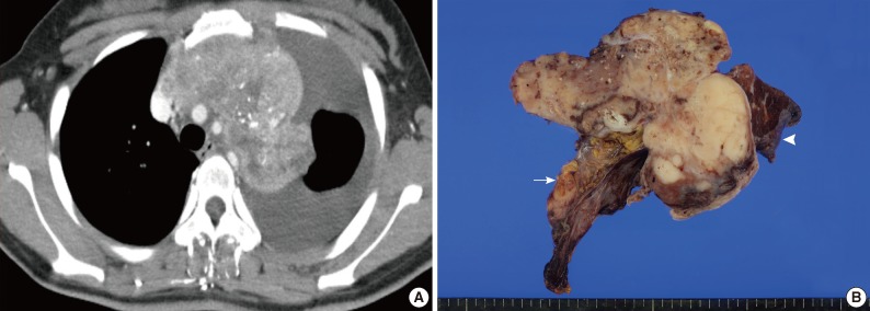
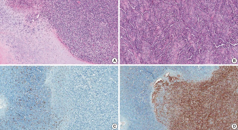
- A 21-year-old woman visited an outside hospital with the chief complaint of intermittent chest discomfort and respiratory difficulty for 2 months. In the medical history, the patient had taken medication for schizophrenia for several years. On the chest computed tomography (CT) scan, the patient had a 13-cm heterogeneous enhancing mass with multifocal calcification in the anterior mediastinum that invaded the pericardium and was associated with massive left pleural effusion. The mass encased the aortic arch and branch along with the left and main pulmonary arteries, but did not appear to be originating from the osteocartilaginous structures of the thoracic region (Fig. 1A). The patient then underwent a CT-guided needle biopsy, for which a diagnosis of solitary fibrous tumor was suggested on a biopsy. Then, the patient was referred to the Dong-A University Medical Center to confirm the diagnosis and undergo further treatment. Although the tumor could not be resected completely due to the extensive invasion into the main vessels, a surgical excision of the mass along with wedge resection of the left upper lung and partial resection of the pericardium was performed. The tumor at the time of surgery was found to invade the pericardium and to be tightly adhered to the left upper lung. However, it could be easily separated from the adjacent ribs. The resected tumor, measuring 13×7.5 cm, was solid, lobulated, and partially covered by a thin fibrous capsule. On the cut section, the tumor was tan white, and soft to fish-flesh, while the lobulated cut surface exhibited focal areas of irregularly sized glistening cartilage and coarse calcification (Fig. 1B). Microscopically, the lesion was composed of a highly cellular proliferation of undifferentiated round, oval, or spindle-shaped cells with an abrupt transition with small islands of well-differentiated hyaline cartilage, frequently associated with central calcification and ossification (Fig. 2A). In some areas, the undifferentiated cells were separated by branching, sinusoidal vascular channels imparting the lesion with a hemangiopericytoma-like pattern (Fig. 2B). Immunohistochemically, the cartilaginous portion of the tumor showed S100 protein positivity, but the undifferentiated cells showed negative immunoreactivity for the S100 protein (Fig. 2C). Undifferentiated cells showed strong membranous immunoreactivity for CD99, whereas only isolated cells in the cartilaginous portion stained for CD99 (Fig. 2D). The tumor cells exhibited negative immunoreactivities for cytokeratin and CD34. The patient made an uneventful postoperative recovery and was discharged on postoperative day 5 with advice to undergo chemotherapy and local radiotherapy. However, the patient refused any further treatment. The patient is still alive with a residual tumor at the time of writing this report (8 months postoperatively).
CASE REPORT
- To date, a total of 11 cases of primary chondrosarcoma of the mediastinum have been reported in the literature. However, 10 cases were found in PubMed and KoreaMed searches of English- and Korean-language articles.1-5 Among the 10 cases, seven cases have presented as EMC and the remaining two had the conventional type with one having the myxoid type. The clinicopathologic features of the seven patients with EMC reported in the literature are summarized in Table 1. The patient in our case report presented with the largest tumor mass (13 cm). Of note, all the tumors except our case have been reported to involve the posterior mediastinum. To our knowledge, this is the first case report of EMC involving the anterior mediastinum.
- Due to an exceedingly rare incidence of EMC presented as mediastinal tumors, the diagnosis of this tumor may be difficult with small biopsy or needle biopsy specimens that demonstrate only one of the two tissue elements. In particular, tumors having a hemangiopericytoma-like pattern without the cartilaginous elements may be mistaken for other tumors having a hemangiopericytoma-like pattern occurring in the anterior mediastinum, such as type A thymoma, solitary fibrous tumor, and synovial sarcoma, as a solitary fibrous tumor was suggested for the diagnosis of this case upon biopsy from an outside hospital. Immunohistochemistry may play an important role in the differential diagnosis of these tumors (Table 2). Although the preoperative diagnosis of EMC is difficult and the incidence is exceedingly rare, it has to be distinguished for the differential diagnosis among the list of anterior mediastinal tumors.
DISCUSSION
- 1. Chetty R. Extraskeletal mesenchymal chondrosarcoma of the mediastinum. Histopathology 1990; 17: 261-263. ArticlePubMed
- 2. Chun CG, Jeon YS, Park YH, Kim WJ, Jun YJ. Extraskeletal mesenchymal chondrosarcoma of the posterior mediastinum: 1 case report. Korean J Thorac Cardiovasc Surg 1995; 28: 1192-1196.
- 3. Suster S, Moran CA. Malignant cartilaginous tumors of the mediastinum: clinicopathological study of six cases presenting as extraskeletal soft tissue masses. Hum Pathol 1997; 28: 588-594. ArticlePubMed
- 4. Hwang EG, Yoon YW, Kim DH, Kim BS, Park JC, Sung DW. Extraskeletal mesenchymal chondrosarcoma of the mediastinum: a case report. Korean J Thorac Cardiovasc Surg 2001; 34: 891-894.
- 5. Jindal T, Chaudhary R, Sharma N, Meena M, Dutta R, Kumar A. Primary mediastinal chondrosarcoma with Horner's syndrome. Gen Thorac Cardiovasc Surg 2011; 59: 145-147. ArticlePubMedPDF
- 6. Shimosato Y, Mukai K, Matsuno Y. Tumors of the mediastinum. In: Silverberg SG, ed. AFIP atlas of tumor pathology, series 4. Fascicle 11. Silverspring: ARP Press, 2010; 19-155.
REFERENCES


| Case No. | Age (yr)/Sex | Location | Size (cm) | Overcome | Reference |
|---|---|---|---|---|---|
| 1 | 25/M | Post | 7.5 | DOD (6 mo) | Chetty [1] |
| 2 | 26/M | Post | 8 | Lost to FU | Chun et al. [2] |
| 3 | 11/M | Post | 5 | FOD (6 yr) | Suster and Moran [3] (case 1) |
| 4 | 15/F | Post | 5.5 | Local recur (7 yr) | Suster and Moran [3] (case 2) |
| 5 | 31/M | Post | 5 | Local recur (3 yr) | Suster and Moran [3] (case 3) |
| 6 | 36/F | Post | 7 | DOD (8 yr) | Suster and Moran [3] (case 4) |
| 7 | 22/M | Post | 7 | FOD (26 mo) | Hwang et al. [4] |
| Present case | 21/F | Ant | 13 | AWD (8 mo) | - |
Figure & Data
References
Citations

- Mesenchymal Tumors of the Mediastinum: An Update on Diagnostic Approach
Joon Hyuk Choi, Jae Y. Ro
Advances in Anatomic Pathology.2021; 28(5): 351. CrossRef - Mesenchymal tumours of the mediastinum—part II
Michael A. den Bakker, Alexander Marx, Kiyoshi Mukai, Philipp Ströbel
Virchows Archiv.2015; 467(5): 501. CrossRef - A chondrosarcoma in the anterior mediastinum mimicking a thymoma
Mia L Østergaard, Rene H Petersen, Anna Kalhauge
Acta Radiologica Open.2015; 4(9): 205846011559565. CrossRef
 PubReader
 ePub Link
-
 Cite this Article
- Cite this Article
-
- Close
- Download Citation
- Close
- Figure
- Related articles


Fig. 1
Fig. 2
| Case No. | Age (yr)/Sex | Location | Size (cm) | Overcome | Reference |
|---|---|---|---|---|---|
| 1 | 25/M | Post | 7.5 | DOD (6 mo) | Chetty [1] |
| 2 | 26/M | Post | 8 | Lost to FU | Chun et al. [2] |
| 3 | 11/M | Post | 5 | FOD (6 yr) | Suster and Moran [3] (case 1) |
| 4 | 15/F | Post | 5.5 | Local recur (7 yr) | Suster and Moran [3] (case 2) |
| 5 | 31/M | Post | 5 | Local recur (3 yr) | Suster and Moran [3] (case 3) |
| 6 | 36/F | Post | 7 | DOD (8 yr) | Suster and Moran [3] (case 4) |
| 7 | 22/M | Post | 7 | FOD (26 mo) | Hwang et al. [4] |
| Present case | 21/F | Ant | 13 | AWD (8 mo) | - |
| Tumor | Cytokeratin | EMA | Vimentin | S100 protein | CD99 | Bcl-2 | CD34 |
|---|---|---|---|---|---|---|---|
| EMC | - | - | + | - or + at isolated cells of undiff, + at cart | + at undiff, - or + at isolated cells of cart | - | - |
| Thymoma, type A | + | + | - | - | - at tumor cells, + at immature T cells | - | - |
| SFT | - | + (30%) | + | - | + (70%) | + (50%) | + |
| SS | + (focal) | + (focal) | + | - | + (focal) | + | - |
M, male; Post, posterior mediastinum; DOD, died of disease; FU, follow-up; FOD, free of disease; F, female; Ant, anterior mediastinum; AWD, alive with disease.
EMA, epithelial membrane antigen; EMC, extraskeletal mesenchymal chondrosarcoma; undiff, undifferentiated area; cart, cartilaginous area; SFT, solitary fibrous tumor; SS, synovial sarcoma.

 E-submission


