Articles
- Page Path
- HOME > J Pathol Transl Med > Volume 47(6); 2013 > Article
-
Case Study
Human Cutaneous Protothecosis: Report of a Case and Literature Review - Jae Yeon Seok, Yoonho Lee1, Hyukmin Lee2, Sang Yeop Yi3, Hwa Eun Oh3, Ji-Sun Song3
-
Korean Journal of Pathology 2013;47(6):575-578.
DOI: https://doi.org/10.4132/KoreanJPathol.2013.47.6.575
Published online: December 24, 2013
Department of Pathology, Gachon University Gil Medical Center, Incheon, Korea.
1Kwandong University College of Medicine, Gangneung, Korea.
2Department of Laboratory Medicine, Kwandong University College of Medicine, Gangneung, Korea.
3Department of Pathology, Kwandong University College of Medicine, Gangneung, Korea.
- Corresponding Author: Ji-Sun Song, M.D. Department of Pathology, Kwandong University College of Medicine, 1 Gangwondaehak-gil, Chuncheon 200-701, Korea. Tel: +82-33-649-7442, Fax: +82-33-641-1074, jssong@kd.ac.kr
© 2013 The Korean Society of Pathologists/The Korean Society for Cytopathology
This is an Open Access article distributed under the terms of the Creative Commons Attribution Non-Commercial License (http://creativecommons.org/licenses/by-nc/3.0/) which permits unrestricted non-commercial use, distribution, and reproduction in any medium, provided the original work is properly cited.
Abstract
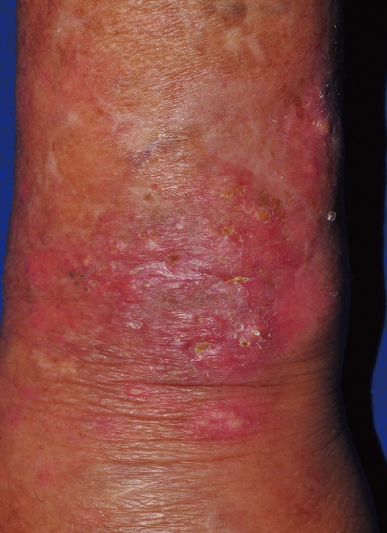
- The Prototheca species is achlorophyllic algae and rarely causes human infection. Human protothecosis presents clinically as a cutaneous infection, olecranon bursitis, and disseminated systemic disease. We report a case of human cutaneous protothecosis involving the left wrist. A 68-year-old man presented with an ill-defined erythematous lesion with crust at the dorsal aspect of his left wrist. A punch biopsy was performed to reveal the histologic features of granulomatous inflammation with necrosis at the upper dermis, containing Prototheca organisms, of which, the characteristic features were highlighted by special staining. Through a molecular study, the Prototheca zopfii species was identified.
- A 68-year-old man presented with an ill-defined erythematous lesion with crust at the dorsal aspect of his left wrist (Fig. 1). The skin lesion developed approximately a month ago without any traumatic history, and the patient complained of poor oral intake and diarrhea for a week. He had been suffering from osteoarthritis and had taken medications for twenty years. He was surgically treated for small bowel perforation, adhesion, and panperitonitis 3 years ago. His occupation was gathering trash for recycling.
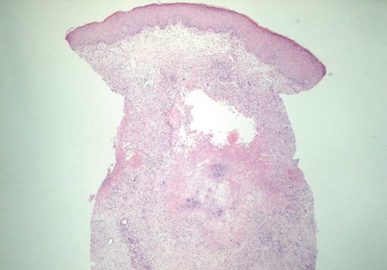
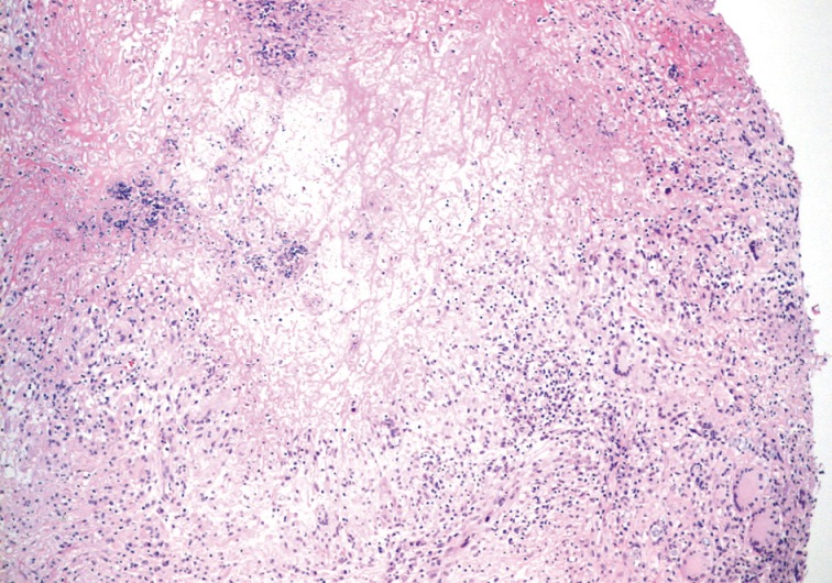
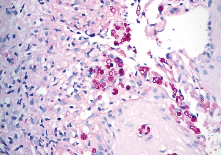
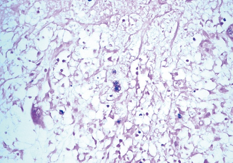
- A punch biopsy was performed to reveal the histologic features of granulomatous inflammation with confluent necrosis at the upper dermis containing many Prototheca organisms (Figs. 2, 3). On periodic acid-Schiff after diastase digestion special staining, non-budding spherical organisms with multiple sporangia containing endospores were observed with a morula- or cartwheel-like appearance, which is the characteristic feature of the Prototheca species (Fig. 4). The organisms were approximately 20 µm in diameter and had a thick cell wall. Upon crystal violet staining, the organisms exhibited multiple small bluish dots (Fig. 5).
- For specification of the species, small subunit ribosomal DNA (SSU rDNA) was amplified by polymerase chain reaction using the universal primers NS3 (5'-GCAAGTCTGGCCAGCAGCC-3') and NS8 (5'-TCCGCAGGTTCACCTACGGA-3'). The sequences were submitted to a BLASTn search by using the National Center for Biotechnology database (NCBI; http://www.ncbi.nlm.nih.gov/BLAST).5 The sequences of the SSU rDNA regions of the isolate were 99% identical with Prototheca zopfii.
- The patient was given antifungal agent itraconazole, and the symptoms were relieved.
CASE REPORT
- Prototheca is ubiquitously isolated from tap water, freshwater streams, swimming pools, soil, stables, and foodstuffs, such as shrimp, cow's milk, butter, potato peel, and bananas.2 A traumatic inoculation plays a role in protothecal infections.2 The major sites of involvement are the exposed areas, including the upper and lower extremities. In the upper extremities, the distal parts are mainly infected, such as fingers, wrists, dorsum of hands, and forearms.6 Other sites of involvement include the forehead, cheek, eyelid, nose,6 scalp, chest, and neck. The patient in this case had no history of trauma, but his occupation involved collecting trash for recycling. The patient's level of hygiene therefore may be poor.
- Cutaneous protothecosis can be associated with an underlying disease or immunosuppression in the form of kidney transplantation, malignancy with chemotherapy or radiotherapy, diabetes mellitus,6 steroid administration, systemic lupus erythematosus, chronic obstructive pulmonary disease, myasthenia gravis, congestive heart failure, acquired immunodeficiency syndrome, gout arthritis,6 and rheumatoid arthritis. Malignancies associated with the protothecal infection involve the hematologic system (acute myelogenous leukemia, Hodgkin lymphoma, and chronic lymphocytic leukemia), breast, and uterus.2
- Cutaneous lesions are described as ulcerations, erythematous plaques, subcutaneous papules, vesicles, erythematous nodules, verrucous or herpetiform lesions,3 infiltrating nodules, and pyoderma-like lesions.6 The tissue reaction can exhibit a minimal cellular response, a chronic granulomatous reaction, or a mixed acute and chronic granulomatous infiltrate with eosinophilic infiltration.1
- The Prototheca species consists of large nonbudding cells with spherical to oval contours and a prominent cell wall. They exhibit the characteristic endospores with a cartwheel-like appearance on a periodic acid-Schiff stain. However, the non-sporulating cells may resemble common pathogens, such as Blastomyces dermatitidis, Cryptococcus neoformans, Paracoccidioides brasiliensis, and Pneumocystis carinii. The different sizes of the sporangia is helpful to discriminate the Prototheca species from these non-sporulating organisms.4 The Prototheca species measures 3 to 30 µm, have double-layered walls, and multiple endospores with a morula-like appearance. The size of Blastomyces species ranges from 8 to 15 µm and exhibits double-refractile cells. The Cryptococcus species is the smallest species (2 to 15 µm) and has a polysaccharide capsule highlighted by mucin stain and demonstrates budding without endospores. The Paracoccidioides species measures 5 to 60 µm and exhibits a mariners-wheel appearance. The sporangia of Coccidioides are larger than the Prototheca species in size, and the endospores are smaller. Electron microscopic findings are helpful in distinguishing the Prototheca species from the Chlorella species which have chloroplasts and a triple-layered cell wall in contrast to the double-layered wall in the Prototheca species.7
- Various species have been classified as Prototheca wickerhamii, Prototheca zopfii, Prototheca stagnora, Prototheca ulmea, and Prototheca blaschkeae.2 The human protothecosis is caused by only two species (Prototheca wickerhamii and Prototheca zopfii), and Prototheca wickerhamii is the most common agent. Prototheca zopfii mainly causes bovine mastitis and canine systemic infection. It infects humans rarely with a relatively poor prognosis. About 10 cases of human protothecosis clarified as Prototheca zopfii have been reported in the English literature (Table 1).8 In Korea, 9 cases of cutaneous protothecosis have been reported and all of them were identified as the Prototheca wickerhamii species.9 This is the first report of human cutaneous infection caused by Prototheca zopfii in Korea.
- Prototheca wickerhamii infections exhibit a susceptibility to antifungal agents and are easily treated. The prognosis of Prototheca zopfii infections is relatively poor. Therefore, the precise identification of this species is challenging and important both pathologically and clinically. The sporangia of Prototheca wickerhamii (7 to 13 µm) are small and round with symmetrical morula formation, while those of Prototheca zopfii (14 to 16 µm) are oval and cylindrical with relatively asymmetrical random internal segmentation.2,7 The confirmative diagnosis on the species can be made by a culture on Sabouraud's dextrose agar, a carbohydrate assimilation test, a yeast biochemical card, or an immunofluorescence study using species-specific antibodies and a molecular study assessing SSU rDNA.2,5,19,20
- The Prototheca species is an uncommon cause of human infection in both immunocompetent and immunocompromised patients. The clinical presentation and tissue reaction of cutaneous infections are similar to the lesions caused by a common deep fungal infection. Although most of the cutaneous protothecosis exhibit an indolent course, disseminated cutaneous lesions can develop under immunosuppressive conditions. The characteristic structures, detected upon histopathologic examination, aid in the precise diagnosis of protothecosis in differential diagnosis with a common cause of fungal infection.
DISCUSSION
- 1. Mayhall CG, Miller CW, Eisen AZ, Kobayashi GS, Medoff G. Cutaneous protothecosis: successful treatment with amphotericin B. Arch Dermatol 1976; 112: 1749-1752. ArticlePubMed
- 2. Lass-Flörl C, Mayr A. Human protothecosis. Clin Microbiol Rev 2007; 20: 230-242. ArticlePubMedPMCPDF
- 3. Davies RR, Spencer H, Wakelin PO. A case of human protothecosis. Trans R Soc Trop Med Hyg 1964; 58: 448-451. ArticlePubMed
- 4. Hillesheim PB, Bahrami S. Cutaneous protothecosis. Arch Pathol Lab Med 2011; 135: 941-944. ArticlePubMedPDF
- 5. Lu S, Xi L, Qin W, Luo Y, Lu C, Li X. Cutaneous protothecosis: two new cases in China and literature review. Int J Dermatol 2012; 51: 328-331. ArticlePubMed
- 6. Chao SC, Hsu MM, Lee JY. Cutaneous protothecosis: report of five cases. Br J Dermatol 2002; 146: 688-693. ArticlePubMed
- 7. Walsh SV, Johnson RA, Tahan SR. Protothecosis: an unusual cause of chronic subcutaneous and soft tissue infection. Am J Dermatopathol 1998; 20: 379-382. ArticlePubMed
- 8. Zhang QQ, Li L, Zhu LP, et al. Cutaneous protothecosis in patient with diabetes mellitus and review of published case reports. Mycopathologia 2012; 173: 163-171. ArticlePubMedPDF
- 9. Moon HS, Lee JH, Park K, Chae JD, Son SJ. A case of cutaneous protothecosis. Korean J Med Mycol 2007; 12: 70-74.
- 10. Kwok N, Schwartz SN. Prototheca sepsis in a lung transplant patient. Clin Microbiol Newsl 1996; 18: 183-184. Article
- 11. Lass-Flörl C, Fille M, Gunsilius E, Gastl G, Nachbaur D. Disseminated infection with Prototheca zopfii after unrelated stem cell transplantation for leukemia. J Clin Microbiol 2004; 42: 4907-4908. ArticlePubMedPMCPDF
- 12. Van den Bossche D, De Bel A, Hendrickx M, et al. Galactomannan enzymatic immunoassay cross-reactivity caused by Prototheca species. J Clin Microbiol 2012; 50: 3371-3373. ArticlePubMedPMCPDF
- 13. Naryshkin S, Frank I, Nachamkin I. Prototheca zopfii isolated from a patient with olecranon bursitis. Diagn Microbiol Infect Dis 1987; 6: 171-174. ArticlePubMed
- 14. Zhang Q, Weng X, Li L, et al. An unusual case of granulomatous lymphadenitis due to Prototheca zopfii var. portoricensis in an immunocompetent man in China. Int J Infect Dis 2010; 14 Suppl 3: e32-e35. ArticlePubMed
- 15. Magerman K, Gordts B, Hindryckx P, Van Landuyt HW. Isolation of Prototheca zopfii from a finger: case report and review of the literature. Acta Clin Belg 1991; 46: 233-236. PubMed
- 16. Shaoxi W, Guixia L, Yongnian S, Ningru G. The first case of Protothecosis zopfii in China. Chin Med Sci J 2000; 15: 121-123. PubMed
- 17. Sidrim JJ, Rocha MF, Camara LM, Brilhante RS, Diogenes MJ, Oliveira AM. Onycholysis caused by a mixed infection of Prototheca zopfii and Candida albicans. Clin Microbiol Newsl 2003; 25: 69-71. Article
- 18. Zhao J, Liu W, Lv G, Shen Y, Wu S. Protothecosis successfully treated with amikacin combined with tetracyclines. Mycoses 2004; 47: 156-158. ArticlePubMed
- 19. Kim JA, Moon SE, Song KY. Two cases of cutaneous protothecosis: unique histopathological findings with crystal violet staining and the therapeutic effect of itraconazole. Ann Dermatol 1997; 9: 201-207. ArticlePDF
- 20. Jun JH, Lee JB, Kim SJ, Lee SC, Won YH. A case of cutaneous prototheocsis. Korean J Med Mycol 2003; 8: 30-34.
REFERENCES

| Reference | Age (yr)/Sex | Medical history | Location | Country |
|---|---|---|---|---|
| Kwok and Schwartz [10] (1996) | 59/F | ESRD, COPD, lung transplantation | Disseminated | USA |
| Lass-Florl et al. [11] (2004) | 58/M | Acute leukemia, BMT | Disseminated (blood, lung, kidney, heart, liver) | Austria |
| Van den Bossche et al. [12] (2012) | 63/M | Follicular lymphoma, BMT | Disseminated (blood, lung) | Belgium |
| Naryshkin et al. [13] (1987) | NA | NA | Olecranon bursitis | USA |
| Zhang et al. [14] (2010) | 39/M | Healthy | Neck lymph node | China |
| Magerman et al. [15] (1991) | 45/F | Healthy | Cutaneous (finger) | Belgium |
| Shaoxi et al. [16] (2000) | 13/F | Healthy | Cutaneous (cheek) | China |
| Sidrim et al. [17] (2003) | 30/M | NA | Cutaneous (foot) | Brazil |
| Zhao et al. [18] (2004) | 18/F | Healthy | Cutaneous (cheek, jaw, nose) | China |
| Zhang et al. [8] (2012) | 66/F | DM | Cutaneous (finger) | China |
| Present case | 68/M | Osteoarthritis | Cutaneous (wrist) | Korea |
Figure & Data
References
Citations

- Successful Treatment of Cutaneous Protothecosis Due to Prototheca wickerhamii with Terbinafine
Yue Chen, Aili Gao, Yanan Ke, Xin Zhou, Li Lin, Sha Lu, Yumei Liu
Clinical, Cosmetic and Investigational Dermatology.2024; Volume 17: 913. CrossRef - Disseminated Protothecosis Due to Prototheca zopfii and Literature Review
Feifan Chen, Mario Saab-Chalhoub, Joy Tao, Amanda T. Harrington, Fritzie S. Albarillo, Andrew S. Crone, Nina M. Clark, Jodi J. Speiser
The American Journal of Dermatopathology.2023; 45(4): 237. CrossRef - The oldest patient infected with cutaneous protothecosis
Gabrielle Perrotti, Christian DiLiberto, Danielle M Pineda
Journal of Wound Care.2023; 32(4): 235. CrossRef - Cutaneous protothecosis: contrasting case presentations
Thomas Orschulok, Joseph Whitfield, Timothy Badrick, Pirathaban Sivabalan, David Sowden, Fiona Lehane
Pathology.2023; 55(7): 1032. CrossRef - Indurated Plaque With Ulceration on the Dorsum of the Left Hand
Juanjuan Li, Xiaomo Wu, Borui Chen
JAMA Dermatology.2022; 158(12): 1442. CrossRef - Cerebral protothecosis mimicking high-grade glioma
James Samarasekara, Soumya Mukherjee, Azzam Ismail, Robert Corns
BMJ Case Reports.2021; 14(1): e235362. CrossRef - Protothecosis algaemia in a patient presenting with septic arthritis: A rare case of Prototheca zopfii isolated from Malaysia
Prem Ananth Palaniappan, Cassandra Anne Abot, Ratna Mohd Tap, Fairuz Amran
IDCases.2021; 24: e01121. CrossRef -
Prototheca bovis, a unicellular achlorophyllous trebouxiophyte green alga in the healthy human intestine
Vasana Jinatham, Diego M. Cantoni, Ian R. Brown, Thanakrit Vichaslip, Picha Suwannahitatorn, Siam Popluechai, Anastasios D. Tsaousis, Eleni Gentekaki
Journal of Medical Microbiology .2021;[Epub] CrossRef - Pseudomicosis por oportunistas: desconocimiento y retos diagnósticos
María Adelaida Garcés-Abad, Nora Cardona Castro
CES Medicina.2021; 35(3): 257. CrossRef - Nanotechnology as a Novel Approach in Combating Microbes Providing an Alternative to Antibiotics
Bismillah Mubeen, Aunza Nayab Ansar, Rabia Rasool, Inam Ullah, Syed Sarim Imam, Sultan Alshehri, Mohammed M. Ghoneim, Sami I. Alzarea, Muhammad Shahid Nadeem, Imran Kazmi
Antibiotics.2021; 10(12): 1473. CrossRef - Prototheca zopfii genotype II induces mitochondrial apoptosis in models of bovine mastitis
Muhammad Shahid, Eduardo R. Cobo, Liben Chen, Paloma A. Cavalcante, Herman W. Barkema, Jian Gao, Siyu Xu, Yang Liu, Cameron G. Knight, John P. Kastelic, Bo Han
Scientific Reports.2020;[Epub] CrossRef - Murine and Human Cathelicidins Contribute Differently to Hallmarks of Mastitis Induced by Pathogenic Prototheca bovis Algae
Muhammad Shahid, Paloma Araujo Cavalcante, Cameron G. Knight, Herman W. Barkema, Bo Han, Jian Gao, Eduardo R. Cobo
Frontiers in Cellular and Infection Microbiology.2020;[Epub] CrossRef - Draining dorsal hand pustules, nodules, and ulcers in a patient with immunosuppression
Michael R. Stephens, Oyinade Aderibigbe, Katherine T. Steele, David E. Elder, Laurel Glaser, Jerry Jacob, Misha Rosenbach
JAAD Case Reports.2019; 5(10): 846. CrossRef - Characterization of the complete mitochondrial genome and phylogenetic analysis of Prototheca stagnorum (chlorellales: Chlorellaceae)
Qian Wu, Yanhong Lan, Hui Xu, Yi Cao
Mitochondrial DNA Part B.2019; 4(2): 4000. CrossRef - Cutaneous protothecosis reminiscent of unilateral solar elastotic bands of forearm in an immunocompromised patient
Yi-Hsin Ho, Yun-Wen Chiu, Chen-Yi Wu, Han-Nan Liu
Dermatologica Sinica.2018; 36(2): 93. CrossRef - A Case of Cutaneous Protothecosis in an Immunocompetent Patient
Ju Yun Woo, Eun Ah Suhng, Ji Yeon Byun, Hae Young Choi, Sun Hee Sung, You Won Choi
Annals of Dermatology.2016; 28(2): 273. CrossRef - Protothecal bursitis after simultaneous kidney/liver transplantation: a case report and review
I. Ramírez, J.F. Nieto‐Ríos, C. Ocampo‐Kohn, A. Aristizábal‐Alzate, G. Zuluaga‐Valencia, O. Muñoz Maya, J.C. Pérez
Transplant Infectious Disease.2016; 18(2): 266. CrossRef - Cutaneous Protothecosis in a Patient with Chronic Lymphocytic Leukemia: A Case Report and Literature Review
Quynh-Giao Nguyen, Ted Rosen
Journal of Fungi.2015; 1(1): 4. CrossRef - Natamycin - antimycotic of polyene macrolides class with unusual properties
I. O. Malova, D. D. Petrunin
Vestnik dermatologii i venerologii.2015; 91(3): 161. CrossRef
 PubReader
 ePub Link
-
 Cite this Article
- Cite this Article
-
- Close
- Download Citation
- Close
- Figure


Fig. 1
Fig. 2
Fig. 3
Fig. 4
Fig. 5
| Reference | Age (yr)/Sex | Medical history | Location | Country |
|---|---|---|---|---|
| Kwok and Schwartz [10] (1996) | 59/F | ESRD, COPD, lung transplantation | Disseminated | USA |
| Lass-Florl et al. [11] (2004) | 58/M | Acute leukemia, BMT | Disseminated (blood, lung, kidney, heart, liver) | Austria |
| Van den Bossche et al. [12] (2012) | 63/M | Follicular lymphoma, BMT | Disseminated (blood, lung) | Belgium |
| Naryshkin et al. [13] (1987) | NA | NA | Olecranon bursitis | USA |
| Zhang et al. [14] (2010) | 39/M | Healthy | Neck lymph node | China |
| Magerman et al. [15] (1991) | 45/F | Healthy | Cutaneous (finger) | Belgium |
| Shaoxi et al. [16] (2000) | 13/F | Healthy | Cutaneous (cheek) | China |
| Sidrim et al. [17] (2003) | 30/M | NA | Cutaneous (foot) | Brazil |
| Zhao et al. [18] (2004) | 18/F | Healthy | Cutaneous (cheek, jaw, nose) | China |
| Zhang et al. [8] (2012) | 66/F | DM | Cutaneous (finger) | China |
| Present case | 68/M | Osteoarthritis | Cutaneous (wrist) | Korea |
F, female; ESRD, end stage renal disease; COPD, chronic obstructive pulmonary disease; M, male; BMT, bone marrow transplantation; NA, not available; DM, diabetes mellitus.

 E-submission


