 E-submission
Articles
- Page Path
- HOME > J Pathol Transl Med > Volume 48(4); 2014 > Article
-
Brief Case Report
Heterotopic Pancreas in Omphalomesenteric Duct Remnant Results in Persistent Umbilical Discharge - Eunhyang Park, Hyojin Kim, Kyu Whan Jung1, Jin-Haeng Chung
-
Korean Journal of Pathology 2014;48(4):323-326.
DOI: https://doi.org/10.4132/KoreanJPathol.2014.48.4.323
Published online: August 26, 2014
Department of Pathology, Seoul National University Bundang Hospital, Seoul National University College of Medicine, Seongnam, Korea.
1Department of Pediatric Surgery, Seoul National University Bundang Hospital, Seoul National University College of Medicine, Seongnam, Korea.
- Corresponding Author: Jin-Haeng Chung, M.D. Department of Pathology, Seoul National University Bundang Hospital, 82 Gumi-ro 173beon-gil, Bundang-gu, Seongnam 463-707, Korea. Tel: +82-31-787-7713, Fax: +82-31-787-4012, chungjh@snu.ac.kr
• Received: July 2, 2013 • Revised: September 3, 2013 • Accepted: September 10, 2013
© 2014 The Korean Society of Pathologists/The Korean Society for Cytopathology
This is an Open Access article distributed under the terms of the Creative Commons Attribution Non-Commercial License (http://creativecommons.org/licenses/by-nc/3.0/) which permits unrestricted non-commercial use, distribution, and reproduction in any medium, provided the original work is properly cited.


Figure & Data
References
Citations
Citations to this article as recorded by 
- Pathology of Rare Umbilical Lesions: A Case Series and Literature Review
Iqbal Singh, Rugvi Patel, Pranjal S Chorya, Karan Patel
Cureus.2026;[Epub] CrossRef - Umbilical heterotopic pancreas in an infant: a case report
Abdulkarim Hasan, Basheer Abdullahi Jabo, Zakaria Elaskary, Khaldon Abdulrahman Alaghbari, Mohamed Abbas Ibrahim, Khalid Nafie
Journal of Pediatric Surgery Case Reports.2025; 120: 103053. CrossRef - Heterotopic pancreas: A diagnosis of exclusion not to ignore
Puneeth Thalasta, Yashwant Singh Rathore, Kamal Kataria, Sunil Chumber, Rajni Yadav, Gopal Puri, Prasanna Ramana Arumugaswamy, Gagan Soni, Ankit Anand
Saudi Surgical Journal.2024; 12(1): 9. CrossRef - Heterotopic Pancreas Mimicking Metastases From Renal Carcinoma
Deepanksha Datta, Rajesh Kumar, Peeyush Varshney, Sudeep Khera, Tanisha Gupta
Clinical Nuclear Medicine.2023; 48(2): e74. CrossRef - Ectopic pancreas, gastric, duodenal and colonic tissue in a case of persistent umbilical discharge
Pavithra Ayyanar, Bikash B. Tripathy, Akash B. Pati, Manoj K. Mohanty, Mukund Sable
Indian Journal of Pathology and Microbiology.2023; 66(2): 403. CrossRef - Amylase Levels Are Useful for Diagnosing Omphalomesenteric Cysts: A Case Report
Hiroko Yoshizawa, Keita Terui, Mitsuyuki Nakata, Tetsuya Mitsunaga, Shugo Komatsu, Takeshi Saito, Tomoro Hishiki
Pediatric Reports.2022; 14(1): 127. CrossRef - Persistent umbilical polyp in a 5-year-old boy – A rare case report with literature review
Asitava Deb Roy, Ritu Roy, Shilpa
IP Archives of Cytology and Histopathology Research.2022; 7(2): 126. CrossRef - Ectopic pancreas
Fang-Chin Hsu, Hai-Ning Hsu, Yi-Jen Peng, Kuo-Feng Hsu
Formosan Journal of Surgery.2021; 54(6): 244. CrossRef - Heterotopic Pancreas Located at the Gastroesophageal Junction in a Hiatal Hernia: A Case Report
Joshua K Jenkins, Forest Smith, Stephen Mularz, Shweta Chaudhary
Cureus.2021;[Epub] CrossRef - Histomorphology of the lesions of the umbilicus
Saranya Singaravel, Poonam C. Yadav
Indian Journal of Pathology and Microbiology.2021; 64(1): 91. CrossRef - Loss of GATA4 causes ectopic pancreas in the stomach
Elisa Rodríguez‐Seguel, Laura Villamayor, Noelia Arroyo, Mónica P De Andrés, Francisco X Real, Franz Martín, David A Cano, Anabel Rojas
The Journal of Pathology.2020; 250(4): 362. CrossRef - Bleeding Umbilical Papule: Answer
Cuong V. Nguyen, Patrick J. McMahon, Ata S. Moshiri, Tricia R. Bhatti, Adam I. Rubin
The American Journal of Dermatopathology.2020; 42(3): 224. CrossRef - Atypical presentations of ectopic pancreatic tissue
P.S. Sulser, S. Azarhoush, D.C. Aronson, S.J. Tharakan, N. Zweifel, U. Moehrlen
Journal of Pediatric Surgery Case Reports.2020; 58: 101450. CrossRef - Intramural ectopic pancreatic tissue of the stomach
Enrica Chiriatti, Paulina Kuczma, Domenico Galasso, E. Koliakos, Edgardo Pezzetta, Olivier Martinet
International Journal of Surgery Case Reports.2020; 73(C): 48. CrossRef - Surgical abdominal exploration in children with umbilical ectopic gastrointestinal tissue
Yi-Li Hou, Jao-Yu Lin
Journal of Pediatric Surgery Case Reports.2019; 49: 101281. CrossRef - Rare case of ectopic pancreas presenting with persistent umbilical discharge
Kazuhiko Nakame, Roko Hamada, Masaya Suzuhigashi, Atsushi Nanashima, Satoshi Ieiri
Pediatrics International.2018; 60(9): 891. CrossRef - Heterotopic Pancreas: Histopathologic Features, Imaging Findings, and Complications
Maryam Rezvani, Christine Menias, Kumaresan Sandrasegaran, Jeffrey D. Olpin, Khaled M. Elsayes, Akram M. Shaaban
RadioGraphics.2017; 37(2): 484. CrossRef - Heterotopic pancreas in the omphalomesenteric duct remnant in a 9-month-old girl: a case report and literature review
Zitong Zhao, Chiang Khi Sim, Sangeeta Mantoo
Diagnostic Pathology.2017;[Epub] CrossRef
 PubReader
 ePub Link
-
 Cite this Article
- Cite this Article
-
- Close
- Download Citation
- Close
- Figure
-

Heterotopic Pancreas in Omphalomesenteric Duct Remnant Results in Persistent Umbilical Discharge


Fig. 1 Pancreatic tissue (upper and lower) with some small intestinal mucosa (middle) and fibrous extracellular components.
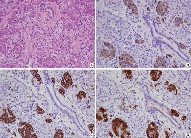
Fig. 2 Acini are separated into lobules by connective tissue, and intercalated ducts are lined with simple low cuboidal epithelium. Pancreatic tissue including acini, ducts, and islets of Langerhans (A). Expression of chromogranin (B), synaptophysin (C), and neuron-specific enolase (D) in islets of Langerhans.
Fig. 1
Fig. 2
Heterotopic Pancreas in Omphalomesenteric Duct Remnant Results in Persistent Umbilical Discharge
| No. | Age/Sex | Mass | Discharge | Size | Site | Reference |
|---|---|---|---|---|---|---|
| 1 | 12 yr/F | N/A | N/A | N/A | Umbilical subcutaneous tissue | Wright (1900), cited by Harris and Wenzl [1] |
| 2 | 22 yr/M | – | + | N/A | Umbilical cyst | Trimingham (1943), cited by Harris and Wenzl [1] |
| 3 | 6 mo/M | – | N/A | 3-mm nodule | Umbilical nodule | Steck and Helwig (1964), cited by Avolio et al. [2] |
| 4 | 13 mo/M | + | + | 12 × 9 × 5 mm | Umbilical mass | Caberwal et al. (1977), cited by Avolio et al. [2] |
| 5 | 60 yr/M | + | – | N/A | Umbilical polyp | Kondoh et al. (1994), cited by Avolio et al. [2] |
| 6 | 8 mo/M | +/– | + | N/A | Umbilical mass | Avolio et al. [2] (1998) |
| 7 | 15 mo/M | – | + | N/A | Umbilical mass | Avolio et al. [2] (1998) |
| 8 | 6 mo/M | – | + | N/A | Urachus | Perez-Martinez et al. (1999), cited by Lee et al. [3] |
| 9 | 3 mo/M | – | + | 1-cm cyst | Umbilical cyst | Tan et al. (2000), cited by Lee et al. [3] |
| 10 | 7 wk/M | – | + | N/A | Umbilical cyst | Tan et al. (2000), cited by Lee et al. [3] |
| 11 | 8 days/M | + | + | 26 × 20 × 7 mm | Umbilical mass | Lee et al. [3] (2005), |
| 12 | 18 mo/M | + | + | N/A | Umbilical mass | Silva et al. (2010), cited by Sharma et al. [4] |
| 13 | 2 yr/M | + | + | 12 × 12 × 10 cm | Umbilical mass | Sharma et al. (2013), cited by Sharma et al. [4] |
| 14 | 3 mo/F | – | + | 7 × 6 × 5 mm | Umbilical cyst | Present case |
Table 1. Cases of heterotopic pancreatic tissue at the umbilicus reported in the English literature
F, female; N/A, not available; M, male; +, present; –, absent.

