Warning: mkdir(): Permission denied in /home/virtual/lib/view_data.php on line 81

Warning: fopen(upload/ip_log/ip_log_2024-04.txt): failed to open stream: No such file or directory in /home/virtual/lib/view_data.php on line 83

Warning: fwrite() expects parameter 1 to be resource, boolean given in /home/virtual/lib/view_data.php on line 84
Cytotoxic Variant of Mycosis Fungoides with CD8+ CD56+ Phenotype: A Case Report and Review of Literature
Skip Navigation
Skip to contents

J Pathol Transl Med : Journal of Pathology and Translational Medicine

OPEN ACCESS
SEARCH
Search

Articles

Page Path
HOME > J Pathol Transl Med > Volume 48(5); 2014 > Article
Brief Case Report
Cytotoxic Variant of Mycosis Fungoides with CD8+ CD56+ Phenotype: A Case Report and Review of Literature
Meeran Kim, Moon Il Park, Myung Lim1, Jinman Kim,
Korean Journal of Pathology 2014;48(5):390-393.
DOI: https://doi.org/10.4132/KoreanJPathol.2014.48.5.390
Published online: October 27, 2014

Departments of Pathology, Chungnam National University Hospital, Daejeon, Korea

1Departments of Dermatology, Chungnam National University Hospital, Daejeon, Korea

Corresponding Author: Jinman Kim, M.D. Department of Pathology, Regional Cancer Center, and Infection Signaling Network Research Center, Chungnam National University School of Medicine, 266 Munhwa-ro,Jung-gu, Daejeon 301-747, Korea Tel: +82-42-580-8237, Fax: +82-42-581-5233, E-mail: 'jinmank@cnu.ac.kr'
• Received: September 9, 2013   • Accepted: October 15, 2013

© 2014 The Korean Society of Pathologists/The Korean Society for Cytopathology

This is an Open Access article distributed under the terms of the Creative Commons Attribution Non-Commercial License (http://creativecommons.org/licenses/by-nc/3.0/) which permits unrestricted noncommercial use, distribution, and reproduction in any medium, provided the original work is properly cited.
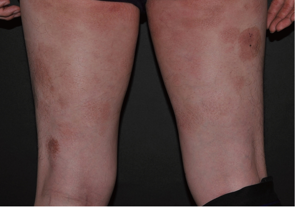
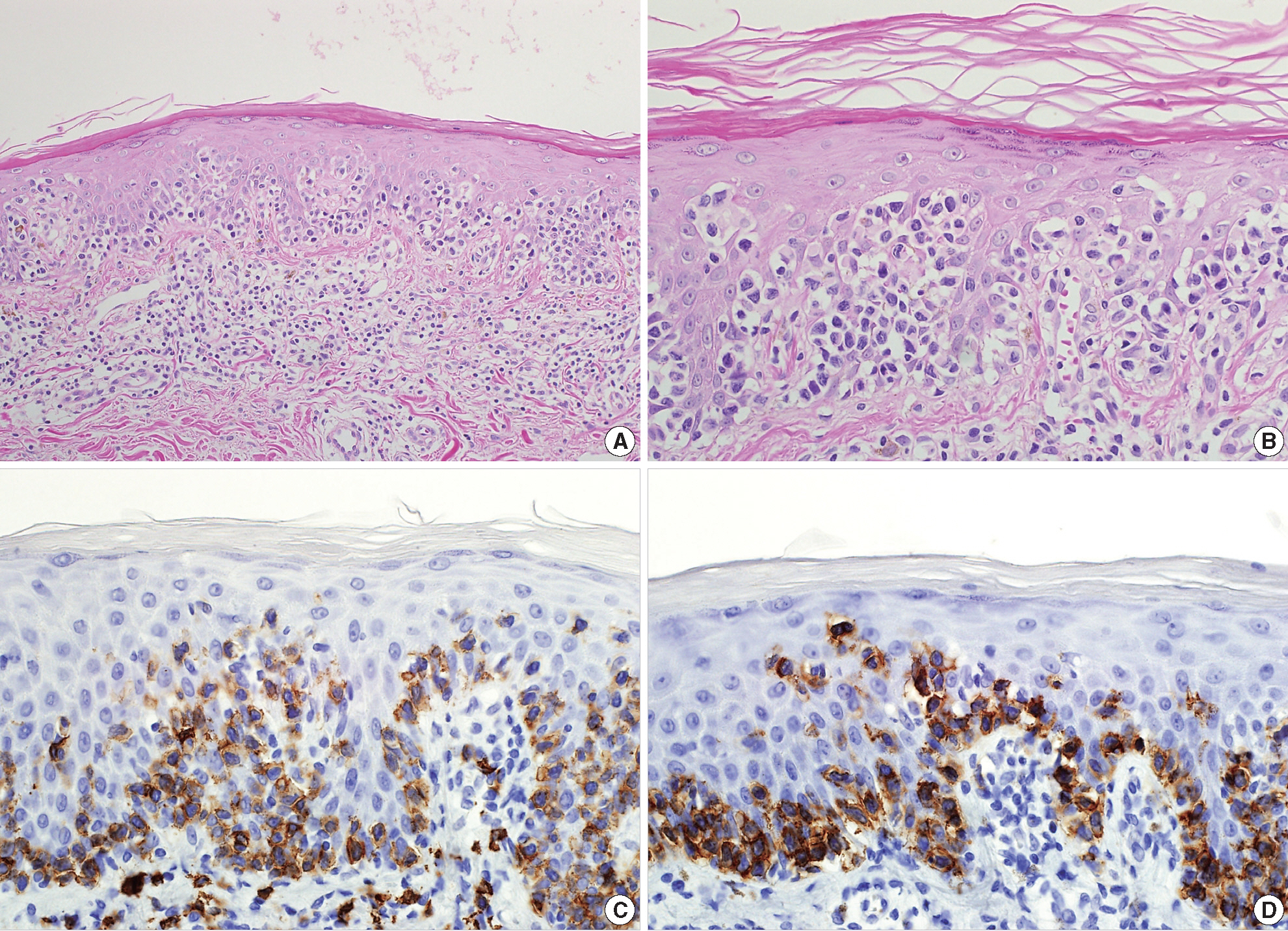
  • 6,055 Views
  • 52 Download
  • 7 Crossref
  • 5 Scopus
Mycosis fungoides (MF) is the most common cutaneous Tcell lymphoma. Approximately 50% of all primary cutaneous lymphomas are MF[1]. These lymphomas are comprised of epidermotropic collections of small- to medium-sized T lymphocytes with cerebriform nuclei. The neoplastic cell shows a mature CD3+, CD4+, CD45RO+, CD8– memory T-cell phenotype. However, rarely, MF with a CD4–, CD8+ cytotoxic T-cell phenotype has been reported[2-5]. The World Health Organization (WHO)-European Organization for Research and Treatment of Cancer (EORTC) classification for cutaneous lymphomas describes three types of cutaneous lymphomas that express CD56: 1) subcutaneous panniculitis-like T-cell lymphoma, 2) extranodal natural killer (NK)/T-cell lymphoma, nasal type, and 3) CD4+/CD56+ hematodermic neoplasm (blastic NKcell lymphoma)[1]. However, the report does not mention, MF with CD56 expression. Earlier in 2003, a report by the EORTC cutaneous lymphoma task force workshop described cytotoxic/natural killer cell cutaneous lymphomas and divided them into eight categories[6]. One of these categories was the CD56+, cytotoxic variant of MF. This report presents a rare CD8+, CD56+ variant of MF having the cytotoxic phenotype.
A 40-year-old male presented with a 5-year history of multiple round erythematous to dusky, brownish scaly patches with mild pruritus (Fig. 1). The skin involvement measured about 30% of total body surface. The lesions appeared first on the buttock and thigh, and lately on the upper arm. The skin lesions did not respond to topical corticosteroid treatment, at which time, he visited our hospital. A skin biopsy was taken from the thigh. The specimen showed a prominent band-like lymphocytic infiltration in the superficial dermis with epidermotropism (Fig. 2A). The epidermotropic lymphocytes were small- to medium-sized with an irregular nuclear membrane and coarse chromatin (Fig. 2B). The immunophenotype of the cells in the epidermis and a few cells in the superficial dermis were CD3+, CD4–, CD8+, CD56+, CD30–, and CD20– (Fig. 2C, D). The majority of lymphocytes in the superficial dermis were not atypical, in contrast to the epidermotropic lymphocytes, and were positive for CD4 and CD8. The neoplastic cells were positive for beta F1 (a marker of alpha/beta T lymphocytes) and granzyme B. An Epstein-Barr virus–encoded small non-polyadenylated RNA-1 (EBER-1) signal was not detected. Lactate dehydrogenase was elevated to 432 IU/L (range, 200 to 400 IU/L). Laboratory blood tests revealed an elevated, total cholesterol level of 235 mg/dL (range, 125 to 220 mg/mL). Complete blood cell count and other results of blood chemistry were within the normal range. The chest X-ray and positron emission tomography–computed tomography revealed no abnormal findings and there was no lymphadenopathy. The patient was diagnosed as having the stage IB, CD8+, CD56+ cytotoxic immunophenotype variant MF. He was treated with narrowband ultraviolet B therapy and his condition was stabilized.
Prognosis of classic MF depends on stage, and, in particular, on the type and extent of skin lesions, as well as the presence of extracutaneous disease[1]. Patients with limited patch/plaque-stage MF have a similar life expectancy to an age, sex, and race-matched control population[1]. However, the prognosis of MF with the cytotoxic phenotype, especially when it express CD56, is not well established.
To date, there are 8 cases of CD56-expressing MF reported in the current literature, and our case is the ninth case report. The immunohistochemical and clinical characteristics of these cases are summarized in Tables 1 and 2. Among the nine cases, six cases co-expressed CD8 in a majority of the neoplastic cells, one co-expressed CD8 in a minority of neoplastic cells, and two were negative for CD8. One out of nine cases was positive for CD4. Age range was broad, from 6- to 85-years-old. Eight cases were females and our case was the only male case. Poikiloderma was the most frequent clinical feature at the time of diagnosis. Seven cases showed good response to the therapy while two cases, including our case, showed limited response. None of the cases, including our case, had an aggressive clinical course. This result suggests that CD56+ MF is a disease with good prognosis similar to classic MF.
Several reports of MF with CD8+ and/or CD56+ expression have suggested that this variant would have no prognostic difference compared with classic MF[2,3,5,7-10]. Massone et al.[9] analyzed 73 biopsy specimens from 68 patients with early MF and divided them into four groups based on the immunophenotype. They stated that there was no statistical difference between the survival curves of the four groups, and therefore concluded that cytotoxic phenotype does not have any prognostic significance.
In our case, with the observed immunophenotype, we considered different diagnoses including primary cutaneous aggressive epidermotropic CD8+ cytotoxic T-cell lymphoma, primary cutaneous gamma/delta T-cell lymphoma, and extranodal NK/T-cell lymphoma, nasal type. We were able to rule out these different diagnoses based on the following: clinical course and presentation, beta F1 positivity, and EBER-1 negativity.
In conclusion, we present a case of a cytotoxic variant of MF with a CD8+, CD56+ immunophenotype. MF with cytotoxic immunophenotype is characterized by a typical clinical presentation, histology, and course of MF. Clinical presentation and course should be a primary consideration in diagnosis. We emphasize that it is important to recognize this rare variant of MF and to distinguish it from other aggressive cutaneous lymphomas to avoid aggressive treatment.

No potential conflict of interest relevant to this article was reported.

This research was supported by Basic Science Research Program through the National Research Foundation of Korea (NRF) funded by the Ministry of Education, Science and Technology (2011-0012351).
Fig. 1.
The skin lesions in the buttocks and both thighs are erythematous to dusky brown.
kjpathol-48-5-390f1.jpg
Fig. 2.
(A) The specimen shows a prominent band-like lymphocytic infiltration with epidermotropism. (B) The epidermotropic lymphocytes are small- to medium-sized with an irregular nuclear membrane and coarse chromatin. These cells display the cytotoxic phenotype, showing CD8 (C) and CD56 (D) immunoreactivity.
kjpathol-48-5-390f2.jpg
Table 1.
Immunohistochemical staining results of the reported cases
Author CD3 CD4 CD8 CD56 Granzyme B CD30
Wain et al.[5] (case No. 1) + - + + - -
Wain et al.[5] (case No. 2) + - - + + -
Wain et al.[5] (case No. 3, poikiloderma) + - + + + -
Wain et al.[5] (case No. 3, tumor) + - + + + +
Sawada et al.[3] + - + + - -
Horst et al.[7] + + +/- + Not performed Not performed
Nikolaou et al.[2] + - + + Not performed -
Klekotka et al.[8] + - - + Not performed Not performed
Shiomi et al.[4] + - + + + -
Present case + - + + + -
Table 2.
Clinical characteristics of the reported case
Author Age (yr)/Sex Clinical pattern Treatment Clinical course EBER-1
Wain et al.[5] (case No. 1) 45/F Poikiloderma Radiotherapy and topical steroid Limited response Not performed
Wain et al.[5] (case No. 2) 6/F Hypo- and hyperpigmentation Topical steroid and nUVB Good response Not performed
Wain et al.[5] (case No. 3) 37/F Poikiloderma, tumor PUVA, excision Good response Not performed
Sawada et al.[3] 68/F Poikiloderma nUVB, oral PUVA Good response Negative
Horst et al.[7] 85/F Erythroderma Topical steroid Good response Not performed
Nikolaou et al.[2] 43/F Poikiloderma PUVA Good response Not performed
Klekotka et al.[8] 33/F Erythroderma Topical steroid and PUVA Good response Not performed
Shiomi et al.[4] 20/F Poikiloderma Topical steroid Good response Negative
Present case 40/M Erythroderma nUVB Limited response Negative

F, female; nUVB, narrowband ultraviolet B; PUVA, psoralen plus ultraviolet A; M, male.

  • 1. Willemze R, Jaffe ES, Burg G, et al. WHO-EORTC classification for cutaneous lymphomas. Blood 2005; 105: 3768–85. ArticlePubMed
  • 2. Nikolaou VA, Papadavid E, Katsambas A, et al. Clinical characteristics and course of CD8+ cytotoxic variant of mycosis fungoides: a case series of seven patients. Br J Dermatol 2009; 161: 826–30. ArticlePubMed
  • 3. Sawada Y, Sugita K, Kabashima R, et al. CD8+ CD56+ mycosis fungoides with an indolent clinical behaviour: case report and literature review. Acta Derm Venereol 2010; 90: 525–6. ArticlePubMed
  • 4. Shiomi T, Monobe Y, Kuwabara C, Hayashi H, Yamamoto T, Sadahira Y. Poikilodermatous mycosis fungoides with a CD8+ CD56+ immunophenotype: a case report and literature review. J Cutan Pathol 2013; 40: 317–20. ArticlePubMed
  • 5. Wain EM, Orchard GE, Mayou S, Atherton DJ, Misch KJ, Russell-Jones R. Mycosis fungoides with a CD56+ immunophenotype. J Am Acad Dermatol 2005; 53: 158–63. ArticlePubMed
  • 6. Santucci M, Pimpinelli N, Massi D, et al. Cytotoxic/natural killer cell cutaneous lymphomas. Report of EORTC Cutaneous Lymphoma Task Force Workshop. Cancer 2003; 97: 610–27. ArticlePubMed
  • 7. Horst BA, Kasper R, LeBoit PE. CD4+, CD56+ mycosis fungoides: case report and review of the literature. Am J Dermatopathol 2009; 31: 74–6. ArticlePubMed
  • 8. Klekotka PA, Faulkner-Jones B, Heffernan MP. A case of CD56+ mycosis fungoides. Arch Dermatol 2006; 142: 1370–2. ArticlePubMed
  • 9. Massone C, Crisman G, Kerl H, Cerroni L. The prognosis of early mycosis fungoides is not influenced by phenotype and T-cell clonality. Br J Dermatol 2008; 159: 881–6. ArticlePubMed
  • 10. Um SH, Oh CW. CD8 expression in mycosis fungoides. Korean J Dermatol 2004; 42: 1525–30.

Figure & Data

References

    Citations

    Citations to this article as recorded by  
    • Chronic Radiation Dermatitis Secondary to Narrow-Band Ultraviolet B Therapy in a Patient With Primary Cutaneous CD8+ T-Cell Lymphoma With Cytotoxic Granules
      Mia P. Edelson, Jane J. Gay, Robert W. Thiel, Douglas J. Grider
      The American Journal of Dermatopathology.2024;[Epub]     CrossRef
    • Primary cutaneous CD8+ cytotoxic T‐cell lymphoma of the face with intraoral involvement, resulting in facial nerve palsy after chemotherapy
      Daphine Caxias Travassos, Heitor Albergoni Silveira, Evânio Vilela Silva, Beatriz Zamboni Martins Panucci, Nilson Coelho da Silva Filho, Paula Verona Ragusa Silva, Andreia Bufalino, Jorge Esquiche León
      Journal of Cutaneous Pathology.2022; 49(6): 560.     CrossRef
    • A Study of Antimicrobial Activity of Herbal Extracts on Clostridium difficile
      Eunhak Seong, Sookyoung Lim, Myeongjong Lee, Hojun Kim
      Journal of Korean Medicine Rehabilitation.2021; 31(1): 47.     CrossRef
    • Rare case of CD8+ CD56+ cytotoxic variant of mycosis fungoides clinically presenting with a combination of hypopigmentation and poikiloderma
      Min‐Young Park, Shinwon Hwang, Jemin Kim, Abdurrahman I. Almurayshid, Sun Och Yoon, Sang Ho Oh
      International Journal of Dermatology.2020;[Epub]     CrossRef
    • Mycosis fungoides in Taiwan shows a relatively high frequency of large cell transformation and CD56 expression
      Ren Ching Wang, Seiji Sakata, Bo-Jung Chen, Sheng-Tsung Chang, Pin-Pen Hsieh, Chi-Shun Yang, Satoko Baba, Kengo Takeuchi, Shih-Sung Chuang
      Pathology.2018; 50(7): 718.     CrossRef
    • CD8 + mycosis fungoides: A low-grade lymphoproliferative disorder
      Maria Estela Martinez-Escala, Robert W. Kantor, Ahuva Cices, Xiaolong A. Zhou, Jason B. Kaplan, Barbara Pro, Jaehyuk Choi, Joan Guitart
      Journal of the American Academy of Dermatology.2017; 77(3): 489.     CrossRef
    • Phenotypic Variation in Different Lesions of Mycosis Fungoides Biopsied Within a Short Period of Time From the Same Patient
      Natalie Kash, Cesare Massone, Regina Fink-Puches, Lorenzo Cerroni
      The American Journal of Dermatopathology.2016; 38(7): 541.     CrossRef

    • PubReader PubReader
    • ePub LinkePub Link
    • Cite this Article
      Cite this Article
      export Copy Download
      Close
      Download Citation
      Download a citation file in RIS format that can be imported by all major citation management software, including EndNote, ProCite, RefWorks, and Reference Manager.

      Format:
      • RIS — For EndNote, ProCite, RefWorks, and most other reference management software
      • BibTeX — For JabRef, BibDesk, and other BibTeX-specific software
      Include:
      • Citation for the content below
      Cytotoxic Variant of Mycosis Fungoides with CD8+ CD56+ Phenotype: A Case Report and Review of Literature
      Korean J Pathol. 2014;48(5):390-393.   Published online October 27, 2014
      Close
    • XML DownloadXML Download
    Figure
    Related articles

    J Pathol Transl Med : Journal of Pathology and Translational Medicine