- Histopathological characteristics of Epstein-Barr virus (EBV)–associated encephalitis and colitis in chronic active EBV infection
-
Betty A Kasimo, James J Yahaya, Sun Och Yoon, Se Hoon Kim, Minsun Jung
-
J Pathol Transl Med. 2025;59(3):188-194. Published online April 16, 2025
-
DOI: https://doi.org/10.4132/jptm.2025.02.21
-
-
 Abstract Abstract
 PDF PDF
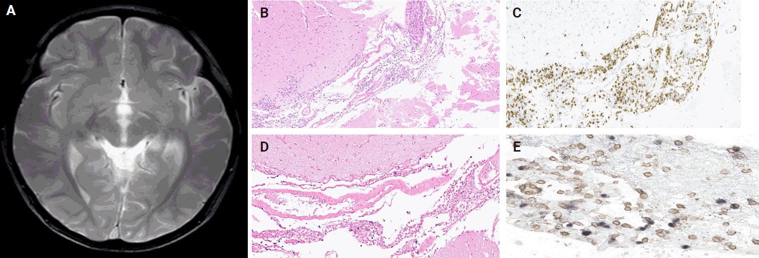
- Chronic active Epstein-Barr virus (CAEBV) can induce complications in various organs, including the brain and gastrointestinal tract. A 3-year-old boy was referred to the hospital with a history of fever and seizures for 15 days. A diagnosis of encephalitis based on computed tomography (CT) and magnetic resonance imaging findings and clinical correlation was made. Laboratory tests showed positive serology for Epstein-Barr virus (EBV) and negative for Rotavirus antigen and IgG and IgM antibodies for cytomegalovirus, herpes simplex virus, and varicella zoster virus, respectively. Abdominal CT showed diffuse wall thickening with fluid distension of small bowel loops, lower abdomen wall thickening, and a small amount of ascites. The biopsy demonstrated positive Epstein-Barr encoding region in situ hybridization in cells within the crypts and lamina propria. The patient was managed with steroids and hematopoietic stem cell transplantation (HSCT). This case showed histopathological characteristics of concurrent EBV-associated encephalitis and colitis in CAEBV infection. The three-step strategy of immunosuppressive therapy, chemotherapy, and allogeneic HSCT should be always be considered for prevention of disease progression.
- Clinicopathological differences in radiation-induced organizing hematomas of the brain based on type of radiation treatment and primary lesions
-
Myung Sun Kim, Se Hoon Kim, Jong-Hee Chang, Mina Park, Yoon Jin Cha
-
J Pathol Transl Med. 2022;56(1):16-21. Published online October 15, 2021
-
DOI: https://doi.org/10.4132/jptm.2021.08.30
-
-
5,657
View
-
236
Download
-
3
Web of Science
-
4
Crossref
-
Abstract
PDF
- Background
Radiation-induced organizing hematoma (RIOH) is a sporadic form of cavernous hemangioma (CH) that occurs after cerebral radiation. RIOH lesions are distinct histologically from de novo CH; however, detailed research on this subject is lacking. In the present study, the clinical and histological features of RIOHs were evaluated based on causative lesions.
Methods
The present study included 37 RIOHs confirmed by surgical excision from January 2009, to May 2020, in Yonsei Severance Hospital. All cases were divided into subgroups based on type of radiation treatment (gamma knife surgery [GKS], n = 24 vs. conventional radiation therapy [RT], n = 13) and pathology of the original lesion (arteriovenous malformation, n = 14; glioma, n = 12; metastasis, n = 4; other tumors, n = 7). The clinicopathological results were compared between the groups.
Results
Clinical data of multiplicity, latency, and size and wall thickness of the original tumors and RIOHs were analyzed. The GKS group showed shorter latency (5.85 ± 4.06 years vs. 11.15 ± 8.27 years, p = .046) and thicker tumor wall (693.7 ± 565.7 μm vs. 406.9 ± 519.7 μm, p = .049) than the conventional RT group. Significant difference was not found based on original pathology.
Conclusions
RIOH is more likely to occur earlier with thick tumor wall in subjects who underwent GKS than in patients who underwent conventional RT. These results indicate the clinical course of RIOH differs based on type of treatment and might help determine the duration of follow-up.
-
Citations
Citations to this article as recorded by  - Radiation-Induced Cavernous Malformation in the Cerebellum: Clinical Features of Two Cases
Hyoung Soo Choi, Chae-Yong Kim, Byung Se Choi, Seung Hyuck Jeon, In Ah Kim, Joo-Young Kim, Kyu Sang Lee, Gheeyoung Choe
Brain Tumor Research and Treatment.2025; 13(2): 58. CrossRef - End-stage ADPKD with a low-frequency PKD1 mosaic variant accelerated by chemoradiotherapy
Hiroaki Hanafusa, Hiroshi Yamaguchi, Naoya Morisada, Ming Juan YE, Riki Matsumoto, Hiroaki Nagase, Kandai Nozu
Human Genome Variation.2024;[Epub] CrossRef - Recapitulating the Key Advances in the Diagnosis and Prognosis of High-Grade Gliomas: Second Half of 2021 Update
Guido Frosina
International Journal of Molecular Sciences.2023; 24(7): 6375. CrossRef - Earlier Age at Surgery for Brain Cavernous Angioma-Related Epilepsy May Achieve Complete Seizure Freedom without Aid of Anti-Seizure Medication
Ayataka Fujimoto, Hideo Enoki, Keisuke Hatano, Keishiro Sato, Tohru Okanishi
Brain Sciences.2022; 12(3): 403. CrossRef
- Adjunctive markers for classification and diagnosis of central nervous system tumors: results of a multi-center neuropathological survey in Korea
-
Yoon Jin Cha, Se Hoon Kim, Na Rae Kim
-
J Pathol Transl Med. 2020;54(2):165-170. Published online February 20, 2020
-
DOI: https://doi.org/10.4132/jptm.2020.02.04
-
-
7,101
View
-
216
Download
-
1
Web of Science
-
1
Crossref
-
Abstract
PDF
 Supplementary Material Supplementary Material
- Background
The revised 4th 2016 World Health Organization (WHO) classification of tumors of the central nervous system (CNS) classification has adopted integrated diagnosis encompassing the histology and molecular features of CNS tumors. We aimed to investigate the immunohistochemistry, molecular testing, and testing methods for diagnosis of CNS tumors in pathological labs of tertiary centers in Korea, and evaluate the adequacy of tests for proper diagnosis in daily practice.
Methods
A survey, composed of eight questions concerning molecular testing for diagnosis of CNS tumors, was sent to 10 neuropathologists working in tertiary centers in Korea.
Results
For diagnosis of astrocytic and oligodendroglial tumors, all 10 centers performed isocitrate dehydrogenase mutations testing and 1p/19q loss of heterozygosity. For glioneuronal tumors, immunohistochemistry (IHC) assays for synaptophysin (n = 9), CD34 (n = 7), BRAF(VE1) (n = 5) were used. For embryonal tumors, particularly in medulloblastoma, four respondents used IHC panel (growth factor receptor bound protein 2-associated protein 1, filamin A, and yes-associated protein 1) for molecular subclassification. Regarding meningioma, all respondents performed Ki-67 IHC and five performed telomerase reverse transcriptase promoter mutation.
Conclusions
Most tertiary centers made proper diagnosis in line with 2016 WHO classification. As classification of CNS tumors has evolved to be more complex and more ancillary tests are required, these should be performed considering the effect of necessity and justification.
-
Citations
Citations to this article as recorded by - Exploring the role of epidermal growth factor receptor variant III in meningeal tumors
Rashmi Rana, Vaishnavi Rathi, Kirti Chauhan, Kriti Jain, Satnam Singh Chhabra, Rajesh Acharya, Samir Kumar Kalra, Anshul Gupta, Sunila Jain, Nirmal Kumar Ganguly, Dharmendra Kumar Yadav, Timir Tripathi
PLOS ONE.2021; 16(9): e0255133. CrossRef
- Hyalinizing Trabecular Tumor of the Thyroid Gland, a Diagnostic Challenge in Fine-Needle Aspiration Cytology: Case Report
-
Ye-Young Rhee, Hong Kyu Jung, Se Hoon Kim, Soo Hee Kim
-
J Pathol Transl Med. 2018;52(4):252-256. Published online June 11, 2018
-
DOI: https://doi.org/10.4132/jptm.2018.04.28
-
-
9,756
View
-
180
Download
-
6
Web of Science
-
8
Crossref
-
Abstract
PDF
- Hyalinizing trabecular tumor (HTT) is a rare thyroid tumor with low to minimal malignant potential. HTT is often misinterpreted as other thyroid tumors, including papillary thyroid carcinoma (PTC) and medullary thyroid carcinoma (MTC), on fine-needle aspiration (FNA) cytology, because of its overlapping cytologic features, such as nuclear grooves and intranulcear pseudoinclusions. Although cytopathologists cannot definitely conclude HTT by FNA cytology, suspicion of HTT is necessary to avoid misdiagnosing HTT as PTC or MTC and to avoid unnecessary aggressive treatment. Here, we report a case of HTT with novel cytologic features in CellPrep liquid based cytology that was diagnosed as suspicious for papillary carcinoma by FNA and finally diagnosed as HTT in the surgical specimen.
-
Citations
Citations to this article as recorded by - Cytomorphological traits of fine-needle aspirates of hyalinizing trabecular tumor of the thyroid gland: A brief report
Fei Wang, Yufei Liu
Indian Journal of Pathology and Microbiology.2024; 67(1): 128. CrossRef - Total thyroidectomy can still remain the method of choice in some Bethesda III cases
Jindrich Lukas, Barbora Hintnausova, Vlasta Sykorova, Martin Syrucek, Marek Maly, Jaroslava Duskova
Biomedical Papers.2023; 167(1): 61. CrossRef - Diagnostic clues for hyalinizing trabecular tumor on fine needle aspiration cytology
Lone Nielsen, Ana María Colino Gallardo, Pablo Pérez Alonso, Luis Ortega Medina, Esthefanía Latorre García, Cristina Díaz del Arco, Reyes Bergillos Jiménez, Lorenzo Alarcón García, Marta Cruz Blanco, Jesús Vega González, Montserrat De la Torre Serrano, Ma
Cytojournal.2023; 20: 19. CrossRef - Clinical Characteristics of the Hyalinizing Trabecular Tumor
Byung-Chang Kim, Shin Jeong Pak, Jae Won Cho, Won Woong Kim, Yu-mi Lee, Tae-Yon Sung, Jung Hwan Baek, Ki-Wook Chung
Journal of Endocrine Surgery.2022; 22(4): 116. CrossRef - A Case of Multifocal Hyalinizing Trabecular Tumors of the Thyroid
Gland
Suhwan Jeong, Hanaro Park
Journal of Clinical Otolaryngology Head and Neck
Surgery.2021; 32(3): 308. CrossRef - The Diagnosis of Hyalinizing Trabecular Tumor: A Difficult and Controversial Thyroid Entity
Esther Diana Rossi, Mauro Papotti, William Faquin, Luigi Maria Larocca, Liron Pantanowitz
Head and Neck Pathology.2020; 14(3): 778. CrossRef - A large series of hyalinizing trabecular tumors: Cytomorphology and ancillary techniques on fine needle aspiration
Marco Dell’Aquila, Carmen Gravina, Alessandra Cocomazzi, Sara Capodimonti, Teresa Musarra, Stefania Sfregola, Vincenzo Fiorentino, Luca Revelli, Maurizio Martini, Guido Fadda, Liron Pantanowitz, Luigi Maria Larocca, Esther Diana Rossi
Cancer Cytopathology.2019; 127(6): 390. CrossRef - GLIS rearrangements in thyroid nodules: A key to preoperative diagnosis of hyalinizing trabecular tumor
Marina N. Nikiforova, Yuri E. Nikiforov, N. Paul Ohori
Cancer Cytopathology.2019; 127(9): 560. CrossRef
- Merkel Cell Carcinoma Metastatic to Pleural Fluid: A Case Report
-
Ye-Young Rhee, Soo Hee Kim, Eun Kyung Kim, Se Hoon Kim
-
J Pathol Transl Med. 2018;52(3):206-209. Published online November 23, 2017
-
DOI: https://doi.org/10.4132/jptm.2017.11.10
-
-
7,175
View
-
130
Download
-
5
Web of Science
-
6
Crossref
-
Abstract
PDF
- Merkel cell carcinoma (MCC) is a rare aggressive neuroendocrine carcinoma of the skin that shows locoregional or distant metastasis. Metastasis of MCC to body cavity effusion is extremely rare; only three cases have been reported so far. Metastatic MCC in effusion cytology shows small blue round cells with fine stippled chromatin like other small blue round cell tumors such as small cell lung carcinoma or lymphoma. The diagnosis of metastatic MCC can grant patients good chances at recently advanced therapeutic options. Here, we present a case of metastatic MCC to pleural effusion with characteristic single file-like pattern.
-
Citations
Citations to this article as recorded by - Pleural Metastasis of Merkel Cell Carcinoma
Sina Maghsoudlou, Marc Pusztaszeri, Mauro Saieg
Diagnostic Cytopathology.2025; 53(6): 308. CrossRef - Merkel cell carcinoma presenting as a malignant pleural effusion post‐COVID‐19 hospitalization: A case report and literature review
Joel Lanceta, Mesut Toprak, Oana C. Rosca
Diagnostic Cytopathology.2022;[Epub] CrossRef - Cytology coupled with immunocytochemistry identifies Merkel cell carcinoma: A rare intruder in the cerebrospinal fluid
Reetu Kundu, Brijdeep Singh, Pranab Dey
Cytopathology.2022; 33(4): 530. CrossRef - Derrame pleural por carcinoma de células de Merkel
María J. Soler-Sempere, María O. Alvárez-Fernández, Isabel Padilla-Navas, María Cabezas-Macián, Jose F. Sánchez-Hernández, Eduardo García-Pachón
Archivos de Bronconeumología.2021; 57(11): 715. CrossRef - A rare case of pleural localisation of both metastatic Merkel cell carcinoma and chronic lymphocytic leukaemia
Elise Kaspi, Shirley Fritz, Julien Colle, Florent Amatore, Diane Frankel, Patrice Roll
Cytopathology.2021; 32(3): 367. CrossRef - Merkel cell carcinoma with pleural effusion
María J. Soler-Sempere, María O. Alvárez-Fernández, Isabel Padilla-Navas, María Cabezas-Macián, Jose F. Sánchez-Hernández, Eduardo García-Pachón
Archivos de Bronconeumología (English Edition).2021; 57(11): 715. CrossRef
- Liquid-Based Cytology of the Cerebrospinal Fluid in a Case of Cryptococcal Meningitis
-
Jiwoon Choi, Se Hoon Kim
-
J Pathol Transl Med. 2018;52(1):61-63. Published online October 26, 2017
-
DOI: https://doi.org/10.4132/jptm.2017.06.13
-
-
8,455
View
-
191
Download
-
5
Web of Science
-
5
Crossref
-
Abstract
PDF
- Cryptococcus neoformans is the most common microorganism found in cerebrospinal fluid (CSF) cytology and causes life-threatening infections in immunocompromised hosts. Although its cytomorphologic features in conventional smear cytology have been well described, those in liquid-based cytology have rarely been. A 73-year-old woman with diffuse large B-cell lymphoma presented with mental confusion and a spiking fever. To rule out infectious conditions, CSF examination was performed. A cytology slide that was prepared using the ThinPrep method showed numerous spherical yeast-form organisms with diameters of 4–11 μm and thick capsules. Occasional asymmetrical, narrow-based budding but no true hyphae or pseudohyphae were observed. Gomori methenamine silver staining was positive. Cryptococcosis was confirmed in blood and CSF through the cryptococcal antigen test and culture. Liquid-based cytology allows for a clean background and additional slides for ancillary testing, facilitating the detection of microorganisms in CSF specimens, particularly when the number of organisms is small.
-
Citations
Citations to this article as recorded by - Unraveling Capsule Biosynthesis and Signaling Networks in Cryptococcus neoformans
Eun-Ha Jang, Ji-Seok Kim, Seong-Ryong Yu, Yong-Sun Bahn, Teresa R. O’Meara
Microbiology Spectrum.2022;[Epub] CrossRef - Cerebrospinal fluid pleocytosis in immunocompromised patients: Can it be Cryptococcus
Ridhi Sood, Ruchita Tyagi, Pavneet Selhi, Harpreet Kaur, Neena Sood
Diagnostic Cytopathology.2020; 48(2): 164. CrossRef - Special Staining of the Liquid-Based Cytopathology Test in Bronchoalveolar Lavage Fluid for Diagnosis of Invasive Pulmonary Aspergillosis with Nonneutropenic Patients
Yue Hu, Lin Zheng, Deng Pan, Lei Shao, Xianfa Xu, Yiming Yu, Qidong Zhuang, Zaichun Deng, Zhongbo Chen
Canadian Respiratory Journal.2020; 2020: 1. CrossRef - Sensitivity of Cerebrospinal Fluid Cytology for the Diagnosis of Cryptococcal Infections
Kelsey E McHugh, Melanie Gersey, Daniel D Rhoads, Gary W Procop, Yaxia Zhang, Christine N Booth, Charles D Sturgis
American Journal of Clinical Pathology.2019; 151(2): 198. CrossRef - Cryptococcal Capsules in Cerebrospinal Fluid Visible on Hemocytometer
Zen Kobayashi, Yuriko Hirota, Shuzo Shintani
Canadian Journal of Neurological Sciences / Journal Canadien des Sciences Neurologiques.2018; 45(6): 700. CrossRef
- A Rare Case of Aggressive Melanotic Schwannoma Occurred in Spinal Nerve of a 59-Year-Old Male
-
Sung-eun Choi, Yoon Jin Cha, Jisup Kim, Hyunseo Cha, Jayeong Seo, Sung-Uk Kuh, Sung-Jun Kim, Se Hoon Kim
-
J Pathol Transl Med. 2017;51(5):505-508. Published online April 4, 2017
-
DOI: https://doi.org/10.4132/jptm.2017.01.04
-
-
13,327
View
-
220
Download
-
21
Web of Science
-
18
Crossref
-
Abstract
PDF
- Melanotic schwannoma (MS) is a rare variant of nerve sheath neoplasm that shows ultrastructural and immunophenotypical features of Schwann cells but also has cytoplasmic melanosomes and is reactive for melanocytic markers as well. Unlike conventional schwannoma, which is totally benign, MS has an unpredictable prognosis and is thought to have low-malignant potential. Herein, we present a rare case of recurrent MS in lumbar spine of a 59-year-old male.
-
Citations
Citations to this article as recorded by - The “Pigmented Side” of Nerve Sheaths: Malignant Melanotic Nerve Sheath Tumor
Raduan Ahmed Franca, Rosa Maria Di Crescenzo, Lorenzo Ugga, Rosa Della Monica, Elena D'Avella
International Journal of Surgical Pathology.2025; 33(4): 1068. CrossRef - Case Report: Cutaneous melanocytic schwannoma with concomitant melanocytoma in a canine
Olwam H. Monakali, Nicolize O'Dell, Louise van der Weyden
Wellcome Open Research.2024; 8: 364. CrossRef - Intradural Melanotic Schwannoma of the Sacral Spine: An Illustrated Case Report of Diagnostic Conundrum
Jiunn-Kai Chong, Navneet Kumar Dubey, Wen-Cheng Lo
Reports.2024; 7(3): 56. CrossRef - Rare giant retroperitoneal melanotic schwannoma: a case report and literature review
Pan Chen, Junfeng Cheng, Lin Zhang
Frontiers in Oncology.2024;[Epub] CrossRef - A Rare Case of Melanotic Schwannoma Occurred Intraosseous of Sacrum: A Literature Review
Xiaobo Yan, Keyi Wang, Nong Lin, Xin Huang, YanBiao Fu, Zhaoming Ye
Orthopaedic Surgery.2023; 15(2): 655. CrossRef - Sporadic spinal psammomatous malignant melanotic nerve sheath tumor: A case report and literature review
Giulio Bonomo, Alessandro Gans, Elio Mazzapicchi, Emanuele Rubiu, Paolo Alimonti, Marica Eoli, Rosina Paterra, Bianca Pollo, Guglielmo Iess, Francesco Restelli, Jacopo Falco, Francesco Acerbi, Marco Paolo Schiariti, Paolo Ferroli, Morgan Broggi
Frontiers in Oncology.2023;[Epub] CrossRef - Case Report: Cutaneous melanocytic schwannoma with concomitant melanocytoma in a canine
Olwam H. Monakali, Nicolize O'Dell, Louise van der Weyden
Wellcome Open Research.2023; 8: 364. CrossRef - Fine‐needle aspiration cytology of melanotic schwannoma in the submandibular gland
Yu‐Hua Huang, Ying‐Chou Lu, Hsuan‐Ying Huang, Chien‐Chin Chen
Diagnostic Cytopathology.2021; 49(1): 142. CrossRef - Checkpoint inhibitors and radiotherapy in refractory malignant melanocytic schwannoma with Carney complex: first evidence of efficacy
Jyoti Bajpai, Akhil Kapoor, Rakesh Jalali, Mrinal M Gounder
BMJ Case Reports.2021; 14(5): e240296. CrossRef - 18F-FDG PET/CT imaging for aggressive melanotic schwannoma of the L3 spinal root
Xun-Ze Shen, Wei Wang, Zhou-Ye Luo
Medicine.2021; 100(8): e24803. CrossRef - Hemorrhagic spinal melanotic schwannoma presenting as acute chest pain: A case report and literature review
Dallas J. Soyland, Dylan R. Goehner, Kayla M. Hoerschgen, Troy D. Gust, Shawn M. Vuong
Surgical Neurology International.2021; 12: 164. CrossRef - Retroperitoneal Recurrence of Melanotic Schwannoma on 18F-FDG PET/CT
Xiangliu OuYang, Lichun Zheng, Xiaoming Zhang
Clinical Nuclear Medicine.2021; 46(12): 991. CrossRef - Schwannoma originating from the common iliac artery: a case report
Seung-Myoung Son, Chang Gok Woo
Journal of International Medical Research.2020;[Epub] CrossRef - Intraosseous Melanotic Schwannoma in the Sacrum Mimicking Primary Bone Tumor
Yoshitaka Nagashima, Yusuke Nishimura, Kaoru Eguchi, Takayuki Awaya, Satoshi Yoshikawa, Shoichi Haimoto, Toshihiko Wakabayashi, Masahito Hara
NMC Case Report Journal.2020; 7(3): 107. CrossRef - Extramedullary melanotic schwannoma recurrence in the cervical vertebral arch: a case report and review of the literature
Zongbin Hou, Teng Shi, Guangrun Li, Lin Tian, Xinna Li, Xiaoyang Liu
Journal of International Medical Research.2020;[Epub] CrossRef - Extramedullary malignant melanotic schwannoma of the spine: Case report and an up to date systematic review of the literature
Georgios Solomou, Adikarige Haritha Dulanka Silva, Adrianna Wong, Ute Pohl, Nikolaos Tzerakis
Annals of Medicine and Surgery.2020; 59: 217. CrossRef - Melanotic Schwannoma of the Vagina: A Report of a Very Rare Tumor and Review of the Literature
Kofi Effah, Stefan Seidl, Edith Gorges, Patrick Kafui Akakpo
Case Reports in Obstetrics and Gynecology.2019; 2019: 1. CrossRef - Melanotic Schwannomas Are Rarely Seen Pigmented Tumors with Unpredictable Prognosis and Challenging Diagnosis
Elif Keskin, Sumeyye Ekmekci, Ozgur Oztekin, Gulden Diniz
Case Reports in Pathology.2017; 2017: 1. CrossRef
- History of the Official Journal Published by the Korean Society of Pathologists: From the Korean Journal of Pathology to the Journal of Pathology and Translational Medicine
-
Se Hoon Kim, Chong Jai Kim, SoonWon Hong
-
J Pathol Transl Med. 2017;51(1):1-6. Published online January 13, 2017
-
DOI: https://doi.org/10.4132/jptm.2017.01.07
-
-
9,776
View
-
130
Download
-
2
Web of Science
-
1
Crossref
-
PDF
-
Citations
Citations to this article as recorded by - A Multistakeholder Approach to the Airport Gate Assignment Problem: Application of Fuzzy Theory for Optimal Performance Indicator Selection
Haonan Li, Xu Wu, Yinghui Liang, Chen Zhang, Yu-Ting Bai
Computational Intelligence and Neuroscience.2021;[Epub] CrossRef
- Diagnostic Accuracy of Cerebrospinal Fluid (CSF) Cytology in Metastatic Tumors: An Analysis of Consecutive CSF Samples
-
Yoon Sung Bae, June-Won Cheong, Won Seok Chang, Sewha Kim, Eun Ji Oh, Se Hoon Kim
-
Korean J Pathol. 2013;47(6):563-568. Published online December 24, 2013
-
DOI: https://doi.org/10.4132/KoreanJPathol.2013.47.6.563
-
-
7,903
View
-
64
Download
-
13
Crossref
-
Abstract
PDF
- Background
Cerebrospinal fluid (CSF) examination can be used to verify the presence of primary malignancies as well as cases of central nervous system (CNS) metastasis. Because of its importance, there have been several studies concerning the sensitivity of CSF cytology. To determine the practical use and reproducibility of diagnoses based on CSF cytology, we evaluated this test by analyzing cytology results from consecutive CSF samples. MethodsBetween July 2010 and June 2013, 385 CSF cytology samples from 42 patients were collected. The samples were gathered using a ventricular catheter and reservoir. CSF cytology of all patients was examined more than two times with immunocytochemistry for cytokeratin. ResultsPrimary neoplastic sites and histologic types of patients' metastatic cancer were diverse. The overall sensitivity for detecting malignancy was 41.3%. Even within short-term intervals, diagnoses frequently changed. ConclusionsOur results were inconsistent, with low sensitivity, when compared to the results of previous studies. However, CSF evaluation can still provide valuable diagnostic and prognostic information because adjuvant treatments are now routinely performed in patients with CNS metastasis. Negative CSF cytology results should not be ignored, and continuous CSF follow-up is essential for following the clinical course of patients with metastatic cancer involving the CNS.
-
Citations
Citations to this article as recorded by - Analytical validation of the Belay Vantage™ assay for evaluation of MGMT promoter methylation using enzymatically converted tumorDNA from cerebrospinal fluid
Kala F Schilter, Qian Nie, Jennifer N Adams, Rakshitha Jagadish, Anthony Acevedo, Alexandra Larson, Samantha A Vo, Brett A Domagala, Kyle M Hernandez, Christopher Douville, Yuxuan Wang, Brian Coe, Chetan Bettegowda, Honey V Reddi
Cancer Genetics.2025; 294-295: 94. CrossRef - Analytical Validation and Clinical Sensitivity of the Belay Summit Assay for the Detection of DNA Variants in Cerebrospinal Fluid of Primary and Metastatic Central Nervous System Cancer
Qian Nie, Kala F. Schilter, Kyle M. Hernandez, Jennifer N. Adams, Rakshitha Jagadish, Anthony Acevedo, Alexandra Larson, Brett A. Domagala, Samantha A. Vo, Sakshi Khurana, Kathleen Mitchell, Dean Ellis, Baymuhammet Muhammedov, Yuxuan Wang, Christopher Dou
The Journal of Molecular Diagnostics.2025; 27(7): 615. CrossRef - Numb cheek syndrome in breast cancer: a case report
Zhibin Tan, Si Ying Tan
Frontiers in Oncology.2024;[Epub] CrossRef - Utility and performance of cell blocks in cerebrospinal fluid cytology: Experience at two teaching hospitals
Hyeji Yoon, Constance V. Chen, Vimal Krishnan, Jill Grochowski, Gioia Iezza, Poonam Vohra, Ronald Balassanian, Nancy Y. Greenland
Cancer Cytopathology.2024;[Epub] CrossRef - Liquid biopsy for evaluating mutations and chromosomal aberrations in cerebrospinal fluid from patients with primary or metastatic CNS tumors
Ahmad Charifa, Sally Agersborg, Arash Mohtashamian, Andrew Ip, Andre Goy, Maher Albitar
The Journal of Liquid Biopsy.2024; 6: 100281. CrossRef - Body fluids
Shyam H. Nemade, Meherbano M. Kamal
Indian Journal of Pathology and Microbiology.2023; 66(1): 75. CrossRef - Standardizing a volume benchmark for cerebrospinal fluids for optimal diagnostic accuracy
David Kim, Susan A. Alperstein, Momin T. Siddiqui
Diagnostic Cytopathology.2021; 49(2): 258. CrossRef - Evaluating Infectious, Neoplastic, Immunological, and Degenerative Diseases of the Central Nervous System with Cerebrospinal Fluid-Based Next-Generation Sequencing
Konstantinos I. Tsamis, Hercules Sakkas, Alexandros Giannakis, Han Suk Ryu, Constantina Gartzonika, Ilias P. Nikas
Molecular Diagnosis & Therapy.2021; 25(2): 207. CrossRef - Imaging of Intraspinal Tumors
Luke N. Ledbetter, John D. Leever
Radiologic Clinics of North America.2019; 57(2): 341. CrossRef - Isolated leptomeningeal carcinomatosis and possible fungal meningitis as late sequelae of oesophageal adenocarcinoma
Richard Dumbill, Sanja Thompson, Heiko Peschl, GDH Turner, Charles Woodrow
BMJ Case Reports.2019; 12(11): e230117. CrossRef - Cytomorphological and immunocytochemical examinations of cerebrospinal fluid in primary and metastatic brain lesions
M. V. Savostikova, L. Ya. Fomina, E. S. Fedoseeva, E. Yu. Furminskaya
Onkologiya. Zhurnal imeni P.A.Gertsena.2018; 7(1): 28. CrossRef - Metastatic Breast Carcinoma in Cerebrospinal Fluid: A Cytopathological Review of 15 Cases
Rema Rao, Syed A. Hoda, Alan Marcus, Rana S. Hoda
The Breast Journal.2017; 23(4): 456. CrossRef - Clinicocytological analysis of cases with positive cerebrospinal fluid in our hospital
Nozomi IWAMOTO, Mitsuaki ISHIDA, Akiko KAGOTANI, Nozomi KASUGA, Muneo IWAI, Yuji HAYASHI, Namie ARITA, Yoshimitsu MIYAHIRA, Ryoji KUSHIMA
The Journal of the Japanese Society of Clinical Cytology.2016; 55(5): 291. CrossRef
- Cytologic Features of Giant Cell Ependymoma: A Case Report and Review of the Literature
-
Myoung Ju Koh, Sun Och Yoon, Hyae Min Jeon, Hyeon Joo Jeong, Soon Won Hong, Se Hoon Kim
-
Korean J Pathol. 2012;46(5):507-513. Published online October 25, 2012
-
DOI: https://doi.org/10.4132/KoreanJPathol.2012.46.5.507
-
-
9,444
View
-
71
Download
-
5
Crossref
-
Abstract
PDF
Here, we present a case of anaplastic giant cell ependymoma (GCE) occurring in a 15-year-old woman. Squash smear slides for intraoperative frozen section diagnosis revealed oval to round cell clusters with a papillary structure in a fibrillary background. This was occasionally accompanied by the presence of bizarre pleomorphic giant cells with hyperchromatic nuclei and prominent intranuclear inclusions. These intranuclear inclusions were a key clue to diagnosis of ependymoma. Histologic analysis revealed features of a high-grade tumor with perivascular pseudorosettes and bizarre pleomorphic giant cells, which established the diagnosis of GCE. We performed a review of literatures about the cytologic features of GCE, including our case, thus proposing that intraoperative frozen diagnosis of GCE would be established by squash smear preparations featuring the mitosis and necrosis, as well as the high cellularity, and the presence of giant cells showing hyperchromatic nuclei with eosinophilic cytoplasm and intranuclear inclusions/pseudoinclusions. -
Citations
Citations to this article as recorded by - A case of myxopapillary ependymoma with predominant giant cell morphology: A rare entity with comprehensive genomic profiling and review of literature
Bryan Morales‐Vargas, Hassan Saad, Daniel Refai, Matthew Schniederjan, Zied Abdullaev, Kenneth Aldape, Malak Abedalthagafi
Neuropathology.2025; 45(1): 13. CrossRef - Report of a case of giant cell ependymoma with unusual clinical and pathological presentation
Mónica B. Mezmezian, Victor Del Caño, Liliana G. Olvi
Neuropathology.2019; 39(4): 313. CrossRef - Giant Cell Ependymoma of Cervicomedullary Junction: A Case Report of a Long-Term Survivor and Literature Review
Martina Cappelletti, Andrea G. Ruggeri, Giorgia Iacopino, Roberto Delfini
World Neurosurgery.2018; 116: 121. CrossRef - Immunohistochemical features of giant cell ependymoma of the filum terminale with unusual clinical and radiological presentation
Fernando Candanedo-Gonzalez, Cindy Sharon Ortiz-Arce, Samuel Rosales-Perez, Ana Lilia Remirez-Castellanos, Candelaria Cordova-Uscanga, Armando Gamboa-Dominguez
Diagnostic Pathology.2017;[Epub] CrossRef - Giant Cell Ependymoma of Lateral Ventricle: Case Report, Literature Review, and Analysis of Prognostic Factors and Genetic Profile
Hirokazu Takami, Christopher S. Graffeo, Avital Perry, Aditya Raghunathan, Robert B. Jenkins, Caterina Giannini, Terry C. Burns
World Neurosurgery.2017; 108: 997.e9. CrossRef
|
 E-submission