 E-submission
Previous issues
- Page Path
- HOME > Articles and issues > Previous issues
- Volume 51(5); September 2017
-


Review
- White Matter Injury of Prematurity: Its Mechanisms and Clinical Features
- Young Ah Lee
- J Pathol Transl Med. 2017;51(5):449-455. Published online August 11, 2017
- DOI: https://doi.org/10.4132/jptm.2017.07.25

- 22,458 View
- 436 Download
- 33 Web of Science
- 34 Crossref
-
 Abstract
 PDF
PDF - A developing central nervous system is vulnerable to various insults such as infection and ischemia. While increased understanding of the dynamic nature of brain development allows a deeper insight into the pathophysiology of perinatal brain injury, the precise nature of specific fetal and neonatal brain injuries and their short- and long-term clinical consequences need special attention and further elucidation. The current review will describe the pathophysiological aspects and clinical significance of white matter injury of prematurity, a main form of perinatal brain injury in premature newborns, with a particular emphasis on its potential antenatal components.
-
Citations
Citations to this article as recorded by
- Early ultrasound-based assessment of preterm white matter injury: association with MRI and neurological outcomes
Janah May Oclaman, Felicia Tang, Natalie Chan, Katelin Kramer, Ari J. Green, Dawn Gano, Fei Jiang, Kayla Cort, Yi Li, Bridget Elaine LaMonica Ostrem
Pediatric Radiology.2026; 56(6): 1308. CrossRef - Therapeutic potential of transcutaneous auricular vagus nerve stimulation in cognitive impairment: insights from preclinical and clinical studies
Di Pan, Jifei Sun, Wenchao Mao, Huaxin Shi
Frontiers in Neurology.2026;[Epub] CrossRef - Perinatal White Matter Injury: Connecting Histology, Pathophysiology and Neurodevelopmental Outcomes
Angela N. Viaene
Neuropathology and Applied Neurobiology.2026;[Epub] CrossRef - Neonatal inflammation impairs developmentally-associated microglia and promotes a highly reactive microglial subset
Adrien Dufour, Ariane Heydari Olya, Sophie Foulon, Clémence Réda, Amazigh Mokhtari, Valérie Faivre, Jennifer Hua, Cindy Bokobza, Andrew D. Griffiths, Philippe Nghe, Pierre Gressens, Andrée Delahaye-Duriez, Juliette Van Steenwinckel
Brain, Behavior, and Immunity.2025; 123: 466. CrossRef - FGF21 Alleviates Hypoxic-Ischemic White Matter Injury in Neonatal Mice by Mediating Inflammation and Oxidative Stress Through PPAR-γ Signaling Pathway
Mingchu Fang, Liying Lu, Jia Lou, Jiahao Ou, Qianqian Yu, Xiaoyue Tao, Jianghu Zhu, Zhenlang Lin
Molecular Neurobiology.2025; 62(4): 4743. CrossRef - Association of the ADRB2 rs1042714 variant with retinopathy of prematurity highlights the importance of the renin-angiotensin-aldosterone system
Anna Chmielarz-Czarnocińska, Anna Durska, Bartosz Skulimowski, Alicja Sobaniec, Anna Gotz-Więckowska, Ewa Strauss
Scientific Reports.2025;[Epub] CrossRef - Imaging of Cerebral Palsy: A Primer for the Radiologist
M.S. Rootman, S. Shinnawi, G. Merhav, B.C. Friedman, L.-t. Pratt
Neurographics.2025; 15(2): 131. CrossRef - The effect of hemoglobin level in early life on periventricular leukomalacia: a case control study
Muchun Yu, Zhihong Sun, Lu He, Caiyu Zhang, Huiqing Sun
Scientific Reports.2025;[Epub] CrossRef - Predictors of poor neurodevelopmental outcomes of very preterm and very low birth weight infants
Rita PISSARRA, Bárbara PEREIRA-NETO, Pedro MIRAGAIA, Sara ALMEIDA, Filipa FLOR-DE-LIMA, Paulo SOARES
Minerva Pediatrics.2025;[Epub] CrossRef - Minimum effective dose of clemastine in a mouse model of preterm white matter injury
Elizabeth P. Odell, Nora Jabassini, Björn Schniedewind, Sarah E. Pease-Raissi, Adam Frymoyer, Uwe Christians, Ari J. Green, Jonah R. Chan, Bridget E. L. Ostrem
Pediatric Research.2024; 96(4): 933. CrossRef - The Neuroprotective Mechanisms of PPAR‐γ: Inhibition of Microglia‐Mediated Neuroinflammation and Oxidative Stress in a Neonatal Mouse Model of Hypoxic‐Ischemic White Matter Injury
Mingchu Fang, Qianqian Yu, Jiahao Ou, Jia Lou, Jianghu Zhu, Zhenlang Lin
CNS Neuroscience & Therapeutics.2024;[Epub] CrossRef - Neurodevelopmental outcome in preterm neonates
Ilija Palić, Ružica Kravljanac
Medicinski podmladak.2024; 75(3): 43. CrossRef - Paediatric cerebral palsy in South Africa: Prevention and care gaps at hospital level
Thembi J. Katangwe, Mariana Kruger, Ronald van Toorn, Jeanetta van Zyl, Sandile Ndlovu, Regan Solomons, Kirsten A. Donald
African Journal of Disability.2024;[Epub] CrossRef - Parsing brain-behavior heterogeneity in very preterm born children using integrated similarity networks
Laila Hadaya, Konstantina Dimitrakopoulou, Lucy D. Vanes, Dana Kanel, Sunniva Fenn-Moltu, Oliver Gale-Grant, Serena J. Counsell, A. David Edwards, Mansoor Saqi, Dafnis Batalle, Chiara Nosarti
Translational Psychiatry.2023;[Epub] CrossRef - Kaempferol improves periventricular white matter injury in premature infants by inhibiting microglial activation
Qiuling Zhuo, Binsha Fu, Liangsun Shi
Materials Express.2023; 13(5): 916. CrossRef - The impact of neonatal morbidities on child growth and developmental outcomes in very low birth weight infants: a nationwide cohort study
Jung Ho Han, So Jin Yoon, Joo Hee Lim, Jeong Eun Shin, Ho Seon Eun, Min Soo Park, Kook In Park, Soon Min Lee
European Journal of Pediatrics.2022; 181(1): 197. CrossRef - Global and Regional White Matter Fractional Anisotropy in Children with Chronic Kidney Disease
Ellen van der Plas, Matthew A. Solomon, Lauren Hopkins, Timothy Koscik, Jordan Schultz, Patrick D. Brophy, Peggy C. Nopoulos, Lyndsay A. Harshman
The Journal of Pediatrics.2022; 242: 166. CrossRef - A Case of Prenatally Diagnosed Congenital Adrenal Hyperplasia With Brain Morphometric Differences
Vidya Rajagopalan, Lloyd Nate Overholtzer, William S. Kim, Jessica L. Wisnowski, David A. Miller, Mitchell E. Geffner, Mimi S. Kim
Journal of Investigative Medicine High Impact Case Reports.2022;[Epub] CrossRef - Role of Vitamin E in Neonatal Neuroprotection: A Comprehensive Narrative Review
Sarah Kolnik, Thomas Wood
Life.2022; 12(7): 1083. CrossRef - Sirt2 promotes white matter oligodendrogenesis during development and in models of neonatal hypoxia
Beata Jablonska, Katrina L. Adams, Panagiotis Kratimenos, Zhen Li, Emma Strickland, Tarik F. Haydar, Katharina Kusch, Klaus-Armin Nave, Vittorio Gallo
Nature Communications.2022;[Epub] CrossRef - PGC-1α activity and mitochondrial dysfunction in preterm infants
Atefeh Mohammadi, Randa Higazy, Estelle B. Gauda
Frontiers in Physiology.2022;[Epub] CrossRef - Adverse Short-Term Outcomes of Preterm Infants Born to Mothers with Preeclampsia by Doppler Cranial Ultrasound Investigation
Qiu Luo, Guixian Chen, Mei Tang
International Journal of Clinical Medicine.2022; 13(03): 157. CrossRef - Intranasal mesenchymal stem cell therapy to boost myelination after encephalopathy of prematurity
Josine E. G. Vaes, Caren M. van Kammen, Chloe Trayford, Annette van der Toorn, Torben Ruhwedel, Manon J. N. L. Benders, Rick M. Dijkhuizen, Wiebke Möbius, Sabine H. van Rijt, Cora H. Nijboer
Glia.2021; 69(3): 655. CrossRef - The impact of trophic and immunomodulatory factors on oligodendrocyte maturation: Potential treatments for encephalopathy of prematurity
Josine E. G. Vaes, Myrna J. V. Brandt, Nikki Wanders, Manon J. N. L. Benders, Caroline G. M. de Theije, Pierre Gressens, Cora H. Nijboer
Glia.2021; 69(6): 1311. CrossRef - Pioglitazone Ameliorates Lipopolysaccharide-Induced Behavioral Impairment, Brain Inflammation, White Matter Injury and Mitochondrial Dysfunction in Neonatal Rats
Jiann-Horng Yeh, Kuo-Ching Wang, Asuka Kaizaki, Jonathan W. Lee, Han-Chi Wei, Michelle A. Tucci, Norma B. Ojeda, Lir-Wan Fan, Lu-Tai Tien
International Journal of Molecular Sciences.2021; 22(12): 6306. CrossRef - Dissecting the Roles of LncRNAs in the Development of Periventricular White Matter Damage
Xinyu Wang, Heng Liu, Xiaoli Liao, Lixing Qiao, Lihua Zhu, Shun Wu, Yan Zhou, Yi Zhang, Bangbang Li, Lili Lin, Jingjing Ma, Qianying Gu, Jiaping Shu
Frontiers in Genetics.2021;[Epub] CrossRef - Targeting Microglial Disturbances to Protect the Brain From Neurodevelopmental Disorders Associated With Prematurity
Andrée Delahaye-Duriez, Adrien Dufour, Cindy Bokobza, Pierre Gressens, Juliette Van Steenwinckel
Journal of Neuropathology & Experimental Neurology.2021; 80(7): 634. CrossRef - Circular RNA expression alteration in whole blood of premature infants with periventricular white matter damage
Lixing Qiao, Sisi Mo, Yan Zhou, Yi Zhang, Bangbang Li, Shun Wu, Lili Lin, Lihua Zhu, Ruibin Zhao
Genomics.2020; 112(4): 2875. CrossRef - Feed-forward neural networks using cerebral MR spectroscopy and DTI might predict neurodevelopmental outcome in preterm neonates
T. Janjic, S. Pereverzyev, M. Hammerl, V. Neubauer, H. Lerchner, V. Wallner, R. Steiger, U. Kiechl-Kohlendorfer, M. Zimmermann, A. Buchheim, A. E. Grams, E. R. Gizewski
European Radiology.2020; 30(12): 6441. CrossRef - White Matter Injury in Early Brain Injury after Subarachnoid Hemorrhage
Jinwei Pang, Jianhua Peng, Ping Yang, Li Kuai, Ligang Chen, John H. Zhang, Yong Jiang
Cell Transplantation.2019; 28(1): 26. CrossRef - The Potential of Stem Cell Therapy to Repair White Matter Injury in Preterm Infants: Lessons Learned From Experimental Models
Josine E. G. Vaes, Marit A. Vink, Caroline G. M. de Theije, Freek E. Hoebeek, Manon J. N. L. Benders, Cora H. A. Nijboer
Frontiers in Physiology.2019;[Epub] CrossRef - Abilitation of Infants with Combined Perinatal Pathology: Capabilities of Approaches and Methods Personalization
Аlexander A. Baranov, Leyla S. Namazova-Baranova, Irina A. Belyaeva, Еlena V. Аntonova, Еlena A. Vishneva, Еlena P. Bombardirova, Vladimir I. Smirnov, Аlexsei I. Molodchenkov, Мariay О. Zubrikhina
Current Pediatrics.2019; 18(2): 91. CrossRef - Advanced nanotherapies to promote neuroregeneration in the injured newborn brain
Olatz Arteaga Cabeza, Alkisti Mikrogeorgiou, Sujatha Kannan, Donna M. Ferriero
Advanced Drug Delivery Reviews.2019; 148: 19. CrossRef - Rapid Postnatal Adaptation of Neurodevelopment in Pigs Born Late Preterm
Charlotte Holme Nielsen, Anne Bladt Brandt, Thomas Thymann, Karina Obelitz-Ryom, Pingping Jiang, Charlotte Vanden Hole, Chris van Ginneken, Stanislava Pankratova, Per Torp Sangild
Developmental Neuroscience.2018; 40(5-6): 586. CrossRef
- Early ultrasound-based assessment of preterm white matter injury: association with MRI and neurological outcomes
Original Articles
- The Potential Roles of MELF-Pattern, Microvessel Density, and VEGF Expression in Survival of Patients with Endometrioid Endometrial Carcinoma: A Morphometrical and Immunohistochemical Analysis of 100 Cases
- Dmitry Aleksandrovich Zinovkin, Md Zahidul Islam Pranjol, Daniil Rudolfovich Petrenyov, Eldar Arkadievich Nadyrov, Oleg Gennadievich Savchenko
- J Pathol Transl Med. 2017;51(5):456-462. Published online September 14, 2017
- DOI: https://doi.org/10.4132/jptm.2017.07.19

- 11,340 View
- 195 Download
- 25 Web of Science
- 21 Crossref
-
Abstract
PDF
- Background
In this study, we hypothesized that microcystic, elongated, fragmented (MELF)-pattern, vascular endothelial growth factor (VEGF) expression by cancer cells and microvessel density of cancer stroma may be associated with progression of endometrioid adenocarcinoma. Methods: The study used data from the Belarus Cancer Registry and archival histological material of 100 patients with retrospectively known good (survival) and poor (disease progression and death) outcomes. All cases were immunohistochemically stained for CD34 and VEGF. Two independent samples were compared for the characteristics of signs, and obtained results were analyzed by receiver operating characteristic analysis, Mann-Whitney U test, χ2 test (Yates correction), and Mantel-Cox test. Multivariate Cox hazard analysis and Spearman correlation test were used. A p-value of less than .05 was considered statistically significant. Results: The observed survival rate of patients with endometrioid adenocarcinoma was significantly lower (p = .002) in MELF-pattern positive patients when compared with MELF-pattern negative patients. The overall survival rate of patients whose tumors had more than 114 vessels/mm2 of tissue was significantly low (p < .001). Interestingly, a similar observation was found in patients with increased vessel area, evidenced by VEGF expression in the glandular tumor component. Conclusions: Our study suggests, for the first time, that these criteria may be used as risk factors of endometrioid adenocarcinoma progression during 5 years after radical surgical treatment. However, a large independent cohort of samples should be considered in the future to validate our findings. -
Citations
Citations to this article as recorded by- Relationship Between Systemic Inflammatory Markers and Histopathological Parameters in Endometrial Adenocarcinoma
Özgecan Gündoğar, Sibel Bektaş, Nilgün Bireroğlu, Süleyman Salman, Deniz Buksur, Fatih İrice, Selçuk Cin
Journal of Clinical Medicine.2026; 15(10): 3840. CrossRef - Association of Local and Distant Organ Metastases With MELF Pattern in Endometrial Cancer
Varol Gülseren, Ertuğrul Şen, Mehmet Dolanbay, Fulya Çağli, Nahit Topaloğlu, Figen Öztürk, Bülent Özçelik, Serdar Serin, Kemal Güngördük
International Journal of Gynecological Pathology.2025; 44(3): 237. CrossRef - Molecular Classification of Endometrial Endometrioid Carcinoma With Microcystic Elongated and Fragmented Pattern
Baohui Ju, Jianghua Wu, Lin Sun, Chunrui Yang, Hu Yu, Quan Hao, Jianmei Wang, Huiying Zhang
International Journal of Gynecological Pathology.2024; 43(3): 233. CrossRef -
The vasculogenic mimicry, CD146
+
and CD105
+
microvessel density in the prognosis of endometrioid endometrial adenocarcinoma: a single-centre immunohistochemical study
Dmitry A. Zinovkin, Hongbo Wang, Zhicheng Yu, Qian Zhang, Yang Zhang, Sitian Wei, Ting Zhou, Qi Zhang, Jun Zhang, Eldar A. Nadyrov, Abdullah Farooq, Yulia Lyzikova, Ilya V. Vejalkin, Irina I. Slepokurova, Md Zahidul Islam Pranjol
Biomarkers.2024; 29(7): 459. CrossRef - Dynamic contrast-enhanced MR imaging of uterine endometrial carcinoma with/without squamous differentiation
Mayumi Takeuchi, Kenji Matsuzaki, Yoshimi Bando, Masafumi Harada
Abdominal Radiology.2023; 48(8): 2494. CrossRef - Role of adipocytokines in endometrial cancer progression
Ran Li, Fang Dong, Ling Zhang, Xiuqin Ni, Guozhi Lin
Frontiers in Pharmacology.2022;[Epub] CrossRef - Endometrial carcinoma: use of tracer kinetic modeling of dynamic contrast-enhanced MRI for preoperative risk assessment
Zhijun Ye, Gang Ning, Xuesheng Li, Tong San Koh, Huizhu Chen, Wanjing Bai, Haibo Qu
Cancer Imaging.2022;[Epub] CrossRef - Microcystic elongated and fragmented (MELF) pattern of invasion: Molecular features and prognostic significance in the PORTEC-1 and -2 trials
A.S.V.M. van den Heerik, K.T.S. Aiyer, E. Stelloo, I.M. Jürgenliemk-Schulz, L.C.H.W. Lutgens, J.J. Jobsen, J.W.M. Mens, E.M. van der Steen-Banasik, C.L. Creutzberg, V.T.H.B.M. Smit, N. Horeweg, T. Bosse
Gynecologic Oncology.2022; 166(3): 530. CrossRef - Pathological features, immunoprofile and mismatch repair protein expression status in uterine endometrioid carcinoma: focus on MELF pattern of myoinvasion
Angela Santoro, Giuseppe Angelico, Frediano Inzani, Saveria Spadola, Damiano Arciuolo, Michele Valente, Teresa Musarra, Giovanni Capelli, Francesco Fanfani, Valerio Gallotta, Giovanni Scambia, Gian Franco Zannoni
European Journal of Surgical Oncology.2021; 47(2): 338. CrossRef - Prognostic impact of tumor budding in endometrial carcinoma within distinct molecular subgroups
Tilman T. Rau, Eva Bettschen, Carol Büchi, Lucine Christe, Amanda Rohner, Michael D. Müller, Joseph W. Carlson, Sara Imboden, Inti Zlobec
Modern Pathology.2021; 34(1): 222. CrossRef - Sentinel Nodal Metastasis Detection in Endometrial Carcinoma With Microcystic, Elongated, and Fragmented (MELF) Pattern by Cytokeratin Immunostaining
Kimmie M Rabe, Molly E Klein, Sayak Ghatak, Irina Stout, Alexandra Schefter, Britt K Erickson, Mahmoud A Khalifa
American Journal of Clinical Pathology.2021; 156(5): 846. CrossRef - Patterns of Myometrial Invasion in Endometrial Adenocarcinoma with Emphasizing on Microcystic, Elongated and Fragmented (MELF) Glands Pattern: A Narrative Review of the Literature
Svetlana Mateva, Margarita Nikolova, Angel Yordanov
Diagnostics.2021; 11(9): 1707. CrossRef - Increase in FoxP3, CD56 immune cells and decrease in glands PGRMC1 expression in the endometrium are associated with recurrent miscarriages
Yulia Anatolievna Lyzikova, Dmitry Aleksandrovich Zinovkin, Md Zahidul Islam Pranjol
European Journal of Obstetrics & Gynecology and Reproductive Biology.2020; 245: 121. CrossRef - Clinicopathologic Association and Prognostic Value of MELF Pattern in Invasive Endocervical Adenocarcinoma (ECA) as Classified by IECC
Sheila E. Segura, Lien Hoang, Monica Boros, Cristina Terinte, Anna Pesci, Sarit Aviel-Ronen, Takako Kiyokawa, Isabel Alvarado-Cabrero, Esther Oliva, Kay J. Park, Robert A. Soslow, Simona Stolnicu
International Journal of Gynecological Pathology.2020; 39(5): 436. CrossRef - Usual-Type Endocervical Adenocarcinoma with a Microcystic, Elongated, and Fragmented Pattern of Stromal Invasion: A Case Report with Emphasis on Ki-67 Immunostaining and Targeted Sequencing Results
Sangjoon Choi, Soohyun Hwang, Sung-Im Do, Hyun-Soo Kim
Case Reports in Oncology.2020; 13(3): 1421. CrossRef - High Expression of Galectin-1, VEGF and Increased Microvessel Density Are Associated with MELF Pattern in Stage I-III Endometrioid Endometrial Adenocarcinoma
Dmitry Aleksandrovich Zinovkin, Sergey Leonidovich Achinovich, Mikhail Grigoryevich Zubritskiy, Jacqueline Linda Whatmore, Md Zahidul Islam Pranjol
Journal of Pathology and Translational Medicine.2019; 53(5): 280. CrossRef - The Roles of Melf Patterns, the Depth of Invasion and Number of Tumor Emboli as the Predictive Factors of the Survival Rate Among Patients with Endometrioid Adenocarcinoma of the Corpus Uterus
D. A. Zinovkin
Health and Ecology Issues.2019; (1): 49. CrossRef - Non-endometrioid and high-grade endometrioid endometrial cancers show DNA fragmentation factor 40 (DFF40) and B-cell lymphoma 2 protein (BCL2) underexpression, which predicts disease-free and overall survival, but not DNA fragmentation factor 45 (DFF45) u
Tomasz Banas, Kazimierz Pitynski, Krzysztof Okon, Aleksandra Winiarska
BMC Cancer.2018;[Epub] CrossRef - Tumor-Associated T-Lymphocytes and Macrophages are Decreased in Endometrioid Endometrial Carcinoma with MELF-Pattern Stromal Changes
Dmitry Aleksandrovich Zinovkin, Md Zahidul Islam Pranjol, Il’ya Andreevich Bilsky, Valeriya Alexandrovna Zmushko
Cancer Microenvironment.2018; 11(2-3): 107. CrossRef - MELF pattern of myometrial invasion and role in possible endometrial cancer diagnostic pathway: A systematic review of the literature
Anastasia Prodromidou, George Vorgias, Konstantinos Bakogiannis, Nikolaos Kalinoglou, Christos Iavazzo
European Journal of Obstetrics & Gynecology and Reproductive Biology.2018; 230: 147. CrossRef - CORRELATIVE INTERRELATIONS OF THE TUMOR MICROENVIRONMENT AND RELATIVE RISK OF UNFAVOURABLE OUTCOME OF ENDOMETRIOID ADENOCARCINOMA OF THE CORPUS UTERI
D. A. Zinovkin
Health and Ecology Issues.2018; (3): 48. CrossRef
- Relationship Between Systemic Inflammatory Markers and Histopathological Parameters in Endometrial Adenocarcinoma
- The Intraoperative Immunohistochemical Staining of CD56 and CK19 Improves Surgical Decision for Thyroid Follicular Lesions
- Ju Yeon Pyo, Sung-eun Choi, Eunah Shin, JaSeung Koo, SoonWon Hong
- J Pathol Transl Med. 2017;51(5):463-470. Published online August 2, 2017
- DOI: https://doi.org/10.4132/jptm.2017.05.25
- 13,003 View
- 161 Download
- 2 Web of Science
- 2 Crossref
-
Abstract
PDF
- Background
When differential diagnosis is difficult in thyroid follicular lesions with overlapping histological features, the immunohistochemical staining can help confirm the diagnosis. We aimed to evaluate the effectiveness of rapid immunohistochemical stains of CD56 and cytokeratin 19 on frozen sections of thyroid follicular lesion and explore the possible gains and limitations of the practice. Methods: Eighty-six nodules of 79 patients whose intraoperative frozen sections were selected as the control group, and 53 nodules of 48 patients whose intraoperative frozen sections were subject to rapid immunohistochemistry were selected as the study group. Results: Five nodules (6%) in the control group were diagnosed as follicular neoplasm and six nodules (7%) were deferred. In the study group, six nodules (11%) were follicular neoplasm and none were deferred. Three nodules (4%) in the control group showed diagnostic discrepancy between the frozen and permanent diagnoses, but none in the study group. The average turnaround time for the frozen diagnosis of the control group was 24 minutes, whereas it was 54 minutes for the study group. Conclusions: Intraoperative rapid immunohistochemical stains significantly decreased the diagnostic discrepancy in this study. Considering the adverse effects of indefinite frozen diagnosis or discrepancy with permanent diagnoses, the intraoperative rapid immunohistochemical stain can help to accurately diagnose and hence provide guidance to surgical treatment. -
Citations
Citations to this article as recorded by- High-Contrast Facile Imaging with Target-Directing Fluorescent Molecular Rotors, the N3-Modified Thioflavin T Derivatives
Yuka Kataoka, Hiroto Fujita, Arina Afanaseva, Chioko Nagao, Kenji Mizuguchi, Yuuya Kasahara, Satoshi Obika, Masayasu Kuwahara
Biochemistry.2019; 58(6): 493. CrossRef - The diagnostic value of TROP-2, SLP-2 and CD56 expression in papillary thyroid carcinoma
Xueyang Yang, Yifang Hu, He Shi, Chengzhou Zhang, Zhixiao Wang, Xiaoyun Liu, Huanhuan Chen, Lijuan Zhang, Dai Cui
European Archives of Oto-Rhino-Laryngology.2018; 275(8): 2127. CrossRef
- High-Contrast Facile Imaging with Target-Directing Fluorescent Molecular Rotors, the N3-Modified Thioflavin T Derivatives
- Diverse Immunoprofile of Ductal Adenocarcinoma of the Prostate with an Emphasis on the Prognostic Factors
- Se Un Jeong, Anuja Kashikar Kekatpure, Ja-Min Park, Minkyu Han, Hee Sang Hwang, Hui Jeong Jeong, Heounjeong Go, Yong Mee Cho
- J Pathol Transl Med. 2017;51(5):471-481. Published online August 9, 2017
- DOI: https://doi.org/10.4132/jptm.2017.06.02
- 12,125 View
- 208 Download
- 15 Web of Science
- 15 Crossref
-
Abstract
PDF
- Background
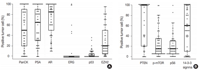
Ductal adenocarcinoma (DAC) of the prostate is an uncommon histologic subtype whose prognostic factors and immunoprofile have not been fully defined. Methods: To define its prognostic factors and immunoprofile, the clinicopathological features, including biochemical recurrence (BCR), of 61 cases of DAC were analyzed. Immunohistochemistry was performed on tissue microarray constructs to assess the expression of prostate cancer-related and mammalian target of rapamycin (mTOR) signaling-related proteins. Results: During the median follow-up period of 19.3 months, BCR occurred in 26 cases (42.6%). DAC demonstrated a wide expression range of prostate cancer-related proteins, including nine cases (14.8%) that were totally negative for pan-cytokeratin (PanCK) immunostaining. The mTOR signaling-related proteins also showed diverse expression. On univariate analysis, BCR was associated with high preoperative serum levels of prostate-specific antigen (PSA), large tumor volume, predominant ductal component, high Gleason score (GS), comedo-necrosis, high tumor stage (pT), lymphovascular invasion, and positive surgical margin. High expressions of phospho-mTOR (p-mTOR) as well as low expressions of PSA, phospho-S6 ribosomal protein (pS6) and PanCK were associated with BCR. On multivariable analysis, GS, pT, and immunohistochemical expressions of PanCK and p-mTOR remained independent prognostic factors for BCR. Conclusions: These results suggest GS, pT, and immunohistochemical expressions of PanCK and p-mTOR as independent prognostic factors for BCR in DAC. Since DAC showed diverse expression of prostate cancer–related proteins, this should be recognized in interpreting the immunoprofile of DAC. The diverse expression of mTOR-related proteins implicates their potential utility as predictive markers for mTOR targeted therapy. -
Citations
Citations to this article as recorded by- True Papillae and Squamous Features in Prostate Carcinoma Represent a Potential Diagnostic Pitfall: Report of 3 Patients
Ioanna-Maria Grypari, Angeliki Pomoni, Dimitra Ganetsou, Olga Kouroukli, Maria Melachrinou, Vasiliki Tzelepi
International Journal of Surgical Pathology.2026;[Epub] CrossRef - Intermediate risk prostate tumors contain lethal subtypes
William L. Harryman, James P. Hinton, Rafael Sainz, Jaime M. C. Gard, John M. Ryniawec, Gregory C. Rogers, Noel A. Warfel, Beatrice S. Knudsen, Raymond B. Nagle, Juan J. Chipollini, Benjamin R. Lee, Belinda L. Sun, Anne E. Cress
Frontiers in Urology.2025;[Epub] CrossRef - High GLUT1 membrane expression and low PSMA membrane expression in Ductal Adenocarcinoma and Intraductal Carcinoma of the prostate
Xingming Wang, Li Zhou, Lin Qi, Ye Zhang, Hongling Yin, Yu Gan, Xiaomei Gao, Yi Cai
Prostate Cancer and Prostatic Diseases.2024; 27(4): 720. CrossRef - Association of Lymphovascular Invasion with Biochemical Recurrence and Adverse Pathological Characteristics of Prostate Cancer: A Systematic Review and Meta-analysis
Jakub Karwacki, Marcel Stodolak, Andrzej Dłubak, Łukasz Nowak, Adam Gurwin, Kamil Kowalczyk, Paweł Kiełb, Nazar Holdun, Wojciech Szlasa, Wojciech Krajewski, Agnieszka Hałoń, Anna Karwacka, Tomasz Szydełko, Bartosz Małkiewicz
European Urology Open Science.2024; 69: 112. CrossRef - Impact of Epithelial Histological Types, Subtypes, and Growth Patterns on Oncological Outcomes for Patients with Nonmetastatic Prostate Cancer Treated with Curative Intent: A Systematic Review
Giancarlo Marra, Geert J.L.H. van Leenders, Fabio Zattoni, Claudia Kesch, Pawel Rajwa, Philip Cornford, Theodorus van der Kwast, Roderick C.N. van den Bergh, Erik Briers, Thomas Van den Broeck, Gert De Meerleer, Maria De Santis, Daniel Eberli, Andrea Faro
European Urology.2023; 84(1): 65. CrossRef - Impact of comedonecrosis on prostate cancer outcome: a systematic review
Kaveri T S Aiyer, Lisa J Kroon, Geert J L H van Leenders
Histopathology.2023; 83(3): 339. CrossRef - Survival after radical prostatectomy vs. radiation therapy in ductal carcinoma of the prostate
Francesco Chierigo, Marco Borghesi, Christoph Würnschimmel, Rocco Simone Flammia, Benedikt Horlemann, Gabriele Sorce, Benedikt Höh, Zhe Tian, Fred Saad, Markus Graefen, Michele Gallucci, Alberto Briganti, Francesco Montorsi, Felix K. H. Chun, Shahrokh F.
International Urology and Nephrology.2022; 54(1): 89. CrossRef - Defining Diagnostic Criteria for Prostatic Ductal Adenocarcinoma at Multiparametric MRI
Weranja K. B. Ranasinghe, Patricia Troncoso, Devaki Shilpa Surasi, Juan José Ibarra Rovira, Priya Bhosale, Janio Szklaruk, Andrea Kokorovic, Xuemei Wang, Mohamed Elsheshtawi, Miao Zhang, Ana Aparicio, Brian F. Chapin, Tharakeswara K. Bathala
Radiology.2022; 303(1): 110. CrossRef - Oncological outcomes of patients with ductal adenocarcinoma of the prostate receiving radical prostatectomy or radiotherapy
Mengzhu Liu, Kun Jin, Shi Qiu, Pengyong Xu, Mingming Zhang, Wufeng Cai, Xiaonan Zheng, Lu Yang, Qiang Wei
Asian Journal of Urology.2021; 8(2): 227. CrossRef - Ductal Prostate Cancers Demonstrate Poor Outcomes with Conventional Therapies
Weranja Ranasinghe, Daniel D. Shapiro, Hyunsoo Hwang, Xuemei Wang, Chad A. Reichard, Mohamed Elsheshtawi, Mary F. Achim, Tharakeswara Bathala, Chad Tang, Ana Aparicio, Shi-Ming Tu, Nora Navone, Timothy C. Thompson, Louis Pisters, Patricia Troncoso, John W
European Urology.2021; 79(2): 298. CrossRef - Optimizing the diagnosis and management of ductal prostate cancer
Weranja Ranasinghe, Daniel D. Shapiro, Miao Zhang, Tharakeswara Bathala, Nora Navone, Timothy C. Thompson, Bradley Broom, Ana Aparicio, Shi-Ming Tu, Chad Tang, John W. Davis, Louis Pisters, Brian F. Chapin
Nature Reviews Urology.2021; 18(6): 337. CrossRef - A first case of ductal adenocarcinoma of the prostate having characteristics of neuroendocrine phenotype with PTEN, RB1 and TP53 alterations
Hiroaki Kobayashi, Takeo Kosaka, Kohei Nakamura, Kazunori Shojo, Hiroshi Hongo, Shuji Mikami, Hiroshi Nishihara, Mototsugu Oya
BMC Medical Genomics.2021;[Epub] CrossRef - Knowing what’s growing: Why ductal and intraductal prostate cancer matter
Mitchell G. Lawrence, Laura H. Porter, David Clouston, Declan G. Murphy, Mark Frydenberg, Renea A. Taylor, Gail P. Risbridger
Science Translational Medicine.2020;[Epub] CrossRef - Integrative Genomic Analysis of Coincident Cancer Foci Implicates CTNNB1 and PTEN Alterations in Ductal Prostate Cancer
Marc Gillard, Justin Lack, Andrea Pontier, Divya Gandla, David Hatcher, Adam G. Sowalsky, Jose Rodriguez-Nieves, Donald Vander Griend, Gladell Paner, David VanderWeele
European Urology Focus.2019; 5(3): 433. CrossRef - Genomic Characterization of Prostatic Ductal Adenocarcinoma Identifies a High Prevalence of DNA Repair Gene Mutations
Michael T. Schweizer, Emmanuel S. Antonarakis, Tarek A. Bismar, Liana B. Guedes, Heather H. Cheng, Maria S. Tretiakova, Funda Vakar-Lopez, Nola Klemfuss, Eric Q. Konnick, Elahe A. Mostaghel, Andrew C. Hsieh, Peter S. Nelson, Evan Y. Yu, R. Bruce Montgomer
JCO Precision Oncology.2019; (3): 1. CrossRef
- True Papillae and Squamous Features in Prostate Carcinoma Represent a Potential Diagnostic Pitfall: Report of 3 Patients
- Acid-Fastness of Histoplasma in Surgical Pathology Practice
- Madhu Rajeshwari, Immaculata Xess, Mehar Chand Sharma, Deepali Jain
- J Pathol Transl Med. 2017;51(5):482-487. Published online September 14, 2017
- DOI: https://doi.org/10.4132/jptm.2017.07.11

- 16,694 View
- 186 Download
- 11 Web of Science
- 10 Crossref
-
Abstract
PDF
- Background
Histoplasmosis (HP) is diagnosed by visualizing intracellular microorganisms in biopsy and/or culture. Periodic-acid Schiff (PAS) and Gomori methenamine silver (GMS) staining methods are routinely used for identification. The acid-fast property of Histoplasma was identified decades ago, but acid-fast staining has not been practiced in current surgical pathology. Awareness of the acid-fast property of Histoplasma, which is due to mycolic acid in the cell wall, is important in distinguishing Histoplasma from other infective microorganisms. Here, we examined acid-fastness in previously diagnosed cases of Histoplasma using the Ziehl-Neelsen (ZN) stain and correlated those findings with other known fungal stains. Methods: All cases diagnosed as HP were retrieved and reviewed along with ZN staining and other fungal stains. We also stained cases diagnosed with Cryptococcus and Leishmania as controls for comparison. Results: A total of 54 patients ranging in age from 11 to 69 years were examined. The most common sites of infection were the skin, adrenal tissue, and respiratory tract. Of the total 43 tissue samples, 20 (46.5%) stained positive with the ZN stain. In viable cases, a significant proportion of microorganisms were positive while necrotic cases showed only rare ZN-positive yeasts. In comparison to PAS and GMS stains, there was a low burden of ZN-positive yeasts. Cryptococcus showed characteristic ZN staining and all cases of Leishmania were negative. Conclusions: Although the morphology of fungal organisms is the foundation of identification, surgical pathologists should be aware of the acid-fast property of fungi, particularly when there is the potential for confusion with other infective organisms. -
Citations
Citations to this article as recorded by-
Development of an
in situ
hybridization assay for the diagnosis of
Mycobacteriaceae
infections of veterinary importance
Agustín Rebollada-Merino, Sean P. McDonough, Francisco A. Uzal, Antonio Rodríguez-Bertos, Rodman G. Getchell, Shotaro Nakagun, Elena A. Demeter
Veterinary Pathology.2026; 63(2): 223. CrossRef - Disseminated histoplasmosis in an HIV/AIDS transgender male‐to‐female with atypical and persistent GI manifestations
Aaron C Yee, Sarah Huang, Ranbir Singh, Dean Rizzi, Naureen Shama, Neil Khoury, Ilan S Weisberg
JGH Open.2024;[Epub] CrossRef - Fungi that are medically relevant to humans and their prospect in a global warming scenario
Bernardo Franco, Naurú Idalia Vargas-Maya, Felipe Padilla-Vaca, Fátima Berenice Ramírez-Montiel, José Ascención Martínez-Álvarez
Academia Molecular Biology and Genomics.2024;[Epub] CrossRef - Emerging Fungal Infections and Cutaneous Manifestations in Immunosuppressed Patients
Jeffrey Alex Varghese, Samantha Guhan, Lida Zheng
Current Dermatology Reports.2023; 12(2): 69. CrossRef - Histoplasma capsulatum var. duboisii: A KwaZulu‐Natal, South Africa public sector perspective
Lerato Claudia Khathali, Gamalenkosi Bonginkosi Nhlonzi, Absalom Mwazha
Journal of Cutaneous Pathology.2022; 49(2): 139. CrossRef - Histologic features of colonic infections
Maria Westerhoff
Der Pathologe.2022; 43(1): 16. CrossRef - Challenge of Ziehl-Neelsen stain for Basidiobolomycosis diagnosis in Indonesia: A unique case report
Metta Octora, Arthur Pohan Kawilarang, Pepy Dwi Endraswari
Annals of Medicine and Surgery.2022; 74: 103278. CrossRef - Pulmonary Histoplasmosis Mimicking Metastatic Lung Cancer: A Case Report
Gion Ruegg, Stefan Zimmerli, Maria Trachsel, Sabina Berezowska, Swantje Engelbrecht, Yonas Martin, Martin Perrig
Diagnostics.2021; 11(2): 328. CrossRef - Cutaneous Histoplasmosis in HIV Seronegative Patients: A Clinicopathological Analysis
Arvind Ahuja, Minakshi Bhardwaj, Poojan Agarwal
Dermatology.2021; 237(6): 934. CrossRef - Challenges in the Diagnosis of Invasive Fungal Infections in Immunocompromised Hosts
Spinello Antinori, Mario Corbellino, Carlo Parravicini
Current Fungal Infection Reports.2018; 12(1): 12. CrossRef
-
Development of an
in situ
hybridization assay for the diagnosis of
Mycobacteriaceae
infections of veterinary importance
- Placental Lesions in Meconium Aspiration Syndrome
- Binnari Kim, Soo-young Oh, Jung-Sun Kim
- J Pathol Transl Med. 2017;51(5):488-498. Published online August 9, 2017
- DOI: https://doi.org/10.4132/jptm.2017.07.20
- 13,700 View
- 226 Download
- 9 Web of Science
- 10 Crossref
-
Abstract
PDF
- Background
Meconium aspiration syndrome (MAS) is defined by respiratory distress requiring supplemental oxygen in a meconium-stained neonate. MAS is clinically subclassified as mild, moderate, and severe according to the oxygen requirement. The aims of this study were to compare the histological findings in the placentas of MAS neonates with those of meconium-stained but non-MAS neonates and to analyze the correlation between the severity of MAS and the grade of its histological parameters. Methods: We collected 160 singleton term placentas from neonates with meconium staining at birth from a tertiary medical center, Seoul, Republic of Korea. We reviewed hematoxylin and eosin sections of tissue samples (full-thickness placental disc, chorioamniotic membranes, and umbilical cord). Results: Funisitis was present more frequently in MAS than in non-MAS (p < .01), of which the stage was correlated with the severity of MAS (p < .001). The histological findings consistent with maternal underperfusion and chronic deciduitis were more frequent in MAS than in non-MAS (p < .05). There was a correlation between the degree of chorionic vascular muscle necrosis and the severity of MAS (p < .05). Conclusions: Our results suggest that fetal inflammatory response evidenced by funisitis occurs prenatally in MAS and that the stage of funisitis and of chorionic vascular muscle necrosis may be a predictive marker of the severity of MAS. -
Citations
Citations to this article as recorded by-
THE EFFECT OF PRECONCEPTION CARE ON PERINATAL OUTCOMES OF THE MOTHER-PLACENTA-FETUS SYSTEM
Y. Podilyakina, L. Stabayeva, D. Kulov, Ye. Kamyshanskiy, Zh. Amirbekova, R. Stundžienė, O. Zhamantayev, K. Nukeshtaeva
Наука и здравоохранение.2026; (6(27)): 67. CrossRef - Impact of meconium-stained amniotic fluid thickness on maternal infectious morbidity: a comprehensive clinical and microbiological analysis
Raneen Abu Shqara, Lior Lowenstein, Maya Frank Wolf
Archives of Gynecology and Obstetrics.2024; 312(1): 59. CrossRef - Machine learning‐based placental clusters and their associations with adverse pregnancy outcomes
Julie M. Petersen, Samantha E. Parker, Kimberly A. Dukes, Jennifer A. Hutcheon, Katherine A. Ahrens, Martha M. Werler
Paediatric and Perinatal Epidemiology.2023; 37(4): 350. CrossRef - The risk of meconium aspiration syndrome (MAS) increases with gestational age at term
Clara Ward, Aaron B. Caughey
The Journal of Maternal-Fetal & Neonatal Medicine.2022; 35(1): 155. CrossRef - Protective placental inflammatory and oxidative stress responses are attenuated in the context of twin pregnancy and chorioamnionitis in assisted reproduction
Hayley R. Price, Nick Pang, Hugh Kim, Michael W. H. Coughtrie, Abby C. Collier
Journal of Assisted Reproduction and Genetics.2022; 39(1): 227. CrossRef - Correlation between Pregnancy Outcome and Placental Pathology in COVID-19 Pregnant Women
Sara A. Al-Rawaf, Enas T. Mousa, Noora M. Kareem, Atif Amin Baig
Infectious Diseases in Obstetrics and Gynecology.2022; 2022: 1. CrossRef - Differential impact of antiretroviral therapy initiated before or during pregnancy on placenta pathology in HIV-positive women
Nadia M. Ikumi, Thokozile R. Malaba, Komala Pillay, Marta C. Cohen, Hlengiwe P. Madlala, Mushi Matjila, Dilly Anumba, Landon Myer, Marie-Louise Newell, Clive M. Gray
AIDS.2021; 35(5): 717. CrossRef - Meconium Aspiration Syndrome: A Narrative Review
Chiara Monfredini, Francesco Cavallin, Paolo Ernesto Villani, Giuseppe Paterlini, Benedetta Allais, Daniele Trevisanuto
Children.2021; 8(3): 230. CrossRef - Isolated acute funisitis in the absence of acute chorioamnionitis: What does it mean?
Tracy B. Grossman, Debra S. Heller, Rebecca N. Baergen
Placenta.2019; 75: 42. CrossRef - Influence of foetal inflammation on the development of meconium aspiration syndrome in term neonates with meconium-stained amniotic fluid
Kyoko Yokoi, Osuke Iwata, Satoru Kobayashi, Kanji Muramatsu, Haruo Goto
PeerJ.2019; 7: e7049. CrossRef
-
THE EFFECT OF PRECONCEPTION CARE ON PERINATAL OUTCOMES OF THE MOTHER-PLACENTA-FETUS SYSTEM
- Intraosseous Hibernoma: A Rare and Unique Intraosseous Lesion
- Boram Song, Hye Jin Ryu, Cheol Lee, Kyung Chul Moon
- J Pathol Transl Med. 2017;51(5):499-504. Published online August 22, 2017
- DOI: https://doi.org/10.4132/jptm.2017.07.28
- 12,168 View
- 139 Download
- 17 Web of Science
- 22 Crossref
-
Abstract
PDF
- Background
Hibernoma is a rare benign tumor of adults that is composed of multivacuolated adipocytes resembling brown fat cells. Hibernoma typically occurs in soft tissue, and intraosseous examples are very rare. Intraosseous hibernomas can radiologically mimic metastatic carcinoma and other tumorous conditions. Methods: To collect the intraosseous hibernomas, we searched the pathologic database and reviewed the hematoxylin and eosin (H&E)–stained slides of bone biopsy samples performed to differentiate radiologically abnormal bone lesions from 2006 to 2016. A total of six intraosseous hibernoma cases were collected, and clinical and radiological information was verified from electronic medical records. H&E slide review and immunohistochemical staining for CD68, pan-cytokeratin, and S-100 protein were performed. Results: Magnetic resonance imaging of intraosseous hibernomas showed low signal intensity with slightly hyperintense foci on T1 and intermediate to high signal intensity on T2 weighted images. Intraosseous hibernomas appeared as heterogeneous sclerotic lesions with trabecular thickening on computed tomography scans and revealed mild hypermetabolism on positron emission tomography scans. Histopathologically, the bone marrow space was replaced by sheets of multivacuolated, foamy adipocytes resembling brown fat cells, without destruction of bone trabeculae. In immunohistochemical analysis, the tumor cells were negative for CD68 and pan-cytokeratin and positive for S-100 protein. Conclusions: Intraosseous hibernoma is very rare. This tumor can be overlooked due to its rarity and resemblance to bone marrow fat. Pathologists need to be aware of this entity to avoid misdiagnosis of this rare lesion. -
Citations
Citations to this article as recorded by- Test yourself answer: 38-year-old female with left hip pain
Ruhaid Khurram, Amar Nitin Kanani, Mohammed Saif Sait, Khamaeel Khaleel Al Lami, Ramanan Rajakulasingam
Skeletal Radiology.2026; 55(6): 1429. CrossRef - Clinical, Radiological, and Pathological Features of Intraosseous Hibernoma: A Systematic Review of Case Reports and Case Series
Jawad Albashri, Ahmed Albashri, Muhannad Alhamrani, Abdulrahman Hassan, Hisham Shamah, Rayan Alhefzi, Najim Z. Alshahrani, Mohammed R. Algethami, Louis-Romée Le Nail, Ramy Samargandi
Current Oncology.2025; 32(10): 535. CrossRef - Imaging of Bone Surface Lesions
Utkarsh Parwal, Allison Khoo, Nicholas G. Rhodes, Patrick G. McEnulty, Eric V. Pang, Jonathan C. Baker, Benjamin E. Northrup, Theodore L. Vander Velde, Mariam A. Malik, Jack W. Jennings, Kelby B. Napier
RadioGraphics.2025;[Epub] CrossRef - Intraosseous hibernoma of the mandible: A case report
Jin-Woo Han
Journal of Korean Dental Association.2025; 63(10): 335. CrossRef - Intraosseous Lipoma of the Maxillary Sinus: First Documented Case in an Asian Patient and Review of the Literature
Eng Seng Yeoh, Tzy Harn Chua, Jacqueline S. G. Hwang, Sathiyamoorthy Selvarajan, Noah B. T. Teo, Kevin Seymour
Case Reports in Dentistry.2025;[Epub] CrossRef - A Rare Case of Large Lateral Chest Wall Hibernoma
Lyubomir Gaydarski, Boycho Landzhov, Ivaylo Kamenov, Julian M Ananiev, Georgi P Georgiev
Cureus.2024;[Epub] CrossRef - Intraosseous hibernoma mimicking sclerotic bone metastasis—a case report
Ali Shaikh, Adil Basha, George Ray, Justin A. Bishop, Avneesh Chhabra
Skeletal Radiology.2024;[Epub] CrossRef - Femoral hibernoma: unique intraosseous tumor
Gökhan Tonkaz, Ertugrul Cakir, Mehmet Tonkaz, Demet Sengul
Wiener klinische Wochenschrift.2024; 136(19-20): 581. CrossRef - Unusual Imaging Findings of Epithelioid Hemangioma: Case Report of Single Intramedullary Sclerotic Bone Lesion
Yun Chul Hwang, Tae Eun Kim, Jae Hyuck Yi
Journal of the Korean Society of Radiology.2024; 85(5): 986. CrossRef - Benign incidental do-not-touch bone lesions
Nuttaya Pattamapaspong, Wilfred CG Peh
The British Journal of Radiology.2023;[Epub] CrossRef - Intraosseous hibernoma: clinicopathologic and imaging analysis of 18 cases
Chiraag N Gangahar, Carina A Dehner, David P Wang, Behrang Amini, Travis Hillen, Christopher O'Conor, Sydney N Jennings, Kathleen Byrnes, Elizabeth A Montgomery, Bogdan A Czerniak, Julia A Bridge, Molly C Schroeder, Jack W Jennings, Wei‐Lien Wang, John S
Histopathology.2023; 83(1): 40. CrossRef - Intraosseous Hibernoma: A Rare Entity in Orthopedics With Peculiar Radiological Features
Ramy Samargandi, Louis-Romée Le Nail, Gonzague de Pinieux, Matthias Tallegas, Elodie Miquelestorena-Standley
Cureus.2023;[Epub] CrossRef - Intraosseous hibernoma of the appendicular skeleton
Salvatore Gitto, Thom Doeleman, Michiel A. J. van de Sande, Kirsten van Langevelde
Skeletal Radiology.2022; 51(6): 1325. CrossRef - Intraosseous hibernoma: Two case reports and a review of the literature
Samantha N. Weiss, Ankit Mohla, Gord Guo Zhu, Christina Gutowski, Tae Won B Kim, Rohan Amin
Radiology Case Reports.2022; 17(7): 2477. CrossRef - Hibernoma of two contiguous vertebrae: uniqueness of a lesion already rare in itself
Donato MASTRANTUONO, Domenico MARTORANO, Guido REGIS, Federica ARABIA, Alessandra LINARI, Federica SANTORO
Journal of Radiological Review.2022;[Epub] CrossRef - Primary extradural tumors of the spinal column
Varun Arvind, Edin Nevzati, Maged Ghaly, Mansoor Nasim, Mazda Farshad, Roman Guggenberger, Daniel Sciubba, Alexander Spiessberger
Journal of Craniovertebral Junction and Spine.2021; 12(4): 336. CrossRef - Spinal Intraosseous Hibernoma: A Case Report and Review of Literature
Mi-Kyung Um, Eugene Lee, Joon Woo Lee, Kyu Sang Lee, Yusuhn Kang, Joong Mo Ahn, Heung Sik Kang
Journal of the Korean Society of Radiology.2020; 81(4): 965. CrossRef - Intraosseous hibernoma: A metastatic mimicker to consider on the differential
Allen Ko, Colin C. Rowell, James B. Vogler, Dmitri E. Samoilov
Radiology Case Reports.2020; 15(12): 2677. CrossRef - A Diagnostic Dilemma of a Subcutaneous Hibernoma: Case Report
Abdullah Saleh AlQattan, Alaa A. Al Abdrabalnabi, Mohammed Abdulrazzaq Al Duhileb, Tarek Ewies, Miral Mashhour, Ahmed Abbas
American Journal of Case Reports.2020;[Epub] CrossRef - Co-expression of MDM2 and CDK4 in transformed human mesenchymal stem cells causes high-grade sarcoma with a dedifferentiated liposarcoma-like morphology
Yu Jin Kim, Mingi Kim, Hyung Kyu Park, Dan Bi Yu, Kyungsoo Jung, Kyoung Song, Yoon-La Choi
Laboratory Investigation.2019; 99(9): 1309. CrossRef - Intraosseous Hibernoma: Five Cases and a Review of the Literature
Francisco A. Myslicki, Andrew E. Rosenberg, Ivan Chaitowitz, Ty K. Subhawong
Journal of Computer Assisted Tomography.2019; 43(5): 793. CrossRef - Hibernoma Mimicking Atypical Lipomatous Tumor
Youssef Al Hmada, Inga-Marie Schaefer, Christopher D.M. Fletcher
American Journal of Surgical Pathology.2018; 42(7): 951. CrossRef
- Test yourself answer: 38-year-old female with left hip pain
Case Studies
- A Rare Case of Aggressive Melanotic Schwannoma Occurred in Spinal Nerve of a 59-Year-Old Male
- Sung-eun Choi, Yoon Jin Cha, Jisup Kim, Hyunseo Cha, Jayeong Seo, Sung-Uk Kuh, Sung-Jun Kim, Se Hoon Kim
- J Pathol Transl Med. 2017;51(5):505-508. Published online April 4, 2017
- DOI: https://doi.org/10.4132/jptm.2017.01.04
- 16,124 View
- 222 Download
- 22 Web of Science
- 18 Crossref
-
Abstract
PDF
- Melanotic schwannoma (MS) is a rare variant of nerve sheath neoplasm that shows ultrastructural and immunophenotypical features of Schwann cells but also has cytoplasmic melanosomes and is reactive for melanocytic markers as well. Unlike conventional schwannoma, which is totally benign, MS has an unpredictable prognosis and is thought to have low-malignant potential. Herein, we present a rare case of recurrent MS in lumbar spine of a 59-year-old male.
-
Citations
Citations to this article as recorded by- The “Pigmented Side” of Nerve Sheaths: Malignant Melanotic Nerve Sheath Tumor
Raduan Ahmed Franca, Rosa Maria Di Crescenzo, Lorenzo Ugga, Rosa Della Monica, Elena D'Avella
International Journal of Surgical Pathology.2025; 33(4): 1068. CrossRef - Case Report: Cutaneous melanocytic schwannoma with concomitant melanocytoma in a canine
Olwam H. Monakali, Nicolize O'Dell, Louise van der Weyden
Wellcome Open Research.2024; 8: 364. CrossRef - Intradural Melanotic Schwannoma of the Sacral Spine: An Illustrated Case Report of Diagnostic Conundrum
Jiunn-Kai Chong, Navneet Kumar Dubey, Wen-Cheng Lo
Reports.2024; 7(3): 56. CrossRef - Rare giant retroperitoneal melanotic schwannoma: a case report and literature review
Pan Chen, Junfeng Cheng, Lin Zhang
Frontiers in Oncology.2024;[Epub] CrossRef - A Rare Case of Melanotic Schwannoma Occurred Intraosseous of Sacrum: A Literature Review
Xiaobo Yan, Keyi Wang, Nong Lin, Xin Huang, YanBiao Fu, Zhaoming Ye
Orthopaedic Surgery.2023; 15(2): 655. CrossRef - Sporadic spinal psammomatous malignant melanotic nerve sheath tumor: A case report and literature review
Giulio Bonomo, Alessandro Gans, Elio Mazzapicchi, Emanuele Rubiu, Paolo Alimonti, Marica Eoli, Rosina Paterra, Bianca Pollo, Guglielmo Iess, Francesco Restelli, Jacopo Falco, Francesco Acerbi, Marco Paolo Schiariti, Paolo Ferroli, Morgan Broggi
Frontiers in Oncology.2023;[Epub] CrossRef - Case Report: Cutaneous melanocytic schwannoma with concomitant melanocytoma in a canine
Olwam H. Monakali, Nicolize O'Dell, Louise van der Weyden
Wellcome Open Research.2023; 8: 364. CrossRef - Fine‐needle aspiration cytology of melanotic schwannoma in the submandibular gland
Yu‐Hua Huang, Ying‐Chou Lu, Hsuan‐Ying Huang, Chien‐Chin Chen
Diagnostic Cytopathology.2021; 49(1): 142. CrossRef - Checkpoint inhibitors and radiotherapy in refractory malignant melanocytic schwannoma with Carney complex: first evidence of efficacy
Jyoti Bajpai, Akhil Kapoor, Rakesh Jalali, Mrinal M Gounder
BMJ Case Reports.2021; 14(5): e240296. CrossRef - 18F-FDG PET/CT imaging for aggressive melanotic schwannoma of the L3 spinal root
Xun-Ze Shen, Wei Wang, Zhou-Ye Luo
Medicine.2021; 100(8): e24803. CrossRef - Hemorrhagic spinal melanotic schwannoma presenting as acute chest pain: A case report and literature review
Dallas J. Soyland, Dylan R. Goehner, Kayla M. Hoerschgen, Troy D. Gust, Shawn M. Vuong
Surgical Neurology International.2021; 12: 164. CrossRef - Retroperitoneal Recurrence of Melanotic Schwannoma on 18F-FDG PET/CT
Xiangliu OuYang, Lichun Zheng, Xiaoming Zhang
Clinical Nuclear Medicine.2021; 46(12): 991. CrossRef - Schwannoma originating from the common iliac artery: a case report
Seung-Myoung Son, Chang Gok Woo
Journal of International Medical Research.2020;[Epub] CrossRef - Intraosseous Melanotic Schwannoma in the Sacrum Mimicking Primary Bone Tumor
Yoshitaka Nagashima, Yusuke Nishimura, Kaoru Eguchi, Takayuki Awaya, Satoshi Yoshikawa, Shoichi Haimoto, Toshihiko Wakabayashi, Masahito Hara
NMC Case Report Journal.2020; 7(3): 107. CrossRef - Extramedullary melanotic schwannoma recurrence in the cervical vertebral arch: a case report and review of the literature
Zongbin Hou, Teng Shi, Guangrun Li, Lin Tian, Xinna Li, Xiaoyang Liu
Journal of International Medical Research.2020;[Epub] CrossRef - Extramedullary malignant melanotic schwannoma of the spine: Case report and an up to date systematic review of the literature
Georgios Solomou, Adikarige Haritha Dulanka Silva, Adrianna Wong, Ute Pohl, Nikolaos Tzerakis
Annals of Medicine and Surgery.2020; 59: 217. CrossRef - Melanotic Schwannoma of the Vagina: A Report of a Very Rare Tumor and Review of the Literature
Kofi Effah, Stefan Seidl, Edith Gorges, Patrick Kafui Akakpo
Case Reports in Obstetrics and Gynecology.2019; 2019: 1. CrossRef - Melanotic Schwannomas Are Rarely Seen Pigmented Tumors with Unpredictable Prognosis and Challenging Diagnosis
Elif Keskin, Sumeyye Ekmekci, Ozgur Oztekin, Gulden Diniz
Case Reports in Pathology.2017; 2017: 1. CrossRef
- The “Pigmented Side” of Nerve Sheaths: Malignant Melanotic Nerve Sheath Tumor
- Cytologic Characteristics of Thymic Adenocarcinoma with Enteric Differentiation: A Study of Four Fine-Needle Aspiration Specimens
- Ah-Young Kwon, Joungho Han, Hae-yon Cho, Seokhwi Kim, Heejin Bang, Jiyeon Hyeon
- J Pathol Transl Med. 2017;51(5):509-512. Published online August 4, 2017
- DOI: https://doi.org/10.4132/jptm.2017.03.22
- 9,942 View
- 126 Download
- 6 Web of Science
- 8 Crossref
-
Abstract
PDF
- Thymic adenocarcinoma is extremely rare. Although its histologic features have been occasionally reported, a lack of description of the cytologic features has hampered the prompt and accurate diagnosis of this condition. Herein, we describe the cytologic findings and histology of four aspiration cytology specimens of thymic adenocarcinoma. The specimens were obtained from primary tumors, metastatic lymph nodes, and pericardial effusions. All four specimens showed three-dimensional glandular clusters with a loss of polarity and nuclear overlapping. One specimen had extensive extracellular mucinous material. Three specimens contained tumor cells with intracytoplasmic vacuoles. While the specimen with extracellular mucin showed relatively mild cytologic atypia, other specimens exhibited more atypical cytologic changes: irregular nuclear membranes, a coarse chromatin pattern, and prominent nucleoli. The cytologic features were correlated with the histologic features in each case of enteric type thymic adenocarcinoma. The differential diagnosis included other thymic carcinomas, yolk sac tumors, and metastatic adenocarcinoma from the lung or colorectum.
-
Citations
Citations to this article as recorded by- Primary thymic adenocarcinoma with signet ring cell-like features: A case report and literature review
Ya Jiang, Ziran Gao, Wenmang Xu, Yuanyuan Wang
Asian Journal of Surgery.2026; 49(1): 589. CrossRef - Case report: Primary adenocarcinoma NOS of the thymus and cytological features
Jonathan Willner, Osvaldo Hernandez, Lea Azour, Andre L. Moreira
Diagnostic Cytopathology.2023;[Epub] CrossRef - Systemic chemotherapy for unresectable or recurrent primary thymic adenocarcinoma of enteric type
Xiaofang Gao
International Cancer Conference Journal.2022; 12(1): 46. CrossRef - Thymic adenocarcinoma accompanied by type A thymoma and pulmonary minimally invasive adenocarcinoma and harboring distinct gene alterations
Yi-Wen Zheng, Lin-Lin Bai, Gui-Yang Jiang, Xu-Yong Lin, Yang Liu, Hong-Tao Xu
Medicine.2021; 100(15): e25254. CrossRef - A case report: primary thymic adenocarcinoma with enteric differentiation
Yuuki Kou, Hirokazu Tanaka, Nobuhisa Yamazaki, Hiroyoshi Watanabe, Makoto Sonobe
The Journal of the Japanese Association for Chest Surgery.2020; 34(2): 107. CrossRef - Primary thymic adenocarcinoma with an aggressive clinical course: An autopsy case showing signet ring cell‐like features
Ayako Shiono, Takashi Fujino, Kyoichi Kaira, Tomomi Kato, Masanori Yasuda, Kunihiko Kobayashi, Hiroshi Kagamu
Thoracic Cancer.2020; 11(12): 3609. CrossRef - Primary Thymic Signet Ring Cell Adenocarcinoma: A Currently Unrecognized Variant
Richard Benedict Supan Roxas, Marie Christine Fajatin Bernardo, Araceli Pacis Jacoba, Janet Lim-Dy, Anarose Cariaga Alvarado, Jasna Metovic, Laura Annaratone, Mauro Papotti
International Journal of Surgical Pathology.2019; 27(3): 315. CrossRef - Disseminated and massive tumor burden in a case of primary thymic mucinous adenocarcinoma
Hui-Wen Liu, Chih-Yi Liu, Yi-Chen Yeh
Journal of Cancer Research and Practice.2019; 6(3): 151. CrossRef
- Primary thymic adenocarcinoma with signet ring cell-like features: A case report and literature review
Brief Case Report
- Iatrogenic Gastric Pseudolipomatosis during Endoscopic Submucosal Dissection
- Sang Wook Kim, Woo Sung Moon
- J Pathol Transl Med. 2017;51(5):513-515. Published online July 31, 2017
- DOI: https://doi.org/10.4132/jptm.2017.04.25
- 8,815 View
- 101 Download
- 3 Web of Science
- 3 Crossref
-
PDF
-
Citations
Citations to this article as recorded by- A Rare Case Report of Colonic Pseudolipomatosis with Distinct Endoscopic and Histological Features
Qi Zhao, Hai-Tao Yu, Qun-Ying Wang, Wei Zhang, Wen-Zhu Dong
Case Reports in Gastroenterology.2025; 19(1): 127. CrossRef - Endoscope disinfectant-induced colonic pseudolipomatosis: case series of a rare condition
Charfeddine Baccouche, Myriam Ayari, Imen Abdelaali, Amen Dhaoui, Taieb Jomni, Mohamed Hedi Douggui
Future Science OA.2024;[Epub] CrossRef - Unusual mucosal lesion: A case of rectal pseudolipomatosis in a 60-year-old patient
Boubacar Efared, Balandougou Sylla, Nawal Hammas, Hinde El Fatemi, Laila Chbani
SAGE Open Medical Case Reports.2019;[Epub] CrossRef
- A Rare Case Report of Colonic Pseudolipomatosis with Distinct Endoscopic and Histological Features
 First
First Prev
Prev


