 E-submission
Search
- Page Path
- HOME > Search
Case Study
- Chronic lymphocytic leukemia and concurrent seminoma in the same testis
- Kosuke Miyai, Fumihisa Kumazawa, Kimiya Sato, Hitoshi Tsuda
- J Pathol Transl Med. 2022;56(1):48-52. Published online October 22, 2021
- DOI: https://doi.org/10.4132/jptm.2021.09.10

- 6,456 View
- 164 Download
-
 Abstract
 PDF
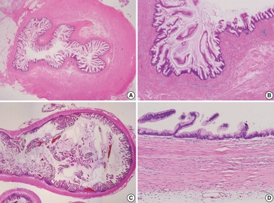
PDF - A 59-year-old man presented with a painless testicular mass and underwent a radical orchiectomy. The resected specimen showed a 5-cm-sized, white-yellow and homogenous solid mass in the testicular parenchyma. Histologically, the central part of the tumor exhibited typical features of seminoma. The peripheral part of the tumor exhibited diffuse infiltration of small, monotonous lymphoid cells involving the tunica albuginea. The monotonous lymphoid cells were immunoreactive for CD20, CD79a, CD5, and CD23, and negative for CD3, CD10, and cyclin D1. Kappa light chain restriction was detected on flow cytometry using the resected specimen. Considering the circulating lymphoid cell count of >5.0×10³/µL, we diagnosed the peripheral component of the tumor as an infiltration of chronic lymphocytic leukemia. This extremely rare combination of seminoma and lymphoid neoplasm should be considered in the differential diagnosis of classic seminoma with extensive lymphoid reaction in tumors arising in elderly patients.
Review
- Standardization of the pathologic diagnosis of appendiceal mucinous neoplasms
- Dong-Wook Kang, Baek-hui Kim, Joon Mee Kim, Jihun Kim, Hee Jin Chang, Mee Soo Chang, Jin-Hee Sohn, Mee-Yon Cho, So-Young Jin, Hee Kyung Chang, Hye Seung Han, Jung Yeon Kim, Hee Sung Kim, Do Youn Park, Ha Young Park, So Jeong Lee, Wonae Lee, Hye Seung Lee, Yoo Na Kang, Younghee Choi
- J Pathol Transl Med. 2021;55(4):247-264. Published online July 8, 2021
- DOI: https://doi.org/10.4132/jptm.2021.05.28
- 25,413 View
- 1,188 Download
- 21 Web of Science
- 20 Crossref
-
Abstract
PDF
 Supplementary Material
 - Although the understanding of appendiceal mucinous neoplasms (AMNs) and their relationship with disseminated peritoneal mucinous disease have advanced, the diagnosis, classification, and treatment of AMNs are still confusing for pathologists and clinicians. The Gastrointestinal Pathology Study Group of the Korean Society of Pathologists (GPSG-KSP) proposed a multicenter study and held a workshop for the “Standardization of the Pathologic Diagnosis of the Appendiceal Mucinous Neoplasm” to overcome the controversy and potential conflicts. The present article is focused on the diagnostic criteria, terminologies, tumor grading, pathologic staging, biologic behavior, treatment, and prognosis of AMNs and disseminated peritoneal mucinous disease. In addition, GPSG-KSP proposes a checklist of standard data elements of appendiceal epithelial neoplasms to standardize pathologic diagnosis. We hope the present article will provide pathologists with updated knowledge on how to handle and diagnose AMNs and disseminated peritoneal mucinous disease.
-
Citations
Citations to this article as recorded by
- The clinical and pathological features of low-grade appendiceal mucinous neoplasm (LAMN)
Omar Hamdy, Gehad A. Saleh, Mona Hany Emile, Ahmed Elhadidy, Ahmed Ibrahim, Ola Elsayed, Ahmed Reda, Yasser Sharaf, Merna M. Hegazi, Osama Bahy, Mahmoud Soliman
Discover Oncology.2026;[Epub] CrossRef - Path beyond the blind end-unravel the imaging spectrum of appendiceal pathologies
Siva K. P. Konduru, Aastha Bhatt, Anil K. Dasyam, Subhash Venigalla, Venkata S. Katabathina, Sriram Jaganathan
Abdominal Radiology.2026;[Epub] CrossRef - When the appendix hides: the radiologist’s guide to appendix variants and mimics
Ana Helena Pavan Amaral, Carolinna Bonetto Nicolau Bignotto, Eduardo Alves Ferreira Martins, Guilherme Baptistella de Napoli, Marco Alexandre Mendes Rodstein, Mariana Marum de Campos, Daniel Lahan-Martins
Abdominal Radiology.2026;[Epub] CrossRef - Intrasplenic metastasis of appendiceal low-grade mucinous neoplasm – A case report and review of the literature
P. Meister, J. Rawitzer, M. Reschke, H.A. Baba, U. Neumann, M. Kaths
Current Problems in Cancer: Case Reports.2025; 18: 100364. CrossRef - Complete laparoscopic resection of giant appendiceal mucinous neoplasm, case report, and literature review
Shatha Awad Althobaiti, Rayan Z. Makeen, Abrar J. Filfilan, Ahmed Abdulaziz Hawsawi
Saudi Surgical Journal.2025; 13(1): 35. CrossRef - Survival Outcomes and Prognostic Factors in Metastatic Unresectable Appendiceal Adenocarcinoma Treated with Palliative Systemic Chemotherapy: A 10-Year Retrospective Analysis from Australia
Jirapat Wonglhow, Hui-Li Wong, Michael Michael, Alexander Heriot, Glen Guerra, Catherine Mitchell, Jeanne Tie
Cancers.2025; 17(20): 3297. CrossRef - Lower Gastrointestinal Bleeding Secondary to Appendiceal Mucinous Neoplasm: A Report of Two Cases and a Review of the Literature
Jesús Omar Soto Llanes, Samanta Kin Dosal Limón, Ana Jimena Iberri Jaime, Mario Zambrano Lara, Billy Jiménez Bobadilla
Cureus.2024;[Epub] CrossRef - Predicting Survival in Mucinous Adenocarcinoma of the Appendix: Demographics, Disease Presentation, and Treatment Methodology
Paul H. McClelland, Stephanie N. Gregory, Shirley K. Nah, Jonathan M. Hernandez, Jeremy L. Davis, Andrew M. Blakely
Annals of Surgical Oncology.2024; 31(9): 6237. CrossRef - Histoséminaire biopsies péritonéales tumorales. Néoplasies mucineuses appendiculaires
Peggy Dartigues
Annales de Pathologie.2024; 44(4): 274. CrossRef - Histoséminaire biopsies péritonéales tumorales. Cas no 2
Peggy Dartigues
Annales de Pathologie.2024; 44(4): 245. CrossRef - A Case of Low-Grade Appendiceal Mucinous Neoplasm: The Role of Preoperative Imaging and Surgical Technique in Achieving Favorable Outcomes
Daniel A Meza-Martinez, Yeudiel Suro Santos, Samantha J Andrade-Ordoñez, Julio A Palomino-Payan, Brando J Fematt-Rodriguez
Cureus.2024;[Epub] CrossRef - Incidental Appendiceal Mucinous Neoplasm Found During Appendectomy in a 15-Year-Old Patient: A Case Report
Fernando Aguilar-Ruiz, Kevin Joseph Fuentes-Calvo, Sara Fernanda Arechavala-Lopez, Irving Fuentes-Calvo, Luis F Arias-Ruiz
Cureus.2024;[Epub] CrossRef - Uncovering the Hidden Threat: Ileocolic Intussusception in an Adult With Appendicular Tumor
Mrunal Panchal, Shishir Kumar, Khushboo Jha, Kaushik Saha, Abhijit Kundu
Cureus.2024;[Epub] CrossRef - Low-Grade Appendiceal Mucinous Neoplasm vs. Appendiceal Diverticulum: Distinction with Histomorphologic Features
Cevriye Cansiz Ersöz, Siyar Ersöz, Berna Savas, Arzu Ensari
Gastrointestinal Disorders.2024; 6(4): 905. CrossRef - Appendiceal perforation secondary to endometriosis with intestinal metaplasia: A case report
Minghua Wang, Jing Liu, Boxin Hu, Simin Wang, Ping Xie, Ping Li
Experimental and Therapeutic Medicine.2023;[Epub] CrossRef - Primary and secondary tumors of the peritoneum: key imaging features and differential diagnosis with surgical and pathological correlation
Javier Miguez González, Francesc Calaf Forn, Laura Pelegrí Martínez, Pilar Lozano Arranz, Rafael Oliveira Caiafa, Jordi Català Forteza, Lina Maria Palacio Arteaga, Ferrán Losa Gaspà, Isabel Ramos Bernadó, Pedro Barrios Sánchez, Juan Ramón Ayuso Colella
Insights into Imaging.2023;[Epub] CrossRef - Muzinöse Tumoren des Peritoneums
Anne Kristin Fischer, Andrea Tannapfel, Alexander Quaas
Die Chirurgie.2023; 94(10): 823. CrossRef - Landscape of Genetic Mutations in Appendiceal Cancers
Marian Constantin, Cristina Mătanie, Livia Petrescu, Alexandra Bolocan, Octavian Andronic, Coralia Bleotu, Mihaela Magdalena Mitache, Sorin Tudorache, Corneliu Ovidiu Vrancianu
Cancers.2023; 15(14): 3591. CrossRef - Delivery of an Incidental Appendiceal Mucinous Neoplasm
Madison Bowles, Jessica Y Ng, Hajir Nabi
Cureus.2022;[Epub] CrossRef - Unearthing novel fusions as therapeutic targets in solid tumors using targeted RNA sequencing
Sungbin An, Hyun Hee Koh, Eun Sol Chang, Juyoung Choi, Ji-Young Song, Mi-Sook Lee, Yoon-La Choi
Frontiers in Oncology.2022;[Epub] CrossRef
- The clinical and pathological features of low-grade appendiceal mucinous neoplasm (LAMN)
Case Study
- Concurrent Anti-glomerular Basement Membrane Nephritis and IgA Nephropathy
- Kwang-Sun Suh, Song-Yi Choi, Go Eun Bae, Dae Eun Choi, Min-kyung Yeo
- J Pathol Transl Med. 2019;53(6):399-402. Published online September 16, 2019
- DOI: https://doi.org/10.4132/jptm.2019.08.05

- 9,048 View
- 186 Download
- 15 Web of Science
- 16 Crossref
-
Abstract
PDFSupplementary Material
- Anti–glomerular basement membrane (GBM) nephritis is characterized by circulating anti-GBM antibodies and crescentic glomerulonephritis (GN) with deposition of IgG along the GBM. In a limited number of cases, glomerular immune complexes have been identified in anti-GBM nephritis. A 38-year-old female presented azotemia, hematuria, and proteinuria without any pulmonary symptoms. A renal biopsy showed crescentic GN with linear IgG deposition along the GBM and mesangial IgA deposition. The patient was diagnosed as concurrent anti-GBM nephritis and IgA nephropathy. Therapies with pulse methylprednisolone and cyclophosphamide administration were effective. Concurrent cases of both anti-GBM nephritis and IgA nephropathy are rare among cases of anti-GBM diseases with deposition of immune complexes. This rare case of concurrent anti-GBM nephritis and IgA nephropathy with literature review is noteworthy.
-
Citations
Citations to this article as recorded by- Correlation of foot process effacement with clinicopathological features and short-term prognosis in IgA nephropathy
Yu Wang, Yang Yang, Xiaoli Wen, Lu Yi, Fang Zeng, Baoqin Zhou, Lijuan Wang, Wenjun Yan, Daijin Ren, Shizhang Xu, Yebei Li, Dehui Liu, Kaiping Luo, Hong Jiang, Gaosi Xu
Annals of Medicine.2026;[Epub] CrossRef - Immunological overlap stratification in anti-GBM disease: prognostic differences and serological correlations—a single-center retrospective cohort study
Yujun Qian, Yuyou Ye, Qian Zhou, Suyan Duan, Chengning Zhang, Yifei Ge, Yanggang Yuan, Changying Xing, Huijuan Mao, Bo Zhang
Frontiers in Immunology.2026;[Epub] CrossRef - A case report of concomitant anti-glomerular basement membrane disease, IgA nephropathy, and membranous nephropathy
Jiajia Zheng, Xiurong Yu, Xiaochun Lin, Hui Guo
BMC Nephrology.2026;[Epub] CrossRef - The importance of the degree of foot process effacement in evaluating the prognosis of IgA nephropathy
Li Gao, Xuan Zhang, Dongrong Yu, Hong Zhu, Qin Zhu
International Urology and Nephrology.2025; 57(10): 3417. CrossRef - Coexistence of anti-glomerular basement membrane disease and IgA nephropathy: an illustrative case and comprehensive literature review
Zewei Chen, Dechao Xu, Fangzheng Cui, Huihui Hou, Zhiguo Mao, Xiang Gao
Renal Failure.2024;[Epub] CrossRef - Clinical features and prognosis of patients with anti-GBM disease combined with mesangial IgA deposition
Wei Ning, Ya-fei Zhao, Ya-ru Liu, Yuan-yuan Qi, Zhan-zheng Zhao
Frontiers in Immunology.2024;[Epub] CrossRef - A Case of Concurrent Anti-Glomerular Basement Membrane Antibody Disease and Immunoglobulin A Nephropathy
Su In Kim, Sung Sun Kim, Chang Seong Kim, Seong Kwon Ma, Soo Wan Kim, Hong Sang Choi
The Korean Journal of Medicine.2024; 99(6): 322. CrossRef - Anti-glomerular basement membrane vasculitis
Claudio Ponticelli, Marta Calatroni, Gabriella Moroni
Autoimmunity Reviews.2023; 22(1): 103212. CrossRef - High-frequency plasma exchange therapy for immunocompromised, type I crescentic glomerulonephritis complicated with IgA nephropathy: A case report and literature review
Huihui Chen, Jingjing Jin, Mei Juan Cheng, Lei He, Wei Zhou, Liping Guo, Zhe Zhe Niu, Xiang Nan Liang, Rong Fang Zhu, Yaling Bai, Jin Sheng Xu
Medicine.2023; 102(3): e32698. CrossRef - Clinical and immunological characteristics of patients with combined anti-glomerular basement membrane disease and IgA nephropathy
Cong-rong Shen, Xiao-yu Jia, Zhao Cui, Xiao-juan Yu, Ming-hui Zhao
Clinical Kidney Journal.2023; 16(9): 1480. CrossRef - Anti-glomerular basement membrane disease with IgA nephropathy: A case report
Chuan Guo, Ming Ye, Shen Li, Ting-Ting Zhu, Xiang-Rong Rao
World Journal of Clinical Cases.2022; 10(12): 3916. CrossRef - Case Report: Coexistence of Anti-Glomerular Basement Membrane Disease, Membranous Nephropathy, and IgA Nephropathy in a Female PatientWith Preserved Renal Function
Wei Qu, Nan Liu, Tianhua Xu, Binyao Tian, Meng Wang, Yanqiu Li, Jianfei Ma, Li Yao
Frontiers in Pharmacology.2022;[Epub] CrossRef - Great prognosis of concurrent anti-GBM disease and IgA nephropathy in a young woman: A case report
Fu Shaojie, Su Sensen, Huang Jingda, Wang Luyu, Zhang Fei, Yu Jinyu, Xu Zhonggao, Wu Hao
Medicine.2022; 101(37): e30686. CrossRef - Serodiagnosis of Anti-glomerular Basement Membrane Disease Using a Newly Developed Chemiluminescence Immunoassay
Alexander Kühnl, Lea Hartwig, Cornelia Dähnrich, Wolfgang Schlumberger
Frontiers in Medicine.2022;[Epub] CrossRef - PATHOLOGY AND RENAL OUTCOME OF THREE UNCOMMON FACES OF CRESCENTRIC GLOMERULONEPHRITIS
Keya Basu, Dipankar Sircar, Manimoy Bandopadhyay
INDIAN JOURNAL OF APPLIED RESEARCH.2021; : 7. CrossRef - Pneumocystis pneumonia secondary to intensive immunosuppression treatment for anti-GBM disease complicated with IgA nephropathy
Manyu Zhang, Dingwei Yang, Weixiu Wang, Fuhao Zhao, Xiaoxiao Zhang, Xue Li
Medicine.2021; 100(45): e27728. CrossRef
- Correlation of foot process effacement with clinicopathological features and short-term prognosis in IgA nephropathy
Original Article
- Current Status of Pathologic Examinations in Korea, 2011–2015, Based on the Health Insurance Review and Assessment Service Dataset
- Sun-ju Byeon
- J Pathol Transl Med. 2017;51(2):137-147. Published online February 22, 2017
- DOI: https://doi.org/10.4132/jptm.2016.12.30
- 9,093 View
- 96 Download
- 1 Web of Science
- 1 Crossref
-
Abstract
PDFSupplementary Material
- Background
Pathologic examinations play an important role in medical services. Until recently, the overall status of pathologic examinations in Korea has not been identified. I conducted a nationwide survey of pathologic examination status using the insurance reimbursements (IRs) dataset from the Health Insurance Review and Assessment Service (HIRA). The aims of this study were to estimate current pathologic examination status in Korea and to provide information for future resource arrangement in the pathology area. Methods: I asked HIRA to provide data on IR requests, including pathologic examinations from 2011 to 2015. Pathologic examination status was investigated according to the following categories: annual statistics, requesting department, type of medical institution, administrative district, and location at which pathologic examinations were performed. Results: Histologic mapping, immunohistochemistry, and cervicovaginal examinations have increased in the last 5 years. Internal medicine, general surgery, obstetrics/gynecology, and urology were the most common medical departments requesting pathologic examinations. The majority of pathologic examinations were frequently performed in tertiary hospitals. About 60.3% of pathologic examinations were requested in medical institutions located in Seoul, Gyeonggi-do, and Busan. More than half of the biopsies and aspiration cytologic examinations were performed using outside services. The mean period between IR requests and 99 percentile IR request completion inspections was 6.2 months. Conclusions: This survey was based on the HIRA dataset, which is one of the largest medical datasets in Korea. The trends of some pathologic examinations were reflected in the policies and needs for detailed diagnosis. The numbers and proportions of pathologic examinations were correlated with the population and medical institutions of the area, as well as patient preference. These data will be helpful for future resource arrangement in the pathology area. -
Citations
Citations to this article as recorded by- Validation of the pathological prognostic staging system proposed in the revised eighth edition of the AJCC staging manual in different molecular subtypes of breast cancer
Nuri Jang, Jung Eun Choi, Su Hwan Kang, Young Kyung Bae
Virchows Archiv.2019; 474(2): 193. CrossRef
- Validation of the pathological prognostic staging system proposed in the revised eighth edition of the AJCC staging manual in different molecular subtypes of breast cancer
Case Study
- Thymoma and Synchronous Primary Mediastinal Seminomas with Florid Follicular Lymphoid Hyperplasia in the Anterior Mediastinum: A Case Report and Review of the Literature
- Hyang-im Lee, In-seok Jang, Kyung Nyeo Jeon, Gyung Hyuck Ko, Jong Sil Lee, Dong Chul Kim, Dae Hyun Song, Jeong-Hee Lee
- J Pathol Transl Med. 2017;51(2):165-170. Published online February 2, 2017
- DOI: https://doi.org/10.4132/jptm.2016.08.24
- 13,237 View
- 144 Download
- 7 Web of Science
- 5 Crossref
-
Abstract
PDF
- Thymoma is the most common neoplasm of the anterior mediastinum and has malignant potential. Germ cell tumors (GCTs) found in the anterior mediastinum are usually benign, and malignant GCTs, such as seminomas, are rare. Histologically, mediastinal seminoma is indistinguishable from testicular seminoma except for site-associated morphological features such as lymphoid follicular hyperplasia. Therefore, excluding metastasis is very important. Recently, we treated a young adult patient with multiple thymic masses that occurred simultaneously. The patient underwent a thymectomy for the removal of the mediastinal masses, one of which was diagnosed as type B2 invasive thymoma, and two of which were diagnosed as primary mediastinal seminomas with massive follicular hyperplasia. The patient received adjuvant chemotherapy after surgical resection. To our knowledge, this is the first description of a thymoma and a mediastinal seminoma occurring simultaneously in the thymus. We present this case along with a literature review.
-
Citations
Citations to this article as recorded by- Primary germ cell tumours of the mediastinum: A review with emphasis on diagnostic challenges
Alexander Fichtner, Alexander Marx, Philipp Ströbel, Felix Bremmer
Histopathology.2024; 84(1): 216. CrossRef - Combined type A thymoma and yolk sac tumour of the mediastinum
Dong Sheng, Yu-Chen Han
Pathology.2024; 56(6): 927. CrossRef - Combined Thymic Epithelial Neoplasms – a Review
Annikka Weissferdt
International Journal of Surgical Pathology.2023; 31(6): 917. CrossRef - Primary mediastinal seminoma presenting with paraneoplastic anti-Hu encephalitis: a case report and literature review
Chelsey M. Williams, Derek B. Allison, Adam B. Coleman, Roshmita Bardhan, Jordan D. Miller, Zin W. Myint
Frontiers in Oncology.2023;[Epub] CrossRef - Primary mediastinal seminoma with florid follicular lymphoid hyperplasia: a case report and review of the literature
Charlotte Holmes, Peh Sun Loo, Sion Barnard
Diagnostic Pathology.2021;[Epub] CrossRef
- Primary germ cell tumours of the mediastinum: A review with emphasis on diagnostic challenges
Review
- Dysembryoplastic Neuroepithelial Tumors
- Yeon-Lim Suh
- J Pathol Transl Med. 2015;49(6):438-449. Published online October 23, 2015
- DOI: https://doi.org/10.4132/jptm.2015.10.05
- 20,525 View
- 305 Download
- 29 Web of Science
- 35 Crossref
-
Abstract
PDF
- Dysembryoplastic neuroepithelial tumor (DNT) is a benign glioneuronal neoplasm that most commonly occurs in children and young adults and may present with medically intractable, chronic seizures. Radiologically, this tumor is characterized by a cortical topography and lack of mass effect or perilesional edema. Partial complex seizures are the most common presentation. Three histologic subtypes of DNTs have been described. Histologically, the recognition of a unique, specific glioneuronal element in brain tumor samples from patients with medically intractable, chronic epilepsy serves as a diagnostic feature for complex or simple DNT types. However, nonspecific DNT has diagnostic difficulty because its histology is indistinguishable from conventional gliomas and because a specific glioneuronal element and/or multinodularity are absent. This review will focus on the clinical, radiographic, histopathological, and immunohistochemical features as well as the molecular genetics of all three variants of DNTs. The histological and cytological differential diagnoses for this lesion, especially the nonspecific variant, will be discussed.
-
Citations
Citations to this article as recorded by- A Case of Dysembryoplastic Neuroepithelial Tumor in an HIV-Positive Adult: Diagnostic Lessons for Clinicians
Mariana Lobo, Susana Viana, Andreia Sá Lima, Isabel Monteiro, Carolina M Cerqueira, Frederico Duarte, Luís M Ribeiro, Sara Camões
Cureus.2026;[Epub] CrossRef - Magnetic resonance imaging findings of dysembryoplastic neuroepithelial tumors and low-grade astrocytomas
Kai-Wei Yu, Shih-Chieh Lin, Hsin-Hung Chen, Chia-Hung Wu, Wei-An Tai, Chung-Han Yang, Te-Ming Lin, Feng-Chi Chang
Journal of the Chinese Medical Association.2026; 89(3): 228. CrossRef - Imaging diagnosis of cystic intraparenchymal brain neoplasms
Sonoko Oshima, Yasutaka Fushimi, Sachi Okuchi, Satoshi Nakajima, Akihiko Sakata, Takayuki Yamamoto, Yuji Nakamoto, Noriko Salamon
Japanese Journal of Radiology.2026;[Epub] CrossRef - Histopathological and molecular heterogeneity of dysembryoplastic neuroepithelial tumors
Yuxiu Wang, Sarra Belakhoua, Yiying Yang, Jonathan Serrano, Craig Horbinski, Daniel R Boué, John C DeWitt, Benjamin Liechty, Declan McGuone, Qinwen Mao, Olga Krasnozhen-Ratush, Stephen Yip, Christopher Dunham, Melissa Umphlett, Seema Shroff, Matija Snuder
Journal of Neuropathology & Experimental Neurology.2026;[Epub] CrossRef - An Imaging Review of Common Pediatric Brain Tumors
Joseph Yang, Brandon Collins, Matthew Beniuk, Alexandra Hodder, Dani Bahnam, Angela Pickles
Roentgen Ray Review.2025;[Epub] CrossRef - Ruptured intratumoral arteriovenous malformation in a patient with dysembryoplastic neuroepithelial tumor: A case report
Takashi Aoka, Masaaki Nishimoto, Hideki Ogiwara
Surgical Neurology International.2025; 16: 375. CrossRef - Pediatric Neuroglial Tumors: A Review of Ependymoma and Dysembryoplastic Neuroepithelial Tumor
Melissa Arfuso, Sandeepkumar Kuril, Harshal Shah, Derek Hanson
Pediatric Neurology.2024; 156: 139. CrossRef - From bedside to bench: New insights in epilepsy‐associated tumors based on recent classification updates and animal models on brain tumor networks
Silvia Cases‐Cunillera, Lea L. Friker, Philipp Müller, Albert J. Becker, Gerrit H. Gielen
Molecular Oncology.2024; 18(12): 2951. CrossRef - Imaging of pediatric glioneuronal and neuronal tumors
Vivek Pai, Suzanne Laughlin, Birgit Ertl-Wagner
Child's Nervous System.2024; 40(10): 3007. CrossRef - Dysembryoplastic Neuroepithelial Tumor: A Case Report of A Benign Intracranial Lesion Masquerading as Seizure Disorder
Garima S Agarwal, Anil K Agrawal, Daksh Singhal, Jayashree Bhawani
Cureus.2024;[Epub] CrossRef - Super T2-FLAIR mismatch sign: a prognostic imaging biomarker for non-enhancing astrocytoma, IDH-mutant
Iori Ozono, Shumpei Onishi, Ushio Yonezawa, Akira Taguchi, Novita Ikbar Khairunnisa, Vishwa Jeet Amatya, Fumiyuki Yamasaki, Yukio Takeshima, Nobutaka Horie
Journal of Neuro-Oncology.2024; 169(3): 571. CrossRef - Genotype-relevant neuroimaging features in low-grade epilepsy-associated tumors
Keiya Iijima, Hiroyuki Fujii, Fumio Suzuki, Kumiko Murayama, Yu-ichi Goto, Yuko Saito, Terunori Sano, Hiroyoshi Suzuki, Hajime Miyata, Yukio Kimura, Takuma Nakashima, Hiromichi Suzuki, Masaki Iwasaki, Noriko Sato
Frontiers in Neurology.2024;[Epub] CrossRef - Extra-temporal pediatric low-grade gliomas and epilepsy
José Hinojosa, Victoria Becerra, Santiago Candela-Cantó, Mariana Alamar, Diego Culebras, Carlos Valencia, Carlos Valera, Jordi Rumiá, Jordi Muchart, Javier Aparicio
Child's Nervous System.2024; 40(10): 3309. CrossRef - Atypical Presentation of Dysembryoplastic Neuroepithelial Tumor
Varis S. Khalilov, Aleksey N. Kislyakov, Natalia A. Medvedeva, Natalia S. Serova
Annals of Clinical and Experimental Neurology.2024; 18(3): 109. CrossRef - Unusual low-grade neuroepithelial tumour in a child
Leia Salongo, Ali Nael, Pournima Navalkele, John Ross Crawford
BMJ Case Reports.2024; 17(10): e262692. CrossRef - Glioneuronal and Neuronal Tumors: Who? When? Where? An Update Based on the 2021 World
Health Organization Classification
A.S. Ayres, G.A. Bandeira, S.F. Ferraciolli, J.T. Takahashi, R.A. Moreno, L.F. de Souza Godoy, Y.R. Casal, L.G.C.A. de Lima, F.P. Frasseto, L.T. Lucato
Neurographics.2023; 13(1): 1. CrossRef - Biological functions of the Olig gene family in brain cancer and therapeutic targeting
Jenny I. Szu, Igor F. Tsigelny, Alexander Wojcinski, Santosh Kesari
Frontiers in Neuroscience.2023;[Epub] CrossRef - Aspekte der Bildgebung des Hippokampus
Isabela S. Alves, Artur M. N. Coutinho, Ana Vieira, Bruno P. Rocha, Ula L. Passos, Vinicius T. Gonçalves, Paulo D. S. Silva, Malia X. Zhan, Paula C. Pinho, Daniel S. Delgado, Marcos F. L. Docema, Hae W. Lee, Bruno A. Policeni, Claudia C. Leite, Maria G. M
Neuroradiologie Scan.2023; 13(03): 197. CrossRef - T2-FLAIR Mismatch Sign in Pediatric Low-Grade Glioma
M.W. Wagner, L. Nobre, K. Namdar, F. Khalvati, U. Tabori, C. Hawkins, B.B. Ertl-Wagner
American Journal of Neuroradiology.2023; 44(7): 841. CrossRef - Clinicopathological features of dysembryoplastic neuroepithelial tumor: a case series
Shabina Rahim, Nasir Ud Din, Jamshid Abdul-Ghafar, Qurratulain Chundriger, Poonum Khan, Zubair Ahmad
Journal of Medical Case Reports.2023;[Epub] CrossRef - Imaging Aspects of the Hippocampus
Isabela S. Alves, Artur M. N. Coutinho, Ana P. F. Vieira, Bruno P. Rocha, Ula L. Passos, Vinicius T. Gonçalves, Paulo D. S. Silva, Malia X. Zhan, Paula C. Pinho, Daniel S. Delgado, Marcos F. L. Docema, Hae W. Lee, Bruno A. Policeni, Claudia C. Leite, Mari
RadioGraphics.2022; 42(3): 822. CrossRef - Dysembryoplastic neuroepithelial tumors: A single-institutional series with special reference to glutamine synthetase expression
Chiara Caporalini, Mirko Scagnet, Selene Moscardi, Gioia Di Stefano, Gianna Baroni, Flavio Giordano, Federico Mussa, Carmen Barba, Iacopo Sardi, Lorenzo Genitori, Anna Maria Buccoliero
Annals of Diagnostic Pathology.2021; 54: 151774. CrossRef - Unusual case of occipital lobe dysembryoplastic neuroepithelial tumour with GNAi1-BRAF fusion
Jennifer H Yang, Denise M Malicki, Michael L Levy, John Ross Crawford
BMJ Case Reports.2021; 14(1): e241440. CrossRef - Malformations of Cortical Development, Cognitive Involvementand Epilepsy: A Single Institution Experience in 19 Young Patients
Valeria Venti, Maria Chiara Consentino, Pierluigi Smilari, Filippo Greco, Claudia Francesca Oliva, Agata Fiumara, Raffaele Falsaperla, Martino Ruggieri, Piero Pavone
Children.2021; 8(8): 637. CrossRef - A Case of Dysembryoplastic Neuroepithelial Tumor in an Adolescent Male
Marcel Yibirin, Diana De Oliveira, Isabella Suarez, Gabriela Lombardo, Carlos Perez
Cureus.2021;[Epub] CrossRef - Clinical and histopathological profile of dysembryoplastic neuroepithelial tumor
Pooja Gupta, Fouzia Siraj, Akanksha Malik, K. B. Shankar
Journal of Cancer Research and Therapeutics.2021; 17(4): 912. CrossRef - Neuroradiological and pathomorphological features of epilepsy associated brain tumors
V. S. Khalilov, A. A. Kholin, A. N. Kisyakov, N. A. Medvedeva, B. R. Bakaeva
Diagnostic radiology and radiotherapy.2021; 12(2): 7. CrossRef - Evaluación prequirúrgica mediante resonancia magnética funcional en pacientes con tumores neuroepiteliales disembrioplásicos: una serie de casos
Natalia García-Casares, Francisco Alfaro-Rubio, José Ramón Ramos-Rodríguez, Álvaro Ocaña-Ledesma, Bernarda Márquez-Márquez, Victoria E. Fernández-Sánchez, Guillermo Ibáñez-Botella, Miguel Ángel Arráez-Sánchez, Pedro J. Serrano-Castro
Neurocirugía.2020; 31(4): 158. CrossRef - Features of the neuroradiological picture of ganglioglioma on the example of 20 clinical cases
V.S. Khalilov, A.A. Kholin, Kh.Sh. Gazdieva, A.N. Kislyakov, N.N. Zavadenko
Zhurnal nevrologii i psikhiatrii im. S.S. Korsakova.2020; 120(11): 90. CrossRef - Malignant Glial Neuronal Tumors After West Nile Virus Neuroinvasive Disease: A Coincidence or a Clue?
Akanksha Sharma, Marie F. Grill, Scott Spritzer, A. Arturo Leis, Mark Anderson, Parminder Vig, Alyx B. Porter
The Neurohospitalist.2019; 9(3): 160. CrossRef - Particularities in differential diagnostics of epileptogenic brain malformations on the low-field MRI-device
V. S. Khalilov, A. A. Kholin, B. R. Bakaeva, M. Yu. Bobylova, Kh. Sh. Gazdieva
Russian Journal of Child Neurology.2019; 13(4): 23. CrossRef - Dysembryoplastic Neuroepithelial Tumors: What You Need to Know
Sabino Luzzi, Angela Elia, Mattia Del Maestro, Samer K. Elbabaa, Sergio Carnevale, Francesco Guerrini, Massimo Caulo, Patrizia Morbini, Renato Galzio
World Neurosurgery.2019; 127: 255. CrossRef - The miR‐139‐5p regulates proliferation of supratentorial paediatric low‐grade gliomas by targeting the PI3K/AKT/mTORC1 signalling
G. Catanzaro, Z. M. Besharat, E. Miele, M. Chiacchiarini, A. Po, A. Carai, C. E. Marras, M. Antonelli, M. Badiali, A. Raso, S. Mascelli, D. Schrimpf, D. Stichel, M. Tartaglia, D. Capper, A. von Deimling, F. Giangaspero, A. Mastronuzzi, F. Locatelli, E. Fe
Neuropathology and Applied Neurobiology.2018; 44(7): 687. CrossRef - Dysembryoplastic Neuroectodermal Tumor: An Analysis from the Surveillance, Epidemiology, and End Results Program, 2004–2013
Ha Son Nguyen, Ninh Doan, Michael Gelsomino, Saman Shabani
World Neurosurgery.2017; 103: 380. CrossRef - Common Histologically Benign Tumors of the Brain
Roy E. Strowd, Jaishri O. Blakeley
Continuum.2017; 23(6): 1680. CrossRef
- A Case of Dysembryoplastic Neuroepithelial Tumor in an HIV-Positive Adult: Diagnostic Lessons for Clinicians
Original Articles
- KIT/PDGFRA Expression and Mutation in Testicular Seminoma and Ovarian Dysgerminoma.
- Song Yi Choi, Kwang Sun Suh, Yong Beom Kim, Hyun Jeong Lee, Eun Sun Kim, Mee Ja Park
- Korean J Pathol. 2009;43(6):528-534.
- DOI: https://doi.org/10.4132/KoreanJPathol.2009.43.6.528
- 5,280 View
- 22 Download
- 1 Crossref
-
Abstract
PDF
- BACKGROUND
KIT and PDGFRA are tyrosine kinase receptors. Stem cell factor/KIT-mediated signaling plays a role in normal spermatogenesis, and the alteration of KIT is important in the pathogenesis of seminomas/dysgerminomas (SD). METHODS: To determine the role of expression and mutation of the KIT and PDGFRA genes, we analyzed 16 seminoma cases, 4 spermatocytic seminoma (SS) cases and 8 dysgerminoma cases for KIT and PDGFRA expression and mutation of KIT (exons 9, 11, 13, and 17) and PDGFRA (exons 12 and 18) using PCR-SSCP methods. RESULTS: KIT was immunohistochemically positive in all 24 SD cases, and one of four (25%) SS cases. PDGFRA was immunohistochemically evident in 16 of the 24 (66.6%) SD cases, and two of the four (50%) SS cases. KIT expression was significantly reduced in SS compared with seminoma (p=0.0035). Four cases (14.3%) displayed mutation in KIT exon 17 or PDGFRA exon 12. Distant metastasis was present in three cases (10.7%), one of which had a nonsense mutation in KIT. CONCLUSIONS: These results indicate that KIT is expressed in the majority of SD cases, but not in most SS cases. However, there was no significant correlation between the clinicopathologic features and mutation or expression of KIT and PDGFRA. -
Citations
Citations to this article as recorded by- Expression of DOG1, PDGFRA, and p16 in Gastrointestinal Stromal Tumors
Sung Hee Jung, Kwang Sun Suh, Dae Young Kang, Dong Wook Kang, Young-Beum Kim, Eun-Sun Kim
Gut and Liver.2011; 5(2): 171. CrossRef
- Expression of DOG1, PDGFRA, and p16 in Gastrointestinal Stromal Tumors
- The Role of MIB-1 Expression and Apoptosis in Experimental Crescentic Glomerulonephritis.
- Nam Hoon Kim, Wan Seop Kim, Jung Woo Noh, Moon Hyang Park
- Korean J Pathol. 1999;33(4):231-242.
- 2,299 View
- 14 Download
-
Abstract
PDF
- It has been postulated that programmed cell death via apoptosis may be critical for remodelling of glomeruli after inflammatory injury. To understand the regulatory mechanism of apoptosis in experimental crescentic glomerulonephritis (CGN), we examined the MIB-1 score (proliferation index, PI) and apoptotic index during the progression of experimental CGN to end-stage renal failure. CGN was induced in New Zealand White rabbits by administration of guinea pig anti-GBM IgG after sensitization with guinea pig IgG and their kidneys were analyzed for the development of crescents through sequential renal biopsies. Serum creatinine levels progressively increased in a time course until day 45. The PI in glomeruli, tubular epithelial cells, and interstitium progressively increased during the progression of experimental CGN. The mean numbers of MIB-1 positive intraglomerular nuclei (PI) were significantly correlated with degrees of crescent formation and the numbers of apoptotic cells in the glomeruli, tubules, and interstitium. Significant apoptosis was present from day 1 (15.8 10.16 cells/glomerular cross section) and increased in number with the proliferative lesions as glomerular inflammation continued. Moreover, apoptosis increased during the resolution of the glomerular inflammation, and many apoptotic cells were present in the sclerotic lesions in day 17 (18.6 12.99 cells/glomerular cross section). As glomerular inflammation subsided, cellular crescents progressed to fibrous crescents with a reduction of cellularity by day 45. On day 45, the glomerular PI and the numbers of apoptotic cells were markedly decreased. The correlations found in CGN between the creatinine level and the percentage of crescents, between the percentage of crescent and PI, and between the PI and number of apoptotic cells support the hypothesis that there is a change in the glomerular and tubulo-interstitial apoptosis under pathologic conditions. These findings indicate that apoptosis plays an essential role in the resolution of intra- and extraglomerular inflammation and in the elimination of glomerular cells within the sclerotic regions for progressive CGN. The regulation of the apoptotic phenomenon and increased PI during CGN may be important in the progression of glomerular inflammation and the development of pathologic glomerular sclerosis.
Case Reports
- Emphysematous Pyelonephritis in Diabetic Nephropathy A report of two cases.
- Jae Ho Han, Lucia Kim, Sung Eun Kim, Soon Won Hong, Hyeon Joo Jeong
- Korean J Pathol. 1999;33(5):367-370.
- 2,767 View
- 15 Download
-
Abstract
PDF
- Diabetic nephropathy is characterized by one or a combination of the following lesions: (1) glomerular involvement with three distinctive patterns: diffuse glomerulosclerosis, nodular glomerulosclerosis, and exudative lesions; (2) arteriolo sclerosis; (3) urinary tract bacterial infection with pyelonephritis and sometimes emphysematous pyelonephritis. Emphysematous pyelonephritis is an uncommon life-threatening and acute suppurative infection of the kidney, and usually occurs in diabetic female patients. It is characterized by the production of intraparenchymal gas. Glucose fermentation has been considered the main cause of the gas formation. We presented two illustrative nephrectomy cases of emphysematous pyelonephritis in addition to the typical pathologic features of diabetic nephropathy.
- Fine Needle Aspiration Cytology of Metastatic Seminoma in Cervical Lymph Node: A Case Report .
- Kee Taek Jang, Hye Rim Park, Jin Seok Ahn
- J Pathol Transl Med. 2001;12(1):57-60.
- 2,621 View
- 45 Download
-
Abstract
PDF
- Fine needle aspiration cytology of the cervical lymph node was performed in a 63-year-old man who had had an orchiectomy for seminoma one year ago. The tumor cells were arranged in loose clusters, occasional sheets, or single cells. The nuclei were round to ovoid with fine or reticular chromatin, and had one or more prominent nucleoli. These cells were intermingled with lymphocytes in a characteristic foamy, lacelike background. Documented reports of the cytologic appearance of the seminoma are rare, especially in the metastatic lesion. The diagnosis of primary gonadal seminoma by fine needle aspiration cytology is probably not indicated since the treatment of primary gonadal tumor requires surgical resection. Because of the characteristic cytologic features, fine needle aspiration cytology may be helpful in evaluation of the extent of tumor spread in the patients with testicular tumors.
Original Article
- The Spontaneously Occurred Apoptosis in Squamous Carcinoams of the Uterine Cervix.
- Chan Hwan Kim, Kwan Kyu Park, Kun Young Kwon, Sang Sook Lee, Eun Sook Chang
- Korean J Pathol. 1990;24(3):254-266.
- 2,067 View
- 12 Download
-
Abstract
PDF
- The apoptosis, a distinctive type of individual cell necrosis, has been considered to play a complementary but opposite role to mitosis in the regulation of animal cell populations. It can be initiated or inhibited by a variety of environmental stimuli, physiologically and pathologically. Apoptosis seems to appear in either non-neoplastic or neoplastic tissues, even malignant tumors in the state of untreatment or irradiation. This study was carried out to investigate the spontaneous occurrence of apoptosis in squamous carcinomas of the uterine cervix and its mechanisms. Light microscopically, noted were the condensation and fragmentation of individual tumor cells with formation of apoptotic bodies that were frequently phagocytosed by nearby intact tumor cells. They were commonly seen in the neighbourhood of coagulative necrosis. Electron microscopically (TEM and SEM), noted were nuclear condensation, margination toward the nuclear membrane and fragmentation of membrane-bounded apoptotic bodies that were well preserved. The intracellular apoptotic bodies were phagosomes and reduced to electron-dense lysosomal residual bodies. The conclusion obtained was as follow: Apoptosis was found in all cases of squamous carcinoma of the uterine cervix, of which the frequency was higher in tumors of poor differentiation than those of well to moderate differentiation. The process of the apoptosis is considered to pass through the step of formation of the apoptotic bodies, phagocytosis by adjoining tumor cells or histiocytes, and then degradation as lysosmal residual bodies.
Case Report
- Pulmonary Adenocarcinoma of Fetal Type: Report of a case.
- Nam Hoon Cho, Kwang Gil Lee
- Korean J Pathol. 1990;24(3):287-293.
- 2,683 View
- 30 Download
-
Abstract
PDF
- Pulmonary adenocarcinoma of fetal type is a very uncommon tumor of the lung which simulates an early stage of lung differentiation. This is a primitive appearing epithelial tumor similar to the epithelial component of pulmonary blastoma but lacking the sarcomatous features. Since the report of Kradin et al, 8 more cases have been reported by a variety of name. These tumors are composed of glycogen-rich, non-ciliated tubular epithelial cells forming irregularly shaped tubules or arranged in a papillary pattern. A very remarkable findings of this tumor is the presence of endocrine cells which is confirmed by argyrophilia, immunohistochemistry or electron microscopy. We experienced a case of this tumor which showed hepatocytoid differentiation in addition to the characteristic histologic findings. Immunohistochemical studies performed on a resected tumor tissue showed immunoreactivity for alpha-fetoprotein, neuron-specific enolase and somatostatin, and endocrine type granules were found ultrastructurally. Although this tumor seems to have a relation with pulmonary blastoma in its histology, immunohistochemistry and ontogeny, a distinction between these should be attained because the average survival of the former group is longer as 23 months, while that of the latter is only 4 months.
Original Article
- An Anion Site Change of the Glomerular Basement Membrane on Various Glomerular Diseases.
- Yu Na Kang, Kwan Kyu Park, Seung Pil Kim, Sung Bae Park, Hyun Chul Kim, Eun Sook Chang, In Soo Suh
- Korean J Pathol. 1997;31(8):765-772.
- 2,412 View
- 14 Download
-
Abstract
PDF
- We studied the ultrastructural alteration of glomerular anionic sites in 6 patients with minimal change nephrotic syndrome, 5 patients with membranous glomerulonephritis, 4 patients with focal segmental glomerulosclerosis, and 4 patients with IgA nephropathy by staining with polyethyleneimine (PEI) as a cationic probe. The control study was examined by using a nephrectomy specimen of non-glomerular disease which had no proteinuria. This method seems to selectively stain heparan sulphate in the basement membranes and has been widely used to evaluate changes in basement membrane charge in various human diseases as well as in experimental studies. The anionic sites in the lamina rara interna and lamina densa of normal glomerular basement membrane were always less numerous and less regularly distributed than those in the lamina rara externa. Characteristic common findings in these glomeruli showed a marked decrease of glomerular anionic sites in the regions with immune-complex deposits and normal distribution in the regions with focally those being absorbed and newly forming glomerular basement membrane. They were not detected in the gap of the basement membrane and on the area of the detached overlying epithelium using the PEI method. But the foot process fusion of epithelial cells seems not to influence the loss of anionic sites on the glomerular basement membrane.
Case Report
- Placental Transmogrification of the Lung: A Brief Case Report.
- Eun Su Park, Joungho Han, Won Jung Koh, Kyung Soo Lee, Jhingook Kim, Jinwon Seo, Jiyoung Kim
- Korean J Pathol. 2008;42(5):308-310.
- 2,791 View
- 52 Download
-
Abstract
PDF
- Placental transmogrification (PT) is an unusual condition in which the alveoli develop a peculiar villous configuration that resembles the placental villi. We report a rare case of pulmonary PT in a 46-year-old man who presented with multiple cystic lesions and nodules on radiography. The patient was treated with a surgical excision. The cut surface of the lung lesion had a villous spongiform manifestation with a partly yellow granular appearance. Microscopically, multiple papillary cores mimicking the villous structures of the placenta were observed within the bullous airspaces. These papillary cores contained many vascular structures, lymphoid aggregates, interstitial clear cells, mature fat and dystrophic calcification. This case was solitary and not associated with other pulmonary or systemic diseases. The etiology is unknown, and further studies will be needed to understand the pathogenesis of the lesion.
Original Article
- Ultrastructural Feature of Proximal Convoluted Tubular Cells of Rat Induced by Gentamicin.
- Byoung Yuk Lee, Tae Jung Shon, Jong Min Chae
- Korean J Pathol. 1998;32(1):43-50.
- 2,084 View
- 14 Download
-
Abstract
PDF
- Myeloid body formation is an ultrastructural feature of gentamicin induced nephrotoxicity in human being and experimental animals. The origin of the myeloid body is not satisfactorily understood and morphological verification of the developing process of this structure is not fully accomplished. We injected 100 mg/kg/12 hour of gentamicin in 20 Spraque-Dawley rats and examined the ultrastructural feature of the proximal convoluted tubular cells of the kidney every 30 minutes in the first 4 hours, and in 5 hours, 6 hours, 12 hours, 24 hours and 48 hours after injection of gentamicin, with a TEM and a SEM. Myeloid bodies were noted as concentric layers of membranous structures of degenerated endoplasmic reticulum and mitochondria in the lysosome. The number and size of the myeloid body containing lysosomes were increased with time. We can deduce from this observation that injured cell organelles by diffusible gentamicin within the cells are autophagocytosed by lysosomes which were also injured by the drug from pinocytotic vesicles, and incompletely digested organellar remnants are retained in the lysosomes as myeloid bodies. So we think that the myeloid body formation is a result of an exaggerated and a pathologic autophagocytic process due to cell injury induced by gentamicin.
Case Report
- Cytologic Features of Metastatic Retroperitoneal Seminoma: A Case Report.
- Mi Seon Kwon, Eun Joo Seo, Young Shin Kim, Chang Suk Kang, Sang In Shim
- J Pathol Transl Med. 1995;6(1):71-75.
- 2,210 View
- 12 Download
-
Abstract
PDF
- A fine needle aspiration biopsy specimen of a retroperitoneal mass in a 26-year-old man who had had an orchiectomy for seminoma was submitted for cytologic evaluation. Cytologic features of the specimen included uniform neoplastic cells found singly or in groups of several cells intermingled with lymphocytes in a characteristic foamy, lacelike background. These cells varied from 10 to 20 m in diameter. The nuclei were round to ovoid with fine or reticular chromatin and one or more prominent nucleoli. The poorly defined cytoplasm stained pale-blue or blue with cytoplasmic vacuoles. The cytologic appearance was consistent with seminoma. Documented reports of the cytological appearance of seminoma are rare. The diagnosis of primary gonadal seminoma by fine needle aspiration biopsy is probably not indicated since the treatment of a primary gonadal tumor, regardless of its histogenesis, requires surgical resection. However, fine needle aspiration biopsy is extremely valuable in the diagnosis of extragonadal as well as metastic and recurrent seminoma.
Original Articles
- Light and Electron Microscopical Studies on the Stroma of Hydatidiform Mole.
- Jong Tae Park, Sang Woo Juhng, Kyu Hyuk Cho
- Korean J Pathol. 1987;21(4):240-248.
- 2,294 View
- 13 Download
-
Abstract
PDF
- Many investigators were interested in the pathogenesis and the relationship between microscopical features and clinical behavior of hydatidiform mole. Trophoblastic cells in the trophoblastic disease were intensively examined histologically, ultrastructurally, immunohistochemically, and with hormone assay method, etc. But ultrastructural study on the stroma of hydatidiform mole was scarcely reported. In this paper, hydatidiform mole was examined at light and electron microscopic levels, with emphasis on the stroma. The results were as follows: 1) Hydropic degeneration of H-mole is more severe in the center of stroma and is not related with the degree of trophoblastic proliferation. Hofbauer cell and vascular structure are extremely rarely observed in the periphery of stroma which has relatively preserved cellular components. 2) Basement membrane is sometimes separated from trophoblastic layer. Degenerated cells in the stroma contain vacuoles, autophagosomes, and lipid droplets. Collagen is abundant in the loose interstitium. Hofbauer cells have no lysosome or phagosome. Vascular lumen is patient and endothelial cells are degenerated. From the above results, H-mole may be produced due to abnormal changes of trophoblasts and stromal changes may be a secondary process, so called autolysis. Hofbauer cells are not engaged in the stromal degeneration and may be different from usual tissue macrophages.
- Morphologic Changes of the Parenchymal-Stromal Junction in Infiltrating Duct Carcinoma of the Breast: Immunohistochemical and Ultrastructural Features of Myoepithelial Cell, Basement Membrane.
- Min Cheol Lee
- Korean J Pathol. 1988;22(1):42-56.
- 2,185 View
- 12 Download
-
Abstract
PDF
- The morphologic study of noninfiltrating and infiltrating duct carcinoma of the breast disclosed profound alterations along the parenchymal-stromal junction. But fate of myoepithelial cell, changes of basement membrane and the relationship of fibroblast to myofibroblast remain uncertain. To study the morphologic changes of myoepithelial cell, basement membane and stromal fibroblast, a series of 32 not otherwise specified (NOS) type of infiltrating duct carcinoma of the breast with regional lymph node metastases was examined light microscopically after S-100 protein immunoperoxidase staining by biotinavidin system (BAS) and ultrastructurally. The results were as follows. 1) In 18 out of 32 cases, S-100 protein positive myoepithelial cells were observed individually in the parenchyma at the periphery of some carcinomatous duct-like structures or cancer cell nests. The cells were noted in 7 cases of metastatic regional lymph nodes. In 5 cases contained with 2 cases of infiltrating duct carcinoma with focal sarcomatous metaplasia, S-100 protein positive cells were seen in fibroblast-like spindle cells in stroma adjacent to cancer nests. 2) Ultrastructural features of myoepithelial cells showed significant loss of fine microfilament and hemides-mosomes and relative imcrease of coarse large filaments. Morphologic transformation of myoepithelial cells to neoplastic epithelial cells or stromal fibroblast-like spindle cells were suggested in 3 NOR type and 2 metaplastic type carcinomas. 3) The ultrastructural changes of basement membrane disclosed some variations from case to case and even within a single tumor if large number of blocks were studied. Focal destruction, splitting, segmentation and extensive loss of basement membrane arround cancer nests were noted. On the other hand, basement membrane material surrounded cancer nests or individual cancer cells irregularly. 4) Most stromal fibroblasts in infiltrating duct carcinoma had abundant rough endoplasmic reticulum with enlarged plump cytoplasm. Some of them were transformed to myofibroblasts which had perinuclear rough endoplasmic reticulum and peripheral microfilaments with dense bodies in their cytoplasm.
- Pulmonary Lymphangioleiomyomatosis: Pathologic Analysis of Eight Korean Cases.
- Seung Sook Lee, Jeong Wook Seo, Eul Keun Ham, Yong Il Kim, Nam Hee Won, Jung Gi Im, Young Soo Shim
- Korean J Pathol. 1994;28(4):358-367.
- 2,237 View
- 15 Download
-
Abstract
PDF
- Histopathology of pulmonary lymphangioleiomyomatosis(LAM) is studied using four new cases and six previously reported cases, which include two cases without definite evidence of LAM. The important diagnostic features of this lesion were nodular proliferation of immature smooth muscle and cleft or cyst formation within the nodules of smooth muscle cells. The nuclei of the smooth muscle cells were bigger than those of blood vessels or fibrotic lung, and the direction of nuclei was irregular. The lung parenchyma showed little inflammatory change but there were multiple air cysts with smooth muscle nodules at their margin. There were two cases with exuberant proliferation of smooth muscle nodules and two cases with papilliferous projections of the cells into lymphatic lumen. Whereas, three cases had only a few small slender nodules of smooth muscle cells at the margin of air cyst. The lymphatic lumen with smooth muscle nodules is dilated in four cases but other four cases show collapsed lumen. Pulmonary hemorrhage and hemosiderosis were prominent in three cases. There were variety of histology in terms of the cellularity of smooth muscle nodules, the size of the lymphatic lumen and the degree of pulmonary destruction, which may have significance on the clinical presentation and prognostication.
Case Reports
- Fine Needle Aspiration Cytology of Metastatic Pulmonary Seminoma: A Cese Report.
- Hwa Sook Jeong, Geon Kook Lee, Wun Jae Kim, Jae Ho Earm, Hyung Geun Song
- J Pathol Transl Med. 1996;7(1):97-102.
- 2,295 View
- 21 Download
-
Abstract
PDF
- Fine needle aspiration cytologyof a pulmonary mass was performed on a 51-year-old man who had a left testicular mass. Cytologic features were composed of a homogeneous population of malignant cells associated with a background of foamy and lacelike material. The cellular features were characterized by monomorphous cell proliferation of relatively regular large cells, generally isolated or grouped. Occasionally, fine branching stroma with large tumor cells and scanty lymphocytes were noted. The tumor cells had a round, regular nucleus, prominent round nucleoli, and a thin rim of cytoplasm containing large vacuoles or lacunae filled with glycogen. The fine needle aspiration cytologic diagnosis was highly consistent with metastatic seminoma from testis and less likely primary or other metastatic carcinoma. The diagnosis of resected testicular mass was classic seminoma. Despite the fact that cytopathologists were not familiar with diagnosis of seminoma due to clinician's lack of interest in fine needle aspiration cytology of germ cell tumors including seminoma, it appears that a diagnosis of this tumor should not be problematic in cytologic material if specific histologic criteria are applied.
- Multicystic Renal Dysplasia with Ipsilateral Ectopic Ureteral Orifice and Seminal Vesicle Cyst: A case report.
- Hyun Jin Son, Joo Heon Kim, Myoung Jae Kang
- Korean J Pathol. 2000;34(4):310-313.
- 2,452 View
- 12 Download
-
Abstract
PDF
- Renal dysplasia results from aberrant metanephric histogenesis caused fundamentally by a defect in inducer tissue or responding tissue. Dysplastic kidneys vary tremendously in gross and microscopic appearance but are characterized by abnormal organization and a mixed population of primitive structures, such as fetal or immature cartilage, dysplastic ducts, immature tubules, and undifferentiated mesenchyme. We report a case of unilateral multicystic renal dysplasia associated with an ipsilateral ectopic ureteral orifice entering a seminal vesicle cyst in a 33-year-old man. He was admitted due to primary infertility which had developed three years ago. The his semen analysis revealed oligospermia. No evidence of a family history of renal dysplasia was reported. Microscopic examination showed that the entire kidney was composed of cysts lined by flattened cells, dysplastic ducts and immature tubules surrounded by collars of spindle cells, primitive mesenchyme, and a few aberrantly formed glomeruli.
- Uterine Tumor Resembling Ovarian Sex-Cord Tumor: A case report.
- Il Seon Lee, Soon Bong Chung, Bang Hur, Man Ha Huh
- Korean J Pathol. 1992;26(2):180-185.
- 2,380 View
- 19 Download
-
Abstract
PDF
- The authors report a case of uterine tumor resembling ovarian sex-cord tumor in a 31-year-old woman with emphasis on immunohistochemistry. Histologically this case showed identical features to a well-recognized endometial stromal tumor except for focal epithelial-like differentiation that resembled sex-cord tumors of the ovary. The sex-cord like differentiation of tumor cells were manifested by trabeculae, plexiform cords, and gland-like pattern. We diagnosed this case, according to the features described by Clement and Scully(1976), as uterine tumor resembling ovarian sex-cord tumor, group I. Although the histogenesis of this tumor is unclarified, most authors believe that this tumor may be originated from multipotent mesenchymal cells of the uterus. On immunohistochemical stains, Desmin was uniformly reactive in epithelial-like cells and in focal areas of endometrial stromal sarcoma-like component. Vimentin was partly reactive in all tumor components, however EMA was non-reactive.
- Down Syndrome Associated with Testicular Seminoma: A Case Report.
- Na Rae Kim, Jae Gul Chung, Hyun Yee Cho
- Korean J Pathol. 2003;37(6):442-445.
- 2,375 View
- 18 Download
-
Abstract
PDF
- Individuals with Down syndrome have a susceptibility to neoplastic transformation, increased risk of chronic leukemia and central nervous system tumors. Recently, an increased number of cases of testicular germ cell tumors have been reported in individuals with Down syndrome, with more than forty cases in the literature. Here we report the first Korean case of seminoma with Down syndrome, in a 19-year-old institutionalized man who presented with painful scrotal swelling. Percutaneous needle biopsy showed histology of seminoma with invasion to the adjacent epididymis. Both testes were orthotopic. He underwent orchiectomy of the affected side, and the surgical staging was stage I seminoma with complete resection. We emphasize that the physician and nursing staff should be aware of the increased incidence of testicular seminoma in Down syndrome, because testicular seminoma might be misinterpreted as a scrotal infection with subsequent needle biopsy, which is contraindicated to avoid possible lymphatic metastasis.
Original Article
- Glomerular Basement Membrane Thickness in Minimal Change Disease.
- Yoon Mee Kim, Soon Hee Jung, Hyeon Joo Jeong
- Korean J Pathol. 2000;34(12):994-1000.
- 3,143 View
- 31 Download
-
Abstract
PDF
- The thickness of the glomerular basement membrane may vary not only in glomerular disease, but also in normal persons according to age and sex. But there has been no data on the normal thickness of the basement membrane in Korea. This study was designed to determine the glomerular basement membrane thickness as a reference value according to age and sex, in 50 cases of minimal change disease obtained from patients aged 2~67 years. Measurement of glomerular basement membrane was made on electron micrograph using an image analyzer. The thickness of each case was estimated by the arithmetic and harmonic mean methods. The mean thickness of the glomerular basement membrane was 291.9 47.9 nm by harmonic mean method and 284.2 43.7 nm by arithmetic mean method. And the harmonic mean thickness of the glomerular basement membrane according to age was 249.1 32.5 nm (1~5 years), 256.6 45.3 nm (6~10 years), 279.2 57.9 nm (11~15 years), 303.2 43.8 nm (16~20 years), 335.3 37.5 nm (21~30 years), and 291.1 22.5 nm (over 30 years), respectively. There was a trend that the thickness of glomerular basement membranes increased with the age till 30 years of age. There was no significant sex-related difference. In conclusion, the mean glomerular basement membrane thickness is comparable to the data from western people and shows a trend of increasing thickness according to the age.
 First
First Prev
Prev


