Articles
- Page Path
- HOME > J Pathol Transl Med > Volume 49(4); 2015 > Article
-
Original Article
Analysis of Histologic Features Suspecting Anaplastic Lymphoma Kinase (ALK)-Expressing Pulmonary Adenocarcinoma - In Ho Choi, Dong Won Kim, Sang Yun Ha1, Yoon-La Choi1, Hee Jeong Lee2, Joungho Han1
-
Journal of Pathology and Translational Medicine 2015;49(4):310-317.
DOI: https://doi.org/10.4132/jptm.2015.05.13
Published online: June 22, 2015
Department of Pathology, Soonchunhyang University Seoul Hospital, Soonchunhyang University College of Medicine, Seoul, Korea
1Department of Pathology, Samsung Medical Center, Sungkyunkwan University School of Medicine, Seoul, Korea
2Department of Pathology, Gwangmyeong Sungae Hospital, Gwangmyeong, Korea
- Corresponding Author Joungho Han, MD, PhD Department of Pathology, Samsung Medical Center, Sungkyunkwan University School of Medicine, 81 Irwon-ro, Gangnam-gu, Seoul 135-710, Korea Tel: +82-2-3410-2800 Fax: +82-2-3410-0025 E-mail: hanjho@skku.edu
© 2015 The Korean Society of Pathologists/The Korean Society for Cytopathology
This is an Open Access article distributed under the terms of the Creative Commons Attribution Non-Commercial License (http://creativecommons.org/licenses/by-nc/3.0/) which permits unrestricted noncommercial use, distribution, and reproduction in any medium, provided the original work is properly cited.
Figure & Data
References
Citations

- Evidential deep learning-based ALK-expression screening using H&E-stained histopathological images
Sai Chandra Kosaraju, Sai Phani Parsa, Dae Hyun Song, Hyo Jung An, Yoon-La Choi, Joungho Han, Jung Wook Yang, Mingon Kang
npj Digital Medicine.2025;[Epub] CrossRef - Clinicopathological significances of cribriform pattern in lung adenocarcinoma
Jung-Soo Pyo, Byoung-Hoon Lee, Kyueng-Whan Min, Nae Yu Kim
Pathology - Research and Practice.2024; 253: 155035. CrossRef - Clinicopathological features and prognostic significance of pulmonary adenocarcinoma with signet ring cell components: meta-analysis and SEER analysis
Yang Tan, Ying-he Huang, Jia-wen Xue, Rui Zhang, Run Liu, Yan Wang, Zhen-Bo Feng
Clinical and Experimental Medicine.2023; 23(8): 4341. CrossRef - Lung-Cancer Risk in Mice after Exposure to Gamma Rays, Carbon Ions or Neutrons: Egfr Pathway Activation and Frequent Nuclear Abnormality
Kenshi Suzuki, Shunsuke Yamazaki, Ken-ichi Iwata, Yutaka Yamada, Takamitsu Morioka, Kazuhiro Daino, Mutsumi Kaminishi, Mari Ogawa, Yoshiya Shimada, Shizuko Kakinuma
Radiation Research.2022;[Epub] CrossRef - Pathological cytomorphologic features and the percentage of ALK FISH-positive cells predict pulmonary adenocarcinoma prognosis: a prospective cohort study
Fenge Jiang, Congcong Wang, Ping Yang, Ping Sun, Jiannan Liu
World Journal of Surgical Oncology.2021;[Epub] CrossRef - Cribriform pattern in lung invasive adenocarcinoma correlates with poor prognosis in a Chinese cohort
Yang Qu, Haifeng Lin, Chen Zhang, Kun Li, Haiqing Zhang
Pathology - Research and Practice.2019; 215(2): 347. CrossRef - Incidence of brain metastasis in lung adenocarcinoma at initial diagnosis on the basis of stage and genetic alterations
Bumhee Yang, Hyun Lee, Sang-Won Um, Kyunga Kim, Jae Il Zo, Young Mog Shim, O Jung Kwon, Kyung Soo Lee, Myung-Ju Ahn, Hojoong Kim
Lung Cancer.2019; 129: 28. CrossRef - Qualitative and quantitative cytomorphological features of primary anaplastic lymphoma kinase‐positive lung cancer
Ryuko Tsukamoto, Hiroyuki Ohsaki, Sho Hosokawa, Yasunori Tokuhara, Shingo Kamoshida, Toshiko Sakuma, Tomoo Itoh, Chiho Ohbayashi
Cytopathology.2019; 30(3): 295. CrossRef - Double Trouble: A Case Series on Concomitant Genetic Aberrations in NSCLC
Nele Van Der Steen, Yves Mentens, Marc Ramael, Leticia G. Leon, Paul Germonpré, Jose Ferri, David R. Gandara, Elisa Giovannetti, Godefridus J. Peters, Patrick Pauwels, Christian Rolfo
Clinical Lung Cancer.2018; 19(1): 35. CrossRef - Update on the potential significance of psammoma bodies in lung adenocarcinoma from a modern perspective
Akio Miyake, Koji Okudela, Mai Matsumura, Mitsui Hideaki, Hiromasa Arai, Shigeaki Umeda, Shoji Yamanaka, Yoshihiro Ishikawa, Michihiko Tajiri, Kenichi Ohashi
Histopathology.2018; 72(4): 609. CrossRef - Integrin β3 Inhibition Enhances the Antitumor Activity of ALK Inhibitor in ALK-Rearranged NSCLC
Ka-Won Noh, Insuk Sohn, Ji-Young Song, Hyun-Tae Shin, Yu-Jin Kim, Kyungsoo Jung, Minjung Sung, Mingi Kim, Sungbin An, Joungho Han, Se-Hoon Lee, Mi-Sook Lee, Yoon-La Choi
Clinical Cancer Research.2018; 24(17): 4162. CrossRef - An anaplastic lymphoma kinase-positive lung cancer microlesion: A case report
Tetsuo Kon, Youichiro Baba, Ichiro Fukai, Gen Watanabe, Tomoko Uchiyama, Tetsuya Murata
Human Pathology: Case Reports.2017; 7: 11. CrossRef - The prevalence of ALK rearrangement in pulmonary adenocarcinomas in an unselected Caucasian population from a defined catchment area: impact of smoking
Birgit G Skov, Paul Clementsen, Klaus R Larsen, Jens B Sørensen, Anders Mellemgaard
Histopathology.2017; 70(6): 889. CrossRef - Ciliated muconodular papillary tumor of the lung harboring ALK gene rearrangement: Case report and review of the literature
Yan Jin, Xuxia Shen, Lei Shen, Yihua Sun, Haiquan Chen, Yuan Li
Pathology International.2017; 67(3): 171. CrossRef - Molecular breakdown: a comprehensive view of anaplastic lymphoma kinase (ALK)‐rearranged non‐small cell lung cancer
Ka‐Won Noh, Mi‐Sook Lee, Seung Eun Lee, Ji‐Young Song, Hyun‐Tae Shin, Yu Jin Kim, Doo Yi Oh, Kyungsoo Jung, Minjung Sung, Mingi Kim, Sungbin An, Joungho Han, Young Mog Shim, Jae Ill Zo, Jhingook Kim, Woong‐Yang Park, Se‐Hoon Lee, Yoon‐La Choi
The Journal of Pathology.2017; 243(3): 307. CrossRef - Anaplastic lymphoma kinase immunohistochemistry in lung adenocarcinomas: Evaluation of performance of standard manual method using D5F3 antibody
D Jain, K Jangra, PS Malik, S Arulselvi, K Madan, S Mathur, MC Sharma
Indian Journal of Cancer.2017; 54(1): 209. CrossRef - Clinicopathological Features and Therapeutic Responses of Chinese Patients with Advanced Lung Adenocarcinoma Harboring an Anaplastic Lymphoma Kinase Rearrangement
Danxia Lin, De Zeng, Chen Chen, Xiao Wu, Miaojun Wang, Jiongyu Chen, Hui Lin, Xihui Qiu
Oncology Research and Treatment.2017; 40(1-2): 27. CrossRef - A Validation Study for the Use of ROS1 Immunohistochemical Staining in Screening for ROS1 Translocations in Lung Cancer
Patrizia Viola, Manisha Maurya, James Croud, Jana Gazdova, Nadia Suleman, Eric Lim, Tom Newsom-Davis, Nick Plowman, Alexandra Rice, M. Angeles Montero, David Gonzalez de Castro, Sanjay Popat, Andrew G. Nicholson
Journal of Thoracic Oncology.2016; 11(7): 1029. CrossRef - Non-small Cell Lung Cancer with Concomitant EGFR, KRAS, and ALK Mutation: Clinicopathologic Features of 12 Cases
Taebum Lee, Boram Lee, Yoon-La Choi, Joungho Han, Myung-Ju Ahn, Sang-Won Um
Journal of Pathology and Translational Medicine.2016; 50(3): 197. CrossRef - ALK gene rearranged lung adenocarcinomas: molecular genetics and morphology in cohort of patients from North India
Amanjit Bal, Navneet Singh, Parimal Agarwal, Ashim Das, Digambar Behera
APMIS.2016; 124(10): 832. CrossRef
 PubReader
 ePub Link
-
 Cite this Article
- Cite this Article
-
- Close
- Download Citation
- Close
- Figure


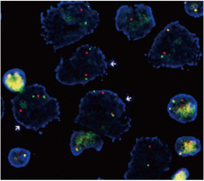
Fig. 1.
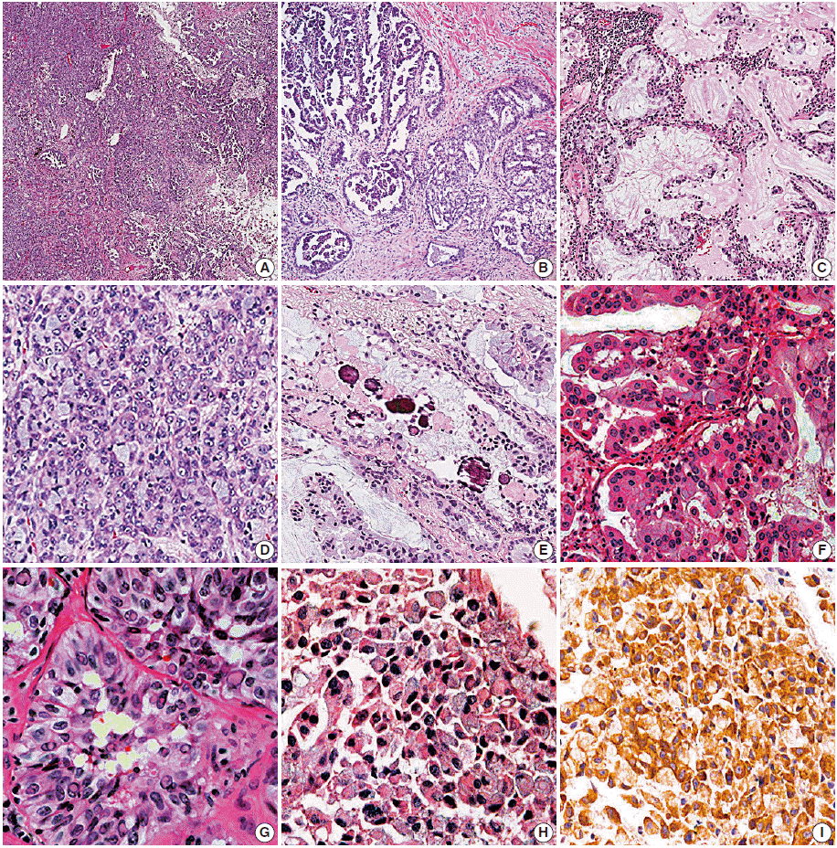
Fig. 2.
| ALK-FISH |
No. of cases (n = 78) | |||
|---|---|---|---|---|
| Rearranged (n = 69) | Not detected (n = 6) | Failed (n = 3) | ||
| ALK-IHC | ||||
| Negative | 1 (1.4) | 0 | 0 | 1 |
| 1+ | 3 (4.3) | 0 | 0 | 3 |
| 2+ | 42 (60.9) | 3 (50.0) | 3 (100) | 48 |
| 3+ | 21 (30.4) | 3 (50.0) | 0 | 24 |
| Failed | 2 (2.9) | 0 | 0 | 2 |
| Sampling method | ||||
| Biopsy | 56 (81.2) | 2 (33.3) | 0 | 58 |
| Resection | 13 (18.8) | 4 (66.7) | 3 (100) | 20 |
| Total cases (n=306) | ALK-positive |
ALK-negative |
p-value | |||
|---|---|---|---|---|---|---|
| Biopsy (n = 129) | Resection (n = 76) | Total (n = 205) | Resection (n = 101) | |||
| Sex (M:F) | 47:82 | 36:40 | 83:122 | 53:48 | .047 |
|
| .501 |
||||||
| Age (yr) | 53.9 ± 11.5 | 55.1 ± 11.7 | 54.4 ± 11.5 | 61.7 ± 8.8 | < .001 |
|
| < .001 |
||||||
| TNM stage, n (%) | a | 1 (0.8) | 29 (38.2) | 30 (14.6) | 20 (19.8) | .234 |
| 1b | 1 (0.8) | 4 (5.3) | 5 (2.4) | 14 (13.9) | ||
| 2a | 4 (3.1) | 8 (10.5) | 12 (5.9) | 12 (11.9) | ||
| 2b | 1 (0.8) | 0 | 1 (0.5) | 8 (7.9) | ||
| 3a | 7 (5.4) | 23 (30.3) | 30 (14.6) | 32 (31.7) | ||
| 3b | 9 (7.0) | 3 (3.9) | 12 (5.9) | 1 (1.0) | ||
| 4 | 106 (82.2) | 9 (11.8) | 115 (56.1) | 14 (13.9) | ||
| ALK-positive resection (n = 76) | ALK-negative resection (n = 101) | p-value | |
|---|---|---|---|
| Histolologic subtype (predominant) | |||
| Acinar | 31 (40.8) | 35 (34.7) | .403 |
| Papillary | 24 (31.6) | 36 (35.6) | .572 |
| Solid | 19 (25.0) | 23 (22.8) | .730 |
| Micropapillary | 0 | 3 (3.0) | .130 |
| Lepidic | 0 | 3 (3.0) | .130 |
| Invasive mucinous | 2 (2.3) | 1 (1.0) | .402 |
| Growth pattern (> 5% of entire tumor) | |||
| Acinar | 65 (85.5) | 73 (72.3) | .035 |
| Papillary | 52 (68.4) | 66 (65.3) | .668 |
| Solid | 45 (59.2) | 36 (35.6) | .002 |
| Micropapillary | 31 (40.8) | 21 (20.8) | .004 |
| Lepidic | 7 (9.2) | 20 (19.8) | .052 |
| Cribriform | 51 (67.1) | 29 (28.7) | < .001 |
| No. of growth patterns |
|||
| 1 | 3 (3.9) | 14 (13.9) | < .001 |
| 2 | 14 (18.4) | 44 (43.6) | |
| 3 | 26 (34.2) | 32 (31.7) | |
| 4 | 23 (30.3) | 9 (8.9) | |
| 5 | 10 (13.2) | 1 (1.0) | |
| 6 | 0 | 1 (1.0) | |
| Microscopic findings | |||
| Presence of signet-ring-cell element | 20 (26.3) | 4 (4.0) | < .001 |
| Extracellular mucin production | 34 (44.7) | 18 (17.8) | < .001 |
| Intracellular mucin content | 58 (76.3) | 57 (56.4) | .006 |
| Hobnail cells with abundant cytoplasm | 4 (5.3) | 35 (34.7) | < .001 |
| Abrupt presence of goblet cell-like cells | 49 (64.5) | 33 (32.7) | < .001 |
| Psammoma body | 33 (43.4) | 5 (5.0) | < .001 |
| Presence of bizarre cells | 19 (25.0) | 43 (42.6) | .015 |
| Presence of multilobated cells | 5 (6.6) | 6 (5.9) | .862 |
| Nuclear inclusion and groove | 16 (21.1) | 10 (9.9) | .038 |
| Prominent macronucleoli | 58 (46.6) | 78 (53.4) | .034 |
| ALK-positive resection (n = 76) | ALK-negative resection (n = 101) | Odds ratio | 95% CI | |
|---|---|---|---|---|
| Growth pattern (> 5% of entire tumor) | ||||
| Acinar | 65 (85.5) | 73 (72.3) | 3.178 | 1.298–7.779 |
| Solid | 45 (59.2) | 36 (35.6) | 2.694 | 1.301–5.577 |
| Micropapillary | 31 (40.8) | 21 (20.8) | 3.414 | 1.581–7.372 |
| Cribriform | 51 (67.1) | 29 (28.7) | 3.771 | 1.877–7.575 |
| Microscopic findings | ||||
| Presence of signet-ring-cell element | 20 (26.3) | 4 (4.0) | 5.466 | 1.488–20.080 |
| Extracellular mucin production | 34 (44.7) | 18 (17.8) | 3.372 | 1.411–8.057 |
| Hobnail cells with abundant cytoplasm | 4 (5.3) | 35 (34.7) | 0.096 | 0.026–0.356 |
| Psammoma body | 33 (43.4) | 5 (5.0) | 16.548 | 5.303–51.641 |
| ALK-positive biopsy (n = 129) | ALK-positive resection (n = 76) | p-value | |
|---|---|---|---|
| Presence of signet-ring-cell element | 40 (31.0) | 20 (26.3) | .476 |
| Extracellular mucin production | 15 (11.6) | 34 (44.7) | < .001 |
| Intracellular mucin content | 101 (78.3) | 58 (76.3) | .743 |
| Hobnail cells with abundant cytoplasm | 5 (3.9) | 4 (5.3) | .640 |
| Abrupt presence of goblet cell-like cells | 69 (53.5) | 49 (64.5) | .124 |
| Psammoma body | 20 (15.5) | 33 (43.4) | < .001 |
| Nuclear inclusion and groove | 7 (5.4) | 16 (21.1) | .001 |
Values are presented as number (%). ALK, anaplastic lymphoma kinase; FISH, fluorescence
ALK, anaplastic lymphoma kinase; M, male; F, female. p-value between the ALK-positive and -negative group; p-value between the ALK-positive and -negative resected group; p-value by Student’s t test; Linear by linear association by chi-square test.
Values are presented as number (%). ALK, anaplastic lymphoma kinase. Number of all presented growth patterns presented in ‘Growth pattern (> 5% of entire tumor)’; p-value by Student’s t-test.
Values are presented as number (%). ALK, anaplastic lymphoma kinase; CI, confidence interval.
Values are presented as number (%). ALK, anaplastic lymphoma kinase.

 E-submission


