Articles
- Page Path
- HOME > J Pathol Transl Med > Volume 51(4); 2017 > Article
-
Case Study
Metaplastic Carcinoma with Chondroid Differentiation Arising in Microglandular Adenosis - Ga-Eon Kim, Nah Ihm Kim, Ji Shin Lee, Min Ho Park1
-
Journal of Pathology and Translational Medicine 2017;51(4):418-421.
DOI: https://doi.org/10.4132/jptm.2016.10.06
Published online: April 4, 2017
Department of Pathology, Chonnam National University Medical School, Gwangju, Korea
1Department of Surgery, Chonnam National University Medical School, Gwangju, Korea
- Corresponding Author Ji Shin Lee, MD Department of Pathology, Chonnam National University Hwasun Hospital, 322 Seoyang-ro, Hwasun 58128, Korea Tel: +82-61-379-7072 Fax: +82-61-379-7079 E-mail: jshinlee@hanmail.net
• Received: August 3, 2016 • Revised: October 3, 2016 • Accepted: October 5, 2016
© 2017 The Korean Society of Pathologists/The Korean Society for Cytopathology
This is an Open Access article distributed under the terms of the Creative Commons Attribution Non-Commercial License (http://creativecommons.org/licenses/by-nc/4.0) which permits unrestricted non-commercial use, distribution, and reproduction in any medium, provided the original work is properly cited.
Abstract
- Microglandular adenosis (MGA) of the breast is a rare, benign proliferative lesion but with a significant rate of associated carcinoma. Herein, we report an unusual case of metaplastic carcinoma with chondroid differentiation associated with typical MGA. Histologically, MGA showed a direct transition to metaplastic carcinoma without an intervening atypical MGA or ductal carcinoma in situ component. The immunohistochemical profile of the metaplastic carcinoma was mostly similar to that of MGA. In both areas, all the epithelial cells were positive for S-100 protein, but negative for estrogen receptor, progesterone receptor, HER2/neu, and epidermal growth factor receptor. An increase in the Ki-67 and p53 labelling index was observed from MGA to invasive carcinoma. To the best of our knowledge, this is the first case of metaplastic carcinoma with chondroid differentiation arising in MGA in Korea. This case supports the hypothesis that a subset of MGA may be a non-obligate morphologic precursor of breast carcinoma, especially the triple-negative subtype.
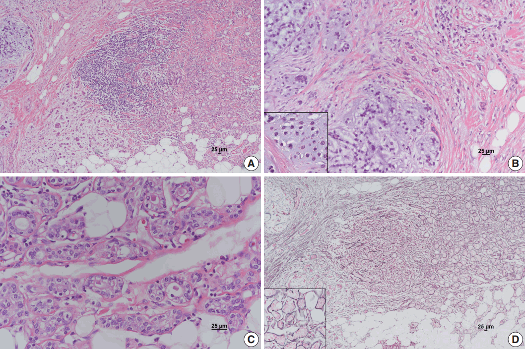
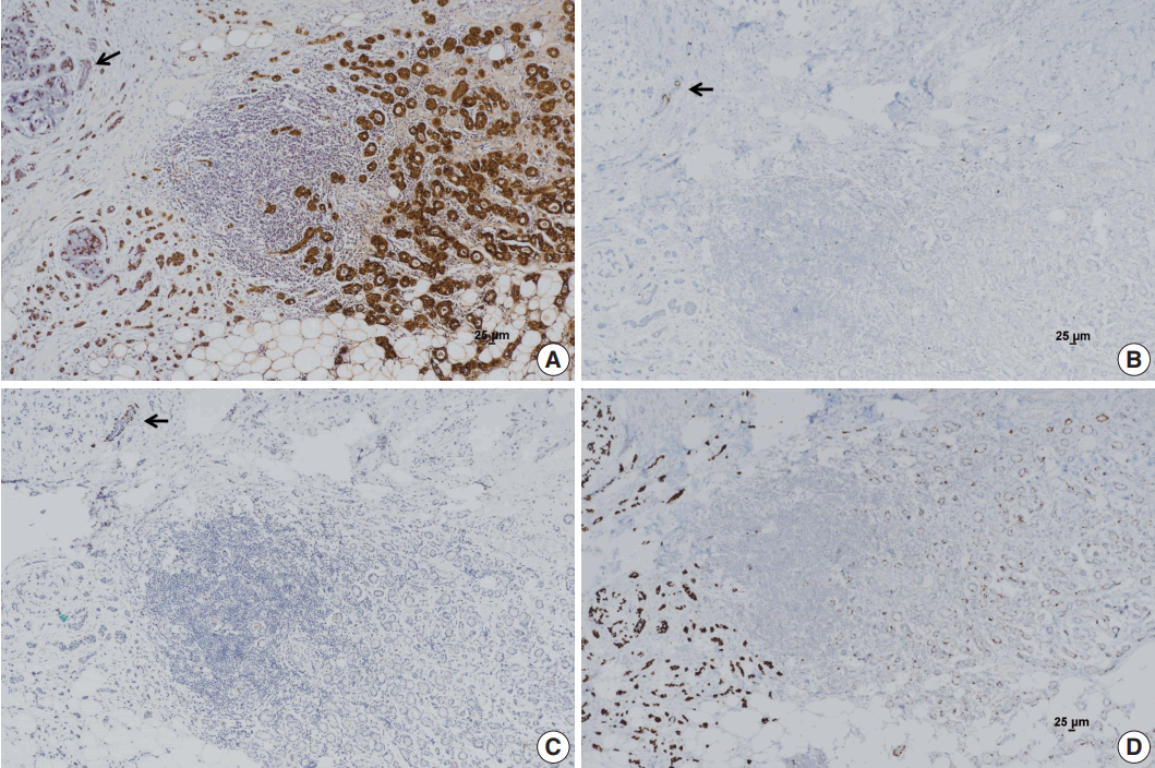
- A 60-year-old female presented with a palpable breast mass in the left breast. Ultrasound-guided core needle biopsy revealed invasive carcinoma. Preoperative breast magnetic resonance imaging showed an irregular solid mass approximately 1.5 cm in size in the upper outer quadrant of the left breast, which showed good contrast enhancement. A conserving operation was performed. Grossly, a grey to white colored, hard mass with ill-defined borders measuring 1.5 cm×1.2 cm×1 cm was observed. Microscopically, the tumor was composed of an invasive carcinoma area and MGA area (Fig. 1A). Invasive carcinoma of no special type, grade 2 with an abrupt transition to chondroid matrix without an intervening spindle cell sarcomatoid component was observed (Fig. 1B). The chondroid matrix comprised 70% of the invasive area. Focal chondroid differentiation in the chondroid matrix area was found, thus the patient was diagnosed with metaplastic carcinoma with chondroid differentiation. In the MGA area, small round glands lined by a single layer of cuboidal epithelial cells were diffused throughout the fibrous or adipose breast tissues (Fig. 1C). Some of the glands were filled with a colloid-like secretion. No obvious architectural complexity or cytological atypia was present. MGA showed direct transition to metaplastic carcinoma without an intervening atypical MGA or ductal carcinoma in situ area. The glands in MGA were surrounded by a basement membrane, which was clearly demonstrated by reticulin staining. However, the basement membrane was disrupted around the invasive nests of the metaplastic carcinoma (Fig. 1D). The immunohistochemical profile of metaplastic carcinoma was nearly identical to that of MGA. In both instances, the epithelial cells were positive for cytokeratin (CK) 7 and S-100 protein, but negative for estrogen receptor (ER), progesterone receptor (PR), HER2/neu, and epidermal growth factor receptor (EGFR) (Fig. 2A, B). Other basal-like markers such as CK 5/6 and nestin were negative. No myoepithelial cells were demonstrated on immunohistochemical stainings for both smooth muscle myosin heavy chain and p63 (Fig. 2C). Ki-67 and p53 were positive in MGA and metaplastic carcinoma but tended to show a more intense staining in metaplastic carcinoma (Fig. 2D). The percentage of p53 and Ki-67 positive cells was 5% and 3% in MGA and 90% and 45% in metaplastic carcinoma, respectively. This case was finally diagnosed as metaplastic carcinoma with chondroid differentiation arising in MGA.
- No metastasis was found in the sentinel lymph nodes. After surgery, the patient received adjuvant chemoradiotherapy. After 14 months of follow-up, no evidence of recurrence was observed.
CASE REPORT
- MGA of the breast is a rare, benign glandular proliferative lesion that mimics invasive carcinoma clinicopathologically [1,2]. Carcinoma arising in MGA has previously been reported [3-8]. The incidence of invasive carcinoma ranges from 23%–64% in patients with MGA [3,8]; the high upper limit may be due to referral bias at institutions [5].
- Invasive breast carcinomas arising in MGA have various histological features [1]. Invasive carcinoma of no special type is the most common type of carcinoma arising in MGA. Specialized variants of carcinoma, including carcinoma with secretory differentiation, carcinoma with squamous differentiation, acinic cell carcinoma, and adenoid cystic carcinoma, have also been reported [3-8]. Metaplastic carcinoma of the breast represents 0.2%–5% of all invasive carcinomas [6]. Metaplastic carcinoma with mesenchymal differentiation is an extremely rare subtype of breast carcinoma arising in MGA [8]. Structural transitions from MGA to atypical MGA and intraductal carcinoma and to invasive carcinoma are usually observed in invasive carcinomas arising in MGA [3-8].
- In the present case, invasive carcinoma showed abrupt transition to chondroid matrix without an intervening spindle cell component. An intervening atypical MGA or ductal carcinoma in situ component between MGA and invasive carcinoma was not observed. Histologically, this case was consistent with metaplastic carcinoma with chondroid differentiation associated with typical MGA.
- The main histological difference between MGA and MGA-associated invasive carcinoma is the presence of a basement membrane. The glands in MGA are surrounded by a basement membrane that can be highlighted using reticulin staining, periodic acid-Schiff staining, and immunohistochemical stains for type IV collagen and laminin. However, the basement membrane in MGA-associated carcinoma is disrupted around invasive nests.
- The immunohistochemical profile of invasive carcinoma arising in MGA is similar to that of MGA [3-6,8]. No myoepithelial cells are found in MGA and related lesions. The epithelial cells are usually positive for CK 7 and S-100 protein, but negative for ER, PR, and HER2/neu. In the present case, all the epithelial cells in MGA and invasive carcinoma were strongly immunoreactive for S-100 protein and negative for ER, PR, and HER2/neu, which was consistent with a triple-negative immunoprofile. Although the positivity for basal-like markers such as EGFR, CK 5/6, and nestin has been described in MGA and associated lesions [8], epithelial cells in the present case lacked these markers. Several studies have reported a trend toward increased positivity for the p53 and proliferation marker Ki-67 together with increased severity of the lesions [4,5]. In the present case, metaplastic carcinoma showed a higher percentage of staining for p53 and Ki-67 than MGA. The identification of MGA and its direct transition to invasive lesions and the maintenance of the characteristic immunoprofile of MGA and invasive carcinoma associated with MGA in this study provided strong evidence suggesting that an MGA subset may constitute a non-obligate morphological precursor of invasive breast carcinoma, especially the triple-negative subtype.
- Recent molecular genetic studies including massively parallel sequencing approaches provide further evidence to support the hypothesis that MGA is, at least in several cases, a neoplastic, clonal lesion and may be a non-obligate precursor for triple-negative or basal-like breast carcinomas [10,11].
- The treatment of carcinomas arising in MGA follows the same general guidelines for breast carcinomas. The prognosis of invasive carcinoma arising in MGA varies in the literature. Some authors reported that patients with invasive carcinoma arising in MGA have a relatively favorable prognosis [3,7]. Another study, however, showed that the outcome for patients with invasive carcinoma arising in MGA ranged from favorable to unfavorable [5]. In our patient, no axillary lymph node metastasis was found. Metaplastic carcinoma, the subtype of invasive carcinoma found in the present case, has lower response rates to conventional adjuvant chemotherapy and a worse clinical outcome than other forms of triple-negative breast cancers. Because metaplastic carcinoma arising in MGA is extremely rare, the prognosis of the lesion requires further characterization with more cases and longer follow-up.
DISCUSSION
Fig. 1.Metaplastic carcinoma with mesenchymal differentiation arising in microglandular adenosis (MGA). (A) MGA on the right shows direct transition to invasive carcinoma on the left. (B) Invasive carcinoma primarily composed of cord-like cells scattered in the chondromyxoid matrix with focal chondroid differentiation (inset). (C) Typical glands in MGA are lined by uniform cuboidal cells regularly spaced around a lumen containing a colloid-like secretion. (D) Basement membranes highlighted by the reticulin stain are preserved in the MGA area (inset) but disrupted in the invasive carcinoma area.


Fig. 2.Immunohistochemical stains. (A) S-100 protein is strongly positive in microglandular adenosis (MGA) and invasive carcinoma. Entrapped normal mammary gland is negative for S-100 protein (arrow). (B) Estrogen receptor (ER) is positive in entrapped normal mammary glands (arrow); however, MGA and invasive carcinoma are negative for ER. (C) No immunoreactivity for p63 is observed in the MGA area or invasive carcinoma area. Normal mammary glands are stained positively in myoepithelial cells (arrow). (D) An increase in p53 labelling index is observed from MGA to invasive carcinoma.


- 1. Shin SJ, Gobbi H. Microglandular adenosis, atypical microglandular adenosis and microglandular adenosis with carcinoma. In: Lakhani SR, Ellis IO, Schnitt SJ, Tan PH, van de Vijver MJ, eds. WHO classification of tumours of the breast. 4th ed. Lyon: IARC Press, 2012; 113-4.
- 2. Brogi E. Adenosis and microglandular adenosis. In: Hoda SA, Brogi E, Koerner FC, Rosen PP, eds. Rosen’s breast pathology. 4th ed. Philadelphia: Lippincott Williams & Wilkins, 2014; 183-212.
- 3. James BA, Cranor ML, Rosen PP. Carcinoma of the breast arising in microglandular adenosis. Am J Clin Pathol 1993; 100: 507-13. ArticlePubMed
- 4. Koenig C, Dadmanesh F, Bratthauer GL, Tavassoli FA. Carcinoma arising in microglandular adenosis: an immunohistochemical analysis of 20 intraepithelial and invasive neoplasms. Int J Surg Pathol 2000; 8: 303-15. ArticlePubMedPDF
- 5. Khalifeh IM, Albarracin C, Diaz LK, et al. Clinical, histopathologic, and immunohistochemical features of microglandular adenosis and transition into in situ and invasive carcinoma. Am J Surg Pathol 2008; 32: 544-52. ArticlePubMed
- 6. Shui R, Bi R, Cheng Y, Lu H, Wang J, Yang W. Matrix-producing carcinoma of the breast in the Chinese population: a clinicopathological study of 13 cases. Pathol Int 2011; 61: 415-22. ArticlePubMed
- 7. Zhong F, Bi R, Yu B, et al. Carcinoma arising in microglandular adenosis of the breast: triple negative phenotype with variable morphology. Int J Clin Exp Pathol 2014; 7: 6149-56. PubMedPMC
- 8. Liu LY, Sheng SH, Zhang ZY, Xu JH. A case of matrix-producing carcinoma of the breast with micoglandular adenosis and review of literature. Int J Clin Exp Pathol 2015; 8: 8568-72. PubMedPMC
- 9. Choi JE, Bae YK. Invasive breast carcinoma arising in microglandular adenosis: two case reports. J Breast Cancer 2013; 16: 432-7. ArticlePubMedPMC
- 10. Shin SJ, Simpson PT, Da Silva L, et al. Molecular evidence for progression of microglandular adenosis (MGA) to invasive carcinoma. Am J Surg Pathol 2009; 33: 496-504. ArticlePubMed
- 11. Guerini-Rocco E, Piscuoglio S, Ng CK, et al. Microglandular adenosis associated with triple-negative breast cancer is a neoplastic lesion of triple-negative phenotype harbouring TP53 somatic mutations. J Pathol 2016; 238: 677-88. PubMedPMC
REFERENCES
Figure & Data
References
Citations
Citations to this article as recorded by 

- Two similar but distinct types of breast acinar cell carcinoma: evidence from histological, immunohistochemical and molecular features
Mingfang Sun, Lin Fu, Hongjiu Ren, Jian Wang, Xuyong Lin, Qingfu Zhang
Histopathology.2025; 87(6): 904. CrossRef - Elucidating the nature of acinic cell carcinoma of the breast with high-grade morphology: evidence from case report
Yunjie Ge, Xianping Wei, Jing-Nan Liu, Ping-Li Sun, Hongwen Gao
Diagnostic Pathology.2024;[Epub] CrossRef - New insights into acinic cell carcinoma of the breast: clinicopathology, origin of histology, molecular features, prognosis, and treatment
Yunjie Ge, Xianping Wei, Jing-Nan Liu, Ping-Li Sun, Hongwen Gao
Frontiers in Oncology.2024;[Epub] CrossRef - Metaplastic Matrix-Producing Carcinoma and Apocrine Lobular Carcinoma In Situ Associated with Microglandular Adenosis: A Unique Case Report
Nektarios Koufopoulos, Dionysios Dimas, Foteini Antoniadou, Kyparissia Sitara, Dimitrios Balalis, Ioannis Boutas, Alina Roxana Gouloumis, Adamantia Kontogeorgi, Lubna Khaldi
Diagnostics.2022; 12(6): 1458. CrossRef - Salivary gland-type mammary carcinoma arising in microglandular adenosis: A case report and clinicopathological review of the literature
Victoria Rico, Yukiko Shibahara, Marjorie Monteiro, Elzbieta Slodkowska, Samantha Tam, Pearl Zaki, Carlo De Angelis, Edward Chow, Katarzyna Joanna Jerzak
Cancer Treatment and Research Communications.2020; 24: 100178. CrossRef - Microglandular adenosis is an advanced precursor breast lesion with evidence of molecular progression to matrix-producing metaplastic carcinoma
Christopher J. Schwartz, Igor Dolgalev, Esther Yoon, Iman Osman, Adriana Heguy, Eleazar C. Vega-Saenz de Miera, Diana Nimeh, George Jour, Farbod Darvishian
Human Pathology.2019; 85: 65. CrossRef
 PubReader
 ePub Link
-
 Cite this Article
- Cite this Article
-
- Close
- Download Citation
- Close
- Figure
Metaplastic Carcinoma with Chondroid Differentiation Arising in Microglandular Adenosis


Fig. 1. Metaplastic carcinoma with mesenchymal differentiation arising in microglandular adenosis (MGA). (A) MGA on the right shows direct transition to invasive carcinoma on the left. (B) Invasive carcinoma primarily composed of cord-like cells scattered in the chondromyxoid matrix with focal chondroid differentiation (inset). (C) Typical glands in MGA are lined by uniform cuboidal cells regularly spaced around a lumen containing a colloid-like secretion. (D) Basement membranes highlighted by the reticulin stain are preserved in the MGA area (inset) but disrupted in the invasive carcinoma area.
Fig. 2. Immunohistochemical stains. (A) S-100 protein is strongly positive in microglandular adenosis (MGA) and invasive carcinoma. Entrapped normal mammary gland is negative for S-100 protein (arrow). (B) Estrogen receptor (ER) is positive in entrapped normal mammary glands (arrow); however, MGA and invasive carcinoma are negative for ER. (C) No immunoreactivity for p63 is observed in the MGA area or invasive carcinoma area. Normal mammary glands are stained positively in myoepithelial cells (arrow). (D) An increase in p53 labelling index is observed from MGA to invasive carcinoma.
Fig. 1.
Fig. 2.
Metaplastic Carcinoma with Chondroid Differentiation Arising in Microglandular Adenosis

 E-submission


