
 E-submission
Articles
- Page Path
- HOME > J Pathol Transl Med > Volume 57(2); 2023 > Article
-
Case Study
Unusual biclonal IgA plasma cell myeloma with aberrant expression of high-risk immunophenotypes: first report of a new diagnostic and clinical challenge -
Carlos A. Monroig-Rivera1
 , Clara N. Finch Cruz2,3
 -
Journal of Pathology and Translational Medicine 2023;57(2):132-137.
DOI: https://doi.org/10.4132/jptm.2023.02.07
Published online: March 14, 2023
1School of Medicine, Ponce Health Sciences University, Ponce, Puerto Rico, USA
2Department of Basic Sciences, School of Medicine - Ponce Health Sciences University, Ponce, Puerto Rico, USA
3Southern Pathology Services, Inc., Ponce, Puerto Rico, USA
- Corresponding Author: Clara N. Finch Cruz, MD, Department of Basic Sciences, School of Medicine - Ponce Health Sciences University, 388 Zona Industrial Reparada 2 Ponce, Puerto Rico 00716-2347, USA Tel: +1-787-840-2575, Fax: +1-787-844-3865, E-mail: cfinch@psm.edu
© 2023The Korean Society of Pathologists/The Korean Society for Cytopathology
This is an Open Access article distributed under the terms of the Creative Commons Attribution Non-Commercial License (http://creativecommons.org/licenses/by-nc/4.0) which permits unrestricted non-commercial use, distribution, and reproduction in any medium, provided the original work is properly cited.


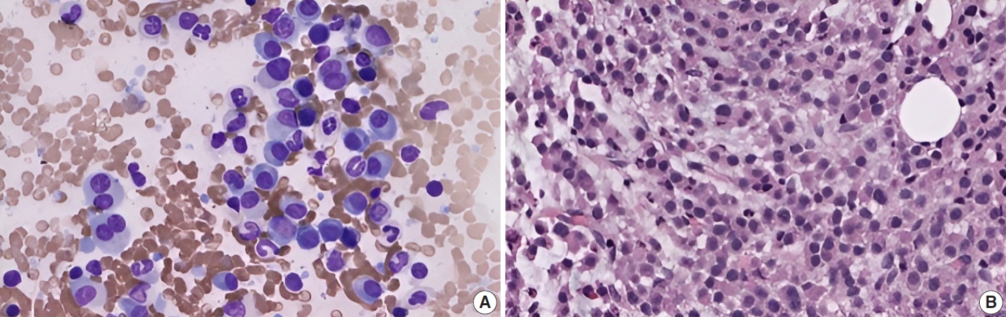

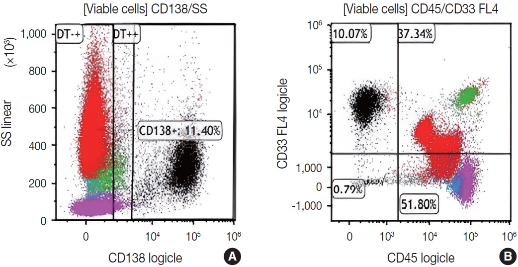
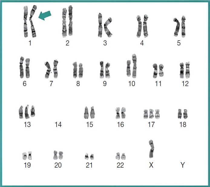
Figure & Data
References
Citations

- Extreme PTH-Independent Hypercalcemia Mediated by a Rare Non-Osteoclastic Osteolysis Mechanism in Newly Diagnosed High-Risk Biclonal IgA Multiple Myeloma: Temporal Association with the Second Dose of Moderna mRNA-1273 COVID-19 Vaccine
Clara N. Finch-Cruz, Serena I. Fazal, Vivianette Fuentes Morales
International Journal of Molecular Sciences.2026; 27(15): 6832. CrossRef
 PubReader
 ePub Link
-
 Cite this Article
- Cite this Article
-
- Close
- Download Citation
- Close
 XML Download
- Figure
-
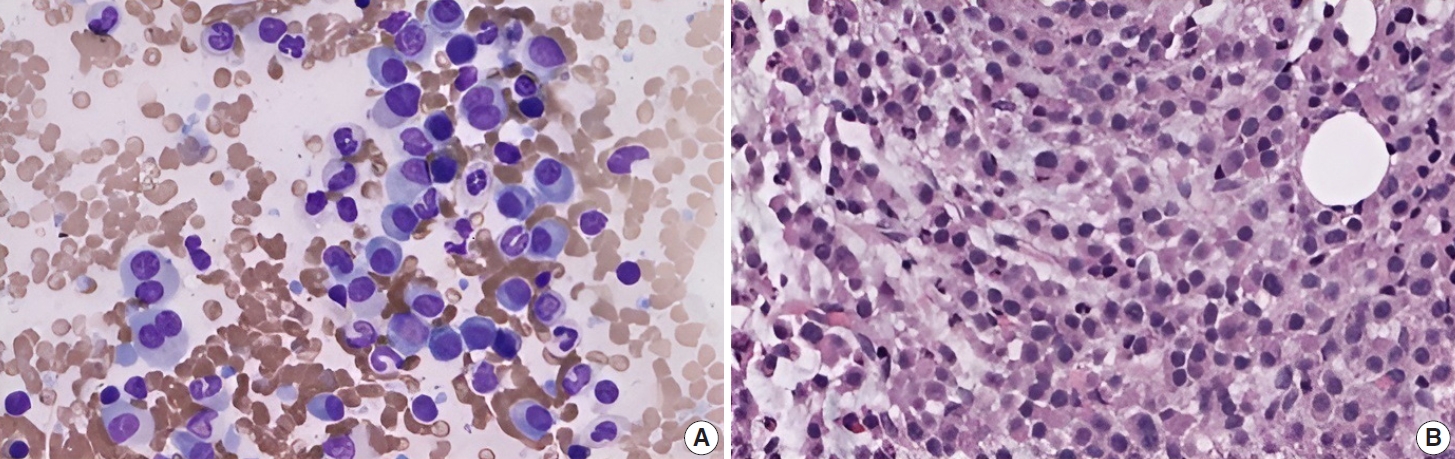
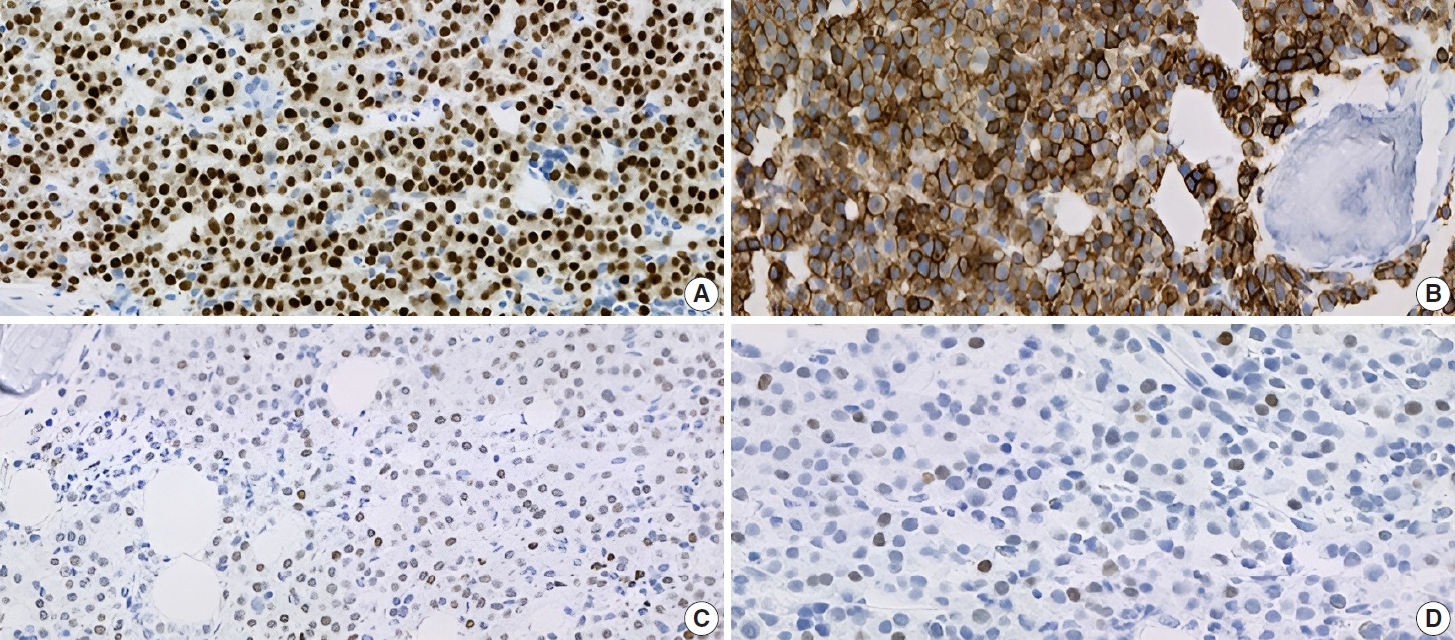
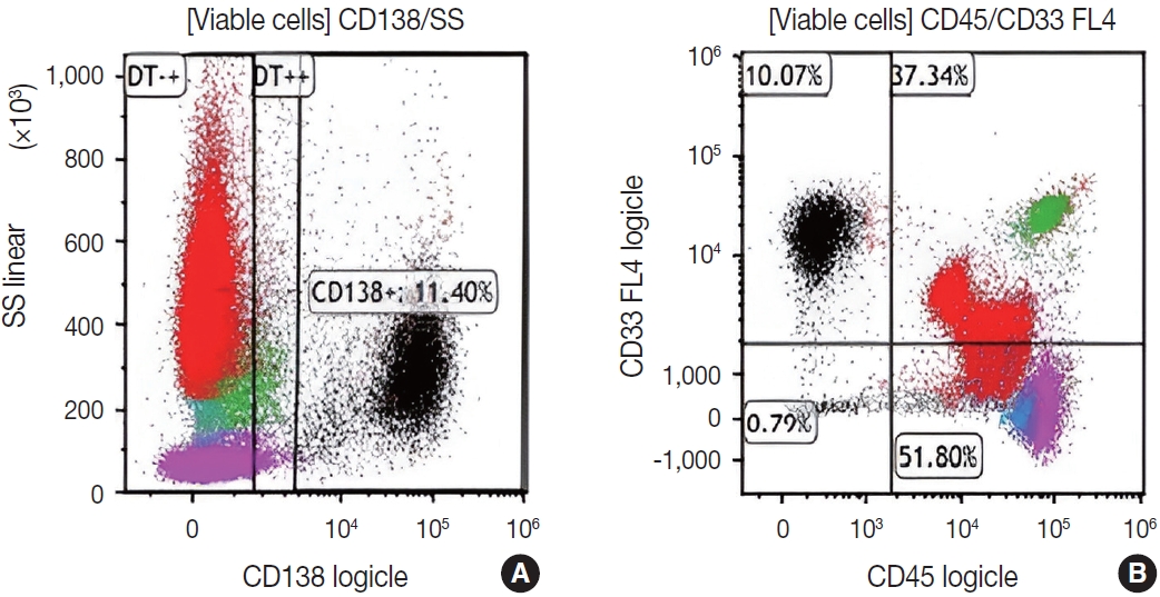
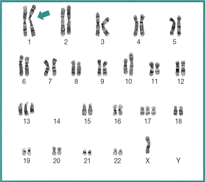

Fig. 1.
Fig. 2.
Fig. 3.
Fig. 4.
| Test | Result | Reference range | Comments |
|---|---|---|---|
| IgG | 465 L | 586–1,602 mg/d | Quantitative immunoglobulin (Ig) serum levels |
| IgM | 7 L | 26–217 mg/dL | |
| IgA | 3410 H | 87–352 mg/dL | |
| Albumin | 3.4 | 2.9–4.4 g/dL | Serum protein electrophoresis |
| Alpha-1-globulin | 0.2 | 0.0–0.4 g/dL | |
| Alpha-2-globulin | 0.7 | 0.4–1.0 g/dL | |
| Beta globulin | 4.5 H | 0.7–1.3 g/dL | |
| Gamma globulin | 0.4 | 0.4–1.8 g/dL | |
| M-spike | 3.9 H | 0.01 g/dL | |
| Beta 1 | 3.2 H | - | |
| Beta 2 | 0.7 H | - | |
| IgA, total | 3.9 H | - | |
| Immunofixation, serum | Biclonal IgA with kappa specificity | - | Confirmatory test |
| Beta-2 microglobulin | 8.4 H | 0.6–2.4 mg/L | High-risk factor, when ≥ 5.5 mg/L |
| Case | Age (yr), sex | Clinical feature | Ig specificity | M-protein clonality | Location | Microscopic findings | Immunophenotyping/Cytogenetics/Molecular studies |
|---|---|---|---|---|---|---|---|
| Annibali et al. (2009) [10] | 39, female | Abdominal pain, and obstructive jaundice 7 yr after ASCT | IgA-lambda | Monoclonal | Head of the pancreas, pleural effusion | US-guided FNA cytology of the pancreatic mass and cytology of pleural effusion revealed myeloma plasma cells | Unknown |
| Age at the time of initial diagnosis | Extramedullary relapse | ||||||
| Cerqueira et al. (2020) [11] | 49, female | Presented with abdominal pain, biliary vomiting of 6 days duration | IgA-kappa | Monoclonal | Kidney, bone marrow | Kidney biopsy demonstrating myeloma kidney | Immunophenotyping: 60% of bone marrow monoclonal plasma cells with 100% CD138+, 100% CD38+, and 45% CD20+ |
| Admitted to ICU for acute kidney failure | |||||||
| Suo et al. (2020) [12] | 57, male | History of liver cirrhosis presenting with abdominal pain and pancytopenia | IgA-kappa | Monoclonal | Liver, MRI- left hepatic mass | Abundant plasmacytoid cells, kappa restricted neoplastic plasma cells | Plasmacytoid cells showed CD138+, kappa+, lambda– |
| Extramedullary involvement | Cytogenetics and FISH suggestive of advanced disease progression | ||||||
| Yamane et al. (2021) [13] | 73, male | Acute left lower abdominal pain | IgA-type | Monoclonal | Left vertebral arch of the 10th thoracic vertebra | Bone marrow biopsy: plasma cell neoplasm with 26.0% of plasma cells | Flow cytometry: CD38+, CD56+, CD138+, MPC-1+. |
| Chromosomal analysis: 45,X,-Y,+5,+6,+7,-8,+9, +11,-13, and +21 | |||||||
| Current case | 65, female | Acute abdominal pain | IgA-kappa | Biclonal | Bone marrow | Hypercellularity (85%–95%), abnormal plasma cells (32%) | Immunophenotyping: CD138+, CD33+, MUM-1+, CD43+, OCT-2+, c-MYC+ |
| Chromosomal analysis: gain of 1q,13,17, loss of 14 |
L, low; H, high. According to the Revised International Staging System for Multiple Myeloma (R-ISS) [
ASCT, autologous stem cell transplant; US, ultrasound; FNA, fine-needle aspiration; ICU; intensive care unit; FISH, fluorescence in situ hybridization.


