Warning: mkdir(): Permission denied in /home/virtual/lib/view_data.php on line 81

Warning: fopen(upload/ip_log/ip_log_2024-11.txt): failed to open stream: No such file or directory in /home/virtual/lib/view_data.php on line 83

Warning: fwrite() expects parameter 1 to be resource, boolean given in /home/virtual/lib/view_data.php on line 84
Prognostic Significance of Absolute Lymphocyte Count/Absolute Monocyte Count Ratio at Diagnosis in Patients with Multiple Myeloma
Skip Navigation
Skip to contents

J Pathol Transl Med : Journal of Pathology and Translational Medicine

OPEN ACCESS
SEARCH
Search

Articles

Page Path
HOME > J Pathol Transl Med > Volume 47(6); 2013 > Article
Original Article
Prognostic Significance of Absolute Lymphocyte Count/Absolute Monocyte Count Ratio at Diagnosis in Patients with Multiple Myeloma
Su-Jin Shin, Jin Roh, Misung Kim, Min Jung Jung, Young Wha Koh, Chan-Sik Park, Dok Hyun Yoon1, Cheolwon Suh1, Chan-Jeong Park2, Hyun Sook Chi2, Jooryung Huh
Korean Journal of Pathology 2013;47(6):526-533.
DOI: https://doi.org/10.4132/KoreanJPathol.2013.47.6.526
Published online: December 24, 2013

Department of Pathology, Asan Medical Center, University of Ulsan College of Medicine, Seoul, Korea.

1Department of Oncology, Asan Medical Center, University of Ulsan College of Medicine, Seoul, Korea.

2Department of Laboratory Medicine, Asan Medical Center, University of Ulsan College of Medicine, Seoul, Korea.

Corresponding Author: Jooryung Huh, M.D. Department of Pathology, Asan Medical Center, University of Ulsan College of Medicine, 88 Olympic-ro 43-gil, Songpa-gu, Seoul 138-736, Korea. Tel: +82-2-3010-4553, Fax: +82-2-472-7898, jrhuh@amc.seoul.kr
• Received: August 19, 2013   • Revised: October 2, 2013   • Accepted: October 10, 2013

© 2013 The Korean Society of Pathologists/The Korean Society for Cytopathology

This is an Open Access article distributed under the terms of the Creative Commons Attribution Non-Commercial License (http://creativecommons.org/licenses/by-nc/3.0/) which permits unrestricted non-commercial use, distribution, and reproduction in any medium, provided the original work is properly cited.

prev next
  • 11,137 Views
  • 86 Download
  • 24 Crossref
  • 28 Scopus
  • Background
    Absolute lymphocyte count (ALC) in peripheral blood has recently been reported to be an independent prognostic factor in multiple myeloma (MM). Previous studies indicated that the absolute monocyte count (AMC) in peripheral blood reflects the state of the tumor microenvironment in lymphomas. Neither the utility of the AMC nor its relationship with ALC has been studied in MM.
  • Methods
    The prognostic value of ALC, AMC, and the ALC/AMC ratio at the time of diagnosis was retrospectively examined in 189 patients with MM.
  • Results
    On univariate analysis, low ALC (<1,400 cells/µL), high AMC (≥490 cells/µL), and low ALC/AMC ratio (<2.9) were correlated with worse overall survival (OS) (p=.002, p=.038, and p=.001, respectively). On multivariate analysis, the ALC/AMC ratio was an independent prognostic factor (p=.047), whereas ALC and AMC were no longer statistical significant. Low ALC, high AMC, and low ALC/AMC ratio were associated with poor prognostic factors such as high International Staging System stage, plasmablastic morphology, hypoalbuminemia, and high β2-microglobulin.
  • Conclusions
    Univariate analysis demonstrated that changes in ALC, AMC, and the ALC/AMC ratio are associated with patient survival in MM. Multivariate analysis showed that, of these factors, the ALC/AMC ratio was an independent prognostic factor for OS.
Absolute lymphocyte count (ALC) in peripheral blood may reflect the body's immune surveillance potential against cancer cells. Recent studies suggest that ALC, as a surrogate marker for host immune status, has prognostic significance in cancer patients. In multiple myeloma (MM), ALC recovery after autologous stem cell transplantation (ASCT) has been reported to be of significant prognostic value.1,2 Furthermore, a recent study showed that ALC at the time of diagnosis was associated with survival in newly diagnosed MM patients.3
In recent years, the importance of the bone marrow (BM) microenvironment in MM has become increasingly apparent. The BM microenvironment provides a 'niche' that supports growth and survival of myeloma cells, and influences their migration and drug resistance.4,5 Among stromal cells in the microenvironment, inflammatory cells play an essential role in tumor progression.6 Tumor-associated macrophages (TAMs), which constitute a significant proportion of tumor-related inflammatory cells, have been linked to the growth, angiogenesis and metastasis of a variety of tumors.7 TAMs secrete factors that directly promote tumor cell proliferation and, by acting on endothelial cells, they also promote tumor neovascularization, which enables tumor progression.8
A prognostic role for TAMs has been reported in patients with classical Hodgkin lymphoma (HL),9,10 diffuse large B-cell lymphoma (DLBCL),11 follicular lymphoma (FL)12 and MM.13 TAMs are derived from circulating monocytes and are recruited to the tumor site by tumor-derived chemotactic factors.14,15 Since the number of circulating monocytes and the TAM content are influenced by tumor-derived chemotactic factors, the peripheral blood absolute monocyte count (AMC) may reflect the level of TAM recruitment, and therefore may have potential value as a surrogate marker for TAMs. In support of this hypothesis, AMC has recently been shown to be a reliable prognostic marker in DLBCL,16 FL,17 and HL.18
The utility of AMC and its relationship with ALC have not been investigated in MM. The aim of this study was therefore to analyze AMC, ALC, and the ALC/AMC ratio, as a simple marker combining an estimate of host immune status and tumor microenvironment, at the time of diagnosis in MM and to correlate the findings with clinical parameters and patient outcome.
Patients
This study included 189 patients who were diagnosed with MM between 2001 and 2011 at the Asan Medical Center, Seoul, Korea. Disease was considered to be plasmablastic when >30% of myeloma cells in the BM biopsy exhibited plasmablastic morphology. All patients met the following criteria: BM involvement; no previous treatment; no previous history of other malignancies, transplantation or immunosuppression; no anti-human immunodeficiency virus antibodies; and availability of laboratory and radiologic data and follow-up information.
AMC and ALC were obtained from routine complete blood count (CBC) with a four-part differential (lymphocytes, monocytes, eosinophils, and neutrophils) using a Sysmex automated hematology analyzer (model E-4000, SE-9000 or XE-2100, Sysmex Co., Kobe, Japan), which was performed at the time of the diagnosis.19
Statistical analysis
Overall survival (OS) was defined as the time between the date of diagnosis and the date of death from any cause. For living patients, OS was defined as the time between diagnosis and the last follow-up date. OS was analyzed using the Kaplan-Meier method and log-rank testing was used to compare groups. Median follow-up with a 95% confidence interval was calculated using the reverse Kaplan-Meier method.20 Multivariate analyses of demographic and clinical characteristics prognostic for OS were performed using the Cox proportional hazards regression model.
The maximal chi-square method was adapted to evaluate the cutoff points in the dataset that best segregated patients into poor and good prognosis subgroups (based on the likelihood of surviving), with the log-rank test as statistic used to measure the strength of the grouping. Maxstat, a maximal chi-square package in R 2.15.2. (R Development Core Team, Vienna, Austria; http://www.R-project.org), was used to identify the optimal cut-off points for ALC, AMC and the ALC/AMC ratio.21,22 Correlations between ALC, AMC, and the ALC/AMC ratio with categorical variables were analyzed using Pearson's chi-square test or Fisher's exact test, and the Mann-Whitney U test was used to evaluate associations with continuous variables. All statistical analyses were performed using the SPSS ver. 18.0 (SPSS Inc., Chicago, IL, USA) or R 2.15.2. A p-value of ≤.05 was considered to be statistically significant.
Ethical permission
The Institutional Review Board (IRB) of Asan Medical Center (Seoul, Korea) approved this study protocol and provided all necessary ethical permissions.
Patient characteristics
The mean age of the 189 patients with MM in the study was 60 years (range, 29 to 84 years), and the male to female ratio was 1.077:1. Paraprotein type was determined by immunoelectrophoresis; IgG was the most common (n=95, 50.3%), followed by light chain (n=48, 25.4%), IgA (n=34, 18%), IgD (n=7, 3.7%), and IgM (n=5, 2.6%). Of the 189 patients, 57 (30.2%) exhibited plasmablastic disease morphology. Punched-out, osteolytic bone lesions (>3 lesions) were identified in 96 patients (50.8%). Following diagnosis, patients were treated with chemotherapy (47.1%), radiotherapy (6.3%), chemotherapy followed by ASCT (35.4%), or conservative treatment (11.1%). At the time of analysis, 146 patients (77.2%) had died of MM, and the estimated 5-year OS was 26%.
At the time of diagnosis, the median AMC was 372 cells/µL (range, 41 to 2,040 cells/µL), the median ALC was 1,581 cells/µL (range, 319 to 5,742 cells/µL) and the median ALC/AMC ratio was 4 (range, 0.83 to 33).
Correlation of ALC, AMC, and the ALC/AMC ratio with clinicopathological variables
The patients were divided into two groups according to ALC, AMC, and the ALC/AMC ratio at diagnosis. Cut-off points for the division of groups, chosen according to the results of maximal chi-square analysis to best segregate patients, were as follows: ALC 1,400 cells/µL, AMC 490 cells/µL, and an ALC/AMC ratio of 2.9. One hundred and fifteen patients (60.8%) belonged to the high ALC (≥1,400 cells/µL) group, 57 patients (30.2%) to the high AMC (≥490 cells/µL) group and 135 patients (71.4%) to the high ALC/AMC ratio (≥2.9) group.
The low ALC, high AMC, and low ALC/AMC ratio groups were associated with poor prognostic factors such as high International Staging System (ISS) stage, plasmablastic morphology, hypoalbuminemia and high β2-microglobulin (β2m). Patients with a low ALC at diagnosis were more likely to present with a high ISS stage (p=.004), plasmablastic morphology (p=.027), hypoalbuminemia (p=.004), anemia (p=.005), low platelets (p=.001), and leukopenia (p=.001). Patients with a high AMC at diagnosis were more likely to be male (p<.0001) and present with leukocytosis (p<.0001) and high β2m (p=.015), serum creatinine (p=.014) and platelets (p=.028). Patients with a low ALC/AMC ratio at diagnosis were more likely to present with plasmablastic morphology (p=.008) and leukocytosis (p=.003), and be male (p=.011) (Table 1).
Prognostic significance of ALC, AMC, and the ALC/AMC ratio
The median follow-up period was 938 days (range, 2 to 5,011 days). Univariate analysis showed that low ALC, high AMC, and low ALC/AMC ratio were correlated with poor OS (p=.002, p=.038, and p=.001, respectively) (Fig. 1). The 5-year OS rate was also shorter in the low ALC (17% vs 33% in the high ALC group), high AMC (18% vs 30% in the low AMC group), and low ALC/AMC ratio groups (13% vs 31% in the high ALC/AMC ratio group).
Univariate analyses also showed that other factors related to poor OS included plasmablastic morphology (p=.016), older age (≥65 years; p=.001), high ISS stage disease (p<.0001), high Durie-Salmon stage (p<.0001), high β2m (p<.0001), hypoalbuminemia (p=.009) and treatment without ASCT (p<.0001) (Table 2).
Multivariate analysis including ALC, AMC, morphology, age, ISS stage, Durie-Salmon stage, and treatment type showed that only treatment type (ASCT vs no ASCT) was an independent prognostic factor for OS (p=.001); ALC and AMC were not statistically significant (p=.106 and p=.107, respectively). Multivariate analysis including the ALC/AMC ratio, morphology, age, ISS stage, Durie-Salmon stage, and treatment type showed that the ALC/AMC ratio and treatment type were both independent prognostic markers (p=.047 and p=.001, respectively) (Table 3).
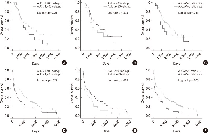
We also assessed ALC, AMC and the ALC/AMC ratio in patients subdivided for factors that can affect prognosis and treatment selection (such as ASCT vs no ASCT, age, sex, and ISS) to determine whether they could predict OS. Among patients who did not undergo ASCT, those with low ALC, high AMC, and low ALC/AMC ratio had significantly shorter OS (p=.029, p=.025, and p=.003, respectively), but patients who received ASCT did not show significant differences in survival (p=.221, p=.323, and p=.343, respectively) (Fig. 2). When we stratified patients according to age, sex, and ISS, low ALC was associated with poor OS in patients who were <65 years-old (p=.009), male (p=.001), or had ISS stage II disease (p=.031). High AMC was associated with poor survival in the ISS stage I group (p<.0001). Low ALC/AMC ratio was associated with poor survival in patients who were <65 years-old (p=.008), male (p=.018), or had ISS stage I (p=.017) or stage III disease (p=.010) (Table 4).
The aim of the present study was to investigate the role of AMC as a prognostic biomarker for MM and to determine its relationship with ALC, an established prognostic factor for MM. To our knowledge, this is the first study to demonstrate the prognostic value of AMC and the ALC/AMC ratio in newly diagnosed MM.
Low ALC (<1,400 cells/µL), high AMC (≥490 cells/µL), and low ALC/AMC ratio (<2.9) were associated with poor OS and poor prognostic factors such as high ISS stage, plasmablastic morphology, hypoalbuminemia, and high β2m. However, on multivariate analysis, only low ALC/AMC ratio was an independent prognostic factor for MM.
Although ALC did not have statistical significance in multivariate analysis due to the relatively small sample size, our results are in agreement with previous reports showing that a high ALC is associated with better prognosis in newly diagnosed MM (cut-off point, 1,400 cells/µL; the same as that used our study),3 relapsed MM after Vel-Dex therapy (cut-off point, 1,100 cells/µL)23 and other lymphomas.24
We also found that shorter OS in MM patients was associated with high AMC and low ALC/AMC ratio, which is similar to the findings of previous studies of malignant lymphomas. AMC has been shown to be a reliable prognostic marker for DLBCL (cut-off point, 630 cells/µL),16 FL (cut-off point, 570 cells/µL),17 and HL (cut-off point, 900 cells/µL),18 and in classical HL, the ALC/AMC ratio at diagnosis is an independent prognostic factor for survival (cut-off point, 1.1).18
Recently it has been reported that TAMs are a prognostic marker for the survival of patients with MM.13 TAMs, which constitute a significant proportion of tumor-infiltrating inflammatory cells, have been linked to the growth, angiogenesis and metastasis of a variety of cancers.7 In MM, macrophages are an abundant and important component of BM stromal cells and contribute to tumor angiogenesis.25,26 TAMs, which are continually being recruited and activated both in an autocrine manner and by cytokines secreted by myeloma cells, adapt functionally, phenotypically and morphologically to collaborate with endothelial cells in vessel formation.27 Moreover, BM macrophages protect myeloma cells from apoptosis.28 TAMs therefore promote tumor growth not only by supporting angiogenesis, but also by protecting tumor cells from apoptosis, via the induction of immunosuppression.29 The important role TAMs play in the biology of MM, thereby affecting patient outcome, makes them a potential target for anti-vascular therapy in MM.26
As TAMs are derived from circulating monocytes and are recruited to the tumor site by tumor-derived chemotactic factors,14,15,30 obtaining an AMC from a CBC at diagnosis is simple and could be more easily applied in clinical practice than counting TAMs. In this study, the ALC/AMC ratio was an independent prognostic factor in MM. Moreover, the ALC/AMC ratio, which may be a reflection of both the tumor microenvironment and host immune status, could provide prognostic information independently.
We found that the patient group with high AMC and low ALC/AMC ratio trended toward worse OS whether or not they received ASCT, although statistical significance was not reached in the ASCT group because of the small sample size. A larger prospective cohort study of uniformly treated patients is required to validate these findings.
In conclusion, the results of the present study suggest that AMC, and, in particular, ALC/AMC ratio, which was found to be an independent prognostic factor for patient survival, could be used to determine the prognosis of MM.
  • 1. Porrata LF, Ristow K, Habermann TM, Witzig TE, Inwards DJ, Markovic SN. Absolute lymphocyte count at the time of first relapse predicts survival in patients with diffuse large B-cell lymphoma. Am J Hematol 2009; 84: 93-97. ArticlePubMed
  • 2. Kim H, Sohn HJ, Kim S, Lee JS, Kim WK, Suh C. Early lymphocyte recovery predicts longer survival after autologous peripheral blood stem cell transplantation in multiple myeloma. Bone Marrow Transplant 2006; 37: 1037-1042. ArticlePubMed
  • 3. Ege H, Gertz MA, Markovic SN, et al. Prediction of survival using absolute lymphocyte count for newly diagnosed patients with multiple myeloma: a retrospective study. Br J Haematol 2008; 141: 792-798. ArticlePubMed
  • 4. Podar K, Chauhan D, Anderson KC. Bone marrow microenvironment and the identification of new targets for myeloma therapy. Leukemia 2009; 23: 10-24. ArticlePubMed
  • 5. Mitsiades CS, Mitsiades NS, Richardson PG, Munshi NC, Anderson KC. Multiple myeloma: a prototypic disease model for the characterization and therapeutic targeting of interactions between tumor cells and their local microenvironment. J Cell Biochem 2007; 101: 950-968. ArticlePubMed
  • 6. Coussens LM, Werb Z. Inflammation and cancer. Nature 2002; 420: 860-867. ArticlePubMedPMC
  • 7. Pollard JW. Tumour-educated macrophages promote tumour progression and metastasis. Nat Rev Cancer 2004; 4: 71-78. ArticlePubMed
  • 8. Lamagna C, Aurrand-Lions M, Imhof BA. Dual role of macrophages in tumor growth and angiogenesis. J Leukoc Biol 2006; 80: 705-713. ArticlePubMed
  • 9. Alvaro-Naranjo T, Lejeune M, Salvadó-Usach MT, et al. Tumor-infiltrating cells as a prognostic factor in Hodgkin's lymphoma: a quantitative tissue microarray study in a large retrospective cohort of 267 patients. Leuk Lymphoma 2005; 46: 1581-1591. ArticlePubMed
  • 10. Steidl C, Lee T, Shah SP, et al. Tumor-associated macrophages and survival in classic Hodgkin's lymphoma. N Engl J Med 2010; 362: 875-885. ArticlePubMedPMC
  • 11. Pusztaszeri MP, Seelentag W, Bosman FT. Immunohistochemical expression of endothelial markers CD31, CD34, von Willebrand factor, and Fli-1 in normal human tissues. J Histochem Cytochem 2006; 54: 385-395. ArticlePubMed
  • 12. Farinha P, Masoudi H, Skinnider BF, et al. Analysis of multiple biomarkers shows that lymphoma-associated macrophage (LAM) content is an independent predictor of survival in follicular lymphoma (FL). Blood 2005; 106: 2169-2174. ArticlePubMed
  • 13. Suyanı E, Sucak GT, Akyürek N, et al. Tumor-associated macrophages as a prognostic parameter in multiple myeloma. Ann Hematol 2013; 92: 669-677. ArticlePubMed
  • 14. Ribatti D, Nico B, Crivellato E, Vacca A. Macrophages and tumor angiogenesis. Leukemia 2007; 21: 2085-2089. ArticlePubMed
  • 15. Green CE, Liu T, Montel V, et al. Chemoattractant signaling between tumor cells and macrophages regulates cancer cell migration, metastasis and neovascularization. PLoS One 2009; 4: e6713.ArticlePubMedPMC
  • 16. Porrata LF, Ristow K, Habermann TM, et al. Absolute monocyte/lymphocyte count prognostic score is independent of immunohistochemically determined cell of origin in predicting survival in diffuse large B-cell lymphoma. Leuk Lymphoma 2012; 53: 2159-2165. ArticlePubMed
  • 17. Wilcox RA, Ristow K, Habermann TM, et al. The absolute monocyte count is associated with overall survival in patients newly diagnosed with follicular lymphoma. Leuk Lymphoma 2012; 53: 575-580. ArticlePubMed
  • 18. Porrata LF, Ristow K, Colgan JP, et al. Peripheral blood lymphocyte/monocyte ratio at diagnosis and survival in classical Hodgkin's lymphoma. Haematologica 2012; 97: 262-269. ArticlePubMedPMC
  • 19. Inoue H. Overview of automated hematology analyzer XE-2100TM. Sysmex J Int 1999; 9: 58-64. Article
  • 20. Clark TG, Bradburn MJ, Love SB, Altman DG. Survival analysis part I: basic concepts and first analyses. Br J Cancer 2003; 89: 232-238. ArticlePubMedPMC
  • 21. Hothorn T, Lausen B. On the exact distribution of maximally selected rank statistics. Comput Stat Data Anal 2003; 43: 121-137. Article
  • 22. Boulesteix AL. Maximally selected chi-square statistics for ordinal variables. Biom J 2006; 48: 451-462. ArticlePubMed
  • 23. Song MK, Chung JS, Joo YD, et al. Clinical value of absolute lymphocyte counts before bortezomib-dexamethasone therapy in relapsed multiple myeloma patients. Acta Haematol 2010; 124: 34-39. ArticlePubMed
  • 24. Le Jeune C, Bertoli S, Elhamri M, et al. Initial absolute lymphocyte count as a prognostic factor for outcome in acute myeloid leukemia. Leuk Lymphoma 2013 8 05 [Epub]. http://dx.doi.org/10.3109/10428194.2013.813504. Article
  • 25. Scavelli C, Nico B, Cirulli T, et al. Vasculogenic mimicry by bone marrow macrophages in patients with multiple myeloma. Oncogene 2008; 27: 663-674. ArticlePubMed
  • 26. Berardi S, Ria R, Reale A, et al. Multiple myeloma macrophages: pivotal players in the tumor microenvironment. J Oncol 2013; 2013: 183602.ArticlePubMedPMCPDF
  • 27. Vacca A, Ribatti D, Presta M, et al. Bone marrow neovascularization, plasma cell angiogenic potential, and matrix metalloproteinase-2 secretion parallel progression of human multiple myeloma. Blood 1999; 93: 3064-3073. ArticlePubMedPDF
  • 28. Zheng Y, Cai Z, Wang S, et al. Macrophages are an abundant component of myeloma microenvironment and protect myeloma cells from chemotherapy drug-induced apoptosis. Blood 2009; 114: 3625-3628. ArticlePubMedPMC
  • 29. Ben-Baruch A. Inflammation-associated immune suppression in cancer: the roles played by cytokines, chemokines and additional mediators. Semin Cancer Biol 2006; 16: 38-52. ArticlePubMed
  • 30. Dirkx AE, Oude Egbrink MG, Wagstaff J, Griffioen AW. Monocyte/macrophage infiltration in tumors: modulators of angiogenesis. J Leukoc Biol 2006; 80: 1183-1196. ArticlePubMed
Fig. 1
Kaplan-Meier curves of overall survival in multiple myeloma patients according to absolute lymphocyte count (ALC) (A), absolute monocyte count (AMC) (B), and the ALC/AMC ratio (C).
kjpathol-47-526-g001.jpg
Fig. 2
Kaplan-Meier curves of overall survival in multiple myeloma patients with (A-C) or without (D-F) autologous stem cell transplantation, according to absolute lymphocyte count (ALC) (A, D), absolute monocyte count (AMC) (B, E), and the ALC/AMC ratio (C, F).
kjpathol-47-526-g002.jpg
Table 1.
Characteristics of multiple myeloma patients according to ALC, AMC, and the ALC/AMC ratio
Characteristic ALC < 1,400 cells/µL ALC ≥ 1,400 cells/µL p-value AMC < 490 cells/µL AMC ≥ 490 cells/µL p-value ALC/AMC < 2.9 ALC/AMC ≥ 2.9 p-value
Age 60 (31-81) 60 (29-84) .939a 60 (29-81) 60 (31-84) .934a 62 (31-84) 59 (29-81) .130a
Sex .459b < .0001b .011b
 Male 41 (41.8) 57 (58.2) 55 (56.1) 43 (43.9) 36 (36.7) 62 (63.3)
 Female 33 (36.3) 58 (63.7) 77 (84.6) 14 (15.4) 18 (19.8) 73 (80.2)
International Staging System (ISS) .004c .195c .142c
 I 8 (22.9) 27 (77.1) 27 (77.1) 8 (22.9) 7 (20.0) 28 (80.0)
 II 29 (33.3) 58 (66.7) 62 (71.3) 25 (28.7) 23 (26.4) 64 (73.6)
 III 33 (54.1) 28 (45.9) 37 (60.7) 24 (39.3) 23 (37.7) 38 (62.3)
Durie-Salmon stage .745c .641c .263c
 I 6 (28.6) 15 (71.4) 15 (71.4) 6 (28.6) 8 (38.1) 13 (61.9)
 II 22 (42.3) 30 (57.7) 37 (71.2) 15 (28.8) 12 (23.1) 40 (76.9)
 IIIA 35 (39.3) 54 (60.7) 64 (71.9) 25 (28.1) 23 (25.8) 66 (74.2)
 IIIB 11 (40.7) 16 (59.3) 16 (59.3) 11 (40.7) 11 (40.7) 16 (59.3)
Morphology .027b .112b .008b
 Plasmablastic 29 (50.9) 28 (49.1) 33 (57.9) 24 (42.1) 26 (45.6) 31 (54.4)
 Plasmacytic 32 (32.3) 67 (67.7) 71 (71.7) 28 (28.3) 24 (24.2) 75 (75.8)
Bone lesion .233b 1.000b .873b
 ≥ 3 lytic bone lesions 42 (43.8) 54 (56.3) 67 (69.8) 29 (30.2) 28 (29.2) 68 (70.8)
 < 3 lytic bone lesions 32 (34.4) 61 (65.6) 65 (69.9) 28 (30.1) 26 (28.0) 67 (72.0)
β2m (mg/L) 8.98 (0.89-82.50) 7.80 (1.40-65.80) .122a 6.47 (0.89-48.40) 12.20 (1.60-82.50) .015a 10.96 (0.89-82.50) 7.14 (1.00-63.80) .087a
Albumin (g/dL) 3.00 (0.90-10.70) 3.22 (1.10-4.60) .004a 3.12 (0.90-10.70) 3.17 (0.90-4.60) .316a 2.99 (0.90-4.50) 3.19 (0.90-10.70) .265a
Cr (mg/dL) 1.70 (0.50-14.40) 1.75 (0.39-18.70) .976a 1.50 (0.50-14.40) 2.27 (0.39-18.70) .014a 2.22 (0.39-18.70) 1.53 (0.50-8.50) .282a
Hb (g/dL) 9.81 (6.40-14.80) 10.68 (5.50-15.40) .005a 10.17 (5.50-15.40) 10.74 (6.30-15.00) .085a 10.48 (6.30-14.80) 10.29 (5.50-15.40) .510a
Ca (mg/dL) 9.13 (4.00-14.60) 9.35 (6.70-15.10) .131a 9.14 (4.00-14.10) 9.56 (7.10–15.10) .098a 9.57 (7.10-15.10) 9.14 (4.00-13.80) .928a
Serum protein (g/dL) 8.22 (3.90-17.00) 8.17 (4.70-13.60) .453a 8.30 (3.90-17.00) 7.93 (4.70-12.50) .280a 7.90 (4.60-12.50) 8.30 (3.90-17.00) .373a
24 hr urine protein (mg/day) 2,092 (11-13,947) 2,123 (9-14,286) .912a 2,049 (9-14,286) 2,261 (11-13,947) .793a 1,749 (11-13,947) 2,250 (9-14,286) .238a
WBC count (× 109/L) 5.72 (2.40-12.70) 6.86 (2.90-17.40) .001a 5.55 (2.40-16.00) 8.40 (4.40-17.40) < .0001a 7.44 (2.40-17.00) 5.99 (2.40-17.40) .003a
Platelets (× 109/L) 176 (23-461) 225 (39-663) .001a 194 (31-523) 231 (23-663) .028a 199 (23-663) 208 (38-584) .448a

Values are presencted as mean (range) or number (%).

ALC, absolute lymphocyte count; AMC, absolute monocyte count; β2m, β2-microglobulin; Cr, creatinine; Hb, hemoglobin; Ca, calcium; WBC, white blood cell.

aMann-Whitney U test;

bTwo-sided Fisher’s exact test;

cTwo-sided Pearson’s chi-square test.

Table 2.
Univariate analyses of ALC, AMC, and the ALC/AMC ratio for OS
Factor HR 95% CI p-value
ALC (cells/µL) ≥ 1,400 vs < 1,400 1.649 1.189-2.286 .002
AMC (cells/µL) < 490 vs ≥ 490 1.443 1.018-2.045 .038
ALC/AMC ratio ≥ 2.9 vs < 2.9 1.772 1.249-2.514 .001
Morphology Plasmacytic vs plasmablastic 1.552 1.081-2.228 .016
Age (yr) < 65 vs ≥ 65 1.793 1.279-2.513 .001
ISS I 1
II 2.148 1.258-3.670 .005
III 4.360 2.526-7.523 < .0001
D-S stage I 1
II 1.982 1.061-3.702 .032
IIIA 1.403 0.769-2.560 .270
IIIB 3.368 1.712-6.627 < .0001
β2-Microglobulin (mg/L) < 3.5 vs ≥ 3.5 2.128 1.468-3.084 < .0001
Albumin (g/dL) < 3.5 vs ≥ 3.5 1.573 1.115-2.219 .009
Bone lesion (lytic bone lesions) < 3 vs ≥ 3 1.108 0.799-1.535 .539
Treatment type ASCT vs no ASCT 2.345 1.637-3.359 < .0001

ALC, absolute lymphocyte count; AMC, absolute monocyte count; OS, overall survival; HR, hazard ratio; CI, confidence interval; ISS, International Staging System; D-S stage, Durie-Salmon stage; ASCT, autologous stem cell transplantation.

Table 3.
Multivariate analyses of ALC, AMC, and the ALC/AMC ratio for OS
Factors HR 95% CI p-value HR 95% CI p-value
ALC (cells/µL) ≥ 1,400 vs < 1,400 1.389 0.933-2.069 .106
AMC (cells/µL) < 490 vs ≥ 490 1.415 0.928-2.157 .107
ALC/AMC ratio ≥ 2.9 vs < 2.9 1.511 1.006-2.272 .047
Morphology Plasmacytic vs plasmablastic 1.396 0.940-2.074 .099 1.366 0.917-2.033 .125
Age (yr) < 65 vs ≥ 65 1.441 0.949-2.189 .087 1.388 0.910-2.116 .128
ISS I 1 1
II 1.129 0.543-2.348 .745 1.122 0.540-2.331 .757
III 1.799 0.824-3.925 .140 2.048 0.947-4.429 .069
D-S stage I 1 1
II 1.089 0.454-2.611 .849 1.151 0.482-2.750 .751
IIIA 0.997 0.427-2.326 .994 1.099 0.479-2.520 .824
IIIB 1.786 0.681-4.683 .239 1.899 0.725-4.975 .192
Treatment type ASCT vs no ASCT 2.252 1.405-3.610 .001 2.131 1.337-3.396 .001

ALC, absolute lymphocyte count; AMC, absolute monocyte count; OS, overall survival; HR, hazard ratio; CI, confidence interval; ISS, International Staging System; D-S stage, Durie-Salmon stage; ASCT, autologous stem cell transplantation.

Table 4.
Stratified univariate analyses for OS
Characteristics ALC < 1,400 cells/µL
AMC ≥ 490 cells/µL
ALC/AMC < 2.9
HR 95% CI p-value HR 95% CI p-value HR 95% CI p-value
Treatment
 ASCT (n = 67) 1.459 0.794-2.679 .221 1.376 0.728-2.600 .323 1.393 0.700-2.769 .343
 No ASCT (n = 122) 1.542 1.043-2.281 .029 1.613 1.058-2.457 .025 1.850 1.227-2.789 .003
Age (yr)
 < 65 (n = 121) 1.728 1.139-2.622 .009 1.517 0.971-2.369 .065 1.843 1.163-2.920 .008
 ≥ 65 (n = 68) 1.606 0.934-2.762 .084 1.323 0.752-2.327 .330 1.408 0.812-2.442 .221
Sex
 Male (n = 98) 2.112 1.351-3.301 .001 1.259 0.806-1.967 .310 1.719 1.091-2.708 .018
 Female (n = 91) 1.245 0.764-2.028 .379 1.433 0.748-2.744 .276 1.559 0.873-2.784 .130
ISS
 I (n = 35) 1.303 0.451-3.761 .624 6.865 2.408-19.576 < .0001 3.169 1.166-8.617 .017
 II (n = 87) 1.720 1.044-2.834 .031 0.982 0.563-1.712 .949 1.209 0.693-2.110 .504
 III (n = 61) 0.936 0.554-1.580 .804 1.171 0.683-2.005 .566 2.011 1.172-3.450 .010

OS, overall survival; ALC, absolute lymphocyte count; AMC, absolute monocyte count; HR, hazard ratio; CI, confidence interval; ASCT, autologous stem cell transplantation; ISS, International Staging System.

Figure & Data

References

    Citations

    Citations to this article as recorded by  
    • Variation of peripheral blood-based biomarkers for response of anti-PD-1 immunotherapy in non-small-cell lung cancer
      Xiaoming Wang, Dayu Chen, Yuyan Ma, Dongping Mo, Feng Yan
      Clinical and Translational Oncology.2024; 26(8): 1934.     CrossRef
    • Descriptive analysis and prognostic factors in cats with myeloma‐related disorders: A multicenter retrospective study of 50 cases
      Lorris Lecot, Isabelle Desmas‐Bazelle, Sarah Benjamin, Pauline De Fornel, Frédérique Ponce, Matthew Kornya, Loïc Desquilbet, Claire Beaudu‐Lange, Catherine Ibisch, David Sayag, Ghita Benchekroun, Jérémy Béguin
      Journal of Veterinary Internal Medicine.2024; 38(3): 1693.     CrossRef
    • Definers and drivers of functional high-risk multiple myeloma: insights from genomic, transcriptomic, and immune profiling
      Rahul Banerjee, Kara I. Cicero, Sarah S. Lee, Andrew J. Cowan
      Frontiers in Oncology.2023;[Epub]     CrossRef
    • Normal Absolute Monocyte Count in Combination with Normal/High Absolute Lymphocyte Count at the Time of Relapse is Associated with Improved Survival in Patients with Early Relapsed Acute Myeloid Leukemia
      Yu Zhang, Kanchun Dai, Qianying Zhang, Yisha Huang, Yiyun Feng, Deeksha Bhardwaj, Kang Yu, Jianhua Feng
      Cancer Investigation.2021; 39(6-7): 550.     CrossRef
    • Real World Experience of Daratumumab: Evaluating Lymphopenia and Adverse Events in Multiple Myeloma Patients
      Francesca Cottini, Ying Huang, Nita Williams, Naresh Bumma, Abdullah M. Khan, Maria Chaudhry, Srinivas Devarakonda, Yvonne A. Efebera, Don M. Benson, Ashley E. Rosko
      Frontiers in Oncology.2021;[Epub]     CrossRef
    • Are the Derived Indexes of Peripheral Whole Blood Cell Counts (NLR, PLR, LMR/MLR) Clinically Significant Prognostic Biomarkers in Multiple Myeloma? A Systematic Review And Meta-Analysis
      Xinwen Zhang, Jialin Duan, Zhenyu Wen, Hao Xiong, Xiaomin Chen, Yang Liu, Kunyu Liao, Chunlan Huang
      Frontiers in Oncology.2021;[Epub]     CrossRef
    • Combined immune score of lymphocyte to monocyte ratio and immunoglobulin levels predicts treatment-free survival of multiple myeloma patients after autologous stem cell transplant
      Karen Sweiss, Jonathan Lee, Nadim Mahmud, Gregory S. Calip, Youngmin Park, Dolores Mahmud, Damiano Rondelli, Pritesh R. Patel
      Bone Marrow Transplantation.2020; 55(1): 199.     CrossRef
    • Low absolute CD4+ T cell counts in peripheral blood predict poor prognosis in patients with newly diagnosed multiple myeloma
      Yan Gu, Yuanyuan Jin, Jie Ding, Wu Yujie, Qinglin Shi, Xiaoyan Qu, Sishu Zhao, Jianyong Li, Chen Lijuan
      Leukemia & Lymphoma.2020; 61(8): 1869.     CrossRef
    • Normal Absolute Monocyte Count at the Time of Relapse is Associated with Improved Survival After First Salvage Therapy in Adult Patients with Early Relapsed B-Lineage Acute Lymphoblastic Leukemia


      Yi-fen Shi, Na Wang, Zi-yang Huang, Rong-rong Chen, Yi-sha Huang, Yi-yi Zhu, Chong-yun Xing, Bin Liang, Kang Yu, Jian-hua Feng
      Cancer Management and Research.2020; Volume 12: 7097.     CrossRef
    • Peripheral blood biomarkers of early immune reconstitution in newly diagnosed multiple myeloma
      Moritz Binder, S. Vincent Rajkumar, Martha Q. Lacy, Morie A. Gertz, Francis K. Buadi, Angela Dispenzieri, Yi L. Hwa, Amie Fonder, Miriam Hobbs, Suzanne R. Hayman, Steven R. Zeldenrust, John A. Lust, Stephen J. Russell, Nelson Leung, Prashant Kapoor, Ronal
      American Journal of Hematology.2019; 94(3): 306.     CrossRef
    • Effect of absolute monocyte count post-transplant on the outcome of patients with acute myeloid leukemia undergoing myeloablative allogeneic hematopoietic stem cell transplant with busulfan and cyclophosphamide conditioning
      Liyuan Tang, Na Wang, Chongyun Xing, Qiang Zhuang, Bin Liang, Lan Sun, Yi Chen, Yan Qian, Zhijian Shen, Songfu Jiang, Kang Yu, Jianhua Feng
      Leukemia Research.2018; 69: 60.     CrossRef
    • A lower ALC/AMC ratio is associated with poor prognosis of peripheral T-cell lymphoma-not otherwise specified
      Qian Li, Shuang Gao, Jing Ma, Su Liu, Yuanfang Yue, Lin Chen, Han Li, Xue Wang, Dongying Li, Zeng Cao, Zhigang Zhao, Xiaofang Wang, Yong Yu, Yizhuo Zhang, Yafei Wang
      Leukemia Research.2018; 73: 5.     CrossRef
    • Peripheral Blood Lymphocyte-to-Monocyte Ratio as a Useful Prognostic Factor in Newly Diagnosed Multiple Myeloma
      Ying Tian, Yue Zhang, Wan-Qiu Zhu, Xiao-Lei Chen, He-Bing Zhou, Wen-Ming Chen
      BioMed Research International.2018; 2018: 1.     CrossRef
    • Significance of the absolute lymphocyte/monocyte ratio as a prognostic immune biomarker in newly diagnosed multiple myeloma
      T Dosani, F Covut, R Beck, J J Driscoll, M de Lima, E Malek
      Blood Cancer Journal.2017; 7(6): e579.     CrossRef
    • Lymphocyte-to-monocyte ratio can predict mortality in pancreatic adenocarcinoma
      Gurshawn Singh, Ammar Nassri, David Kim, Hong Zhu, Zeeshan Ramzan
      World Journal of Gastrointestinal Pharmacology and Therapeutics.2017; 8(1): 60.     CrossRef
    • Bone marrow microenvironmental CD4 + and CD8 + lymphocyte infiltration patterns define overall- and progression free survival in standard risk multiple myeloma – an analysis from the Austrian Myeloma Registry
      Wolfgang Willenbacher, Ella Willenbacher, Claudia Zelle-Rieser, Rainer Biedermann, Roman Weger, Karin Jöhrer, Andrea Brunner
      Leukemia & Lymphoma.2016; 57(6): 1478.     CrossRef
    • Absolute lymphocyte count as a prognostic marker in newly diagnosed multiple myeloma patients
      C. Suriu, L. Akria, D. Azoulay, E. Shaoul, M. Barhoum, A. Braester
      International Journal of Laboratory Hematology.2016;[Epub]     CrossRef
    • The Peripheral Blood Mononuclear Cell Count Is Associated With Bone Health in Elderly Men
      Xianfeng Lin, Hejun Yu, Chenchen Zhao, Yu Qian, Dun Hong, Kangmao Huang, Jian Mo, An Qin, Xiangqian Fang, Shunwu Fan
      Medicine.2016; 95(15): e3357.     CrossRef
    • Prognostic role of lymphocyte to monocyte ratio for patients with cancer: evidence from a systematic review and meta-analysis
      Liangyou Gu, Hongzhao Li, Luyao Chen, Xin Ma, Xintao Li, Yu Gao, Yu Zhang, Yongpeng Xie, Xu Zhang
      Oncotarget.2016; 7(22): 31926.     CrossRef
    • Do lymphocytes count in myeloma? Are we absolutely sure?
      Tamar Tadmor
      Leukemia & Lymphoma.2015; 56(5): 1193.     CrossRef
    • Absolute lymphocyte count is unrelated to overall survival in newly diagnosed elderly patients with multiple myeloma treated with immunomodulatory drugs
      Mariasanta Napolitano, Giorgia Saccullo, Roberto Bono, Antonio Branca, Clotilde Cangialosi, Salvatrice Mancuso, Simona Raso, Gerlando Quintini, Maria Grazia Lipari, Francesco Fabbiano, Giorgina Specchia, Alberto Dolce, Francesco Di Raimondo, Sergio Siragu
      Leukemia & Lymphoma.2015; 56(5): 1507.     CrossRef
    • Distinct Transcriptional and Anti-Mycobacterial Profiles of Peripheral Blood Monocytes Dependent on the Ratio of Monocytes: Lymphocytes
      Vivek Naranbhai, Helen A. Fletcher, Rachel Tanner, Matthew K. O'Shea, Helen McShane, Benjamin P. Fairfax, Julian C. Knight, Adrian V.S. Hill
      EBioMedicine.2015; 2(11): 1619.     CrossRef
    • Prognostic value of absolute monocyte count in chronic lymphocytic leukaemia
      László Szerafin, János Jakó, Ferenc Riskó
      Orvosi Hetilap.2015; 156(15): 592.     CrossRef
    • The lymphocyte to monocyte ratio in peripheral blood represents a novel prognostic marker in patients with pancreatic cancer
      Michael Stotz, Joanna Szkandera, Tatjana Stojakovic, Julia Seidel, Hellmut Samonigg, Peter Kornprat, Renate Schaberl-Moser, Fridericke Seggewies, Gerald Hoefler, Armin Gerger, Martin Pichler
      Clinical Chemistry and Laboratory Medicine (CCLM).2015;[Epub]     CrossRef

    • PubReader PubReader
    • ePub LinkePub Link
    • Cite this Article
      Cite this Article
      export Copy Download
      Close
      Download Citation
      Download a citation file in RIS format that can be imported by all major citation management software, including EndNote, ProCite, RefWorks, and Reference Manager.

      Format:
      • RIS — For EndNote, ProCite, RefWorks, and most other reference management software
      • BibTeX — For JabRef, BibDesk, and other BibTeX-specific software
      Include:
      • Citation for the content below
      Prognostic Significance of Absolute Lymphocyte Count/Absolute Monocyte Count Ratio at Diagnosis in Patients with Multiple Myeloma
      Korean J Pathol. 2013;47(6):526-533.   Published online December 24, 2013
      Close
    • XML DownloadXML Download
    Figure
    • 0
    • 1
    Prognostic Significance of Absolute Lymphocyte Count/Absolute Monocyte Count Ratio at Diagnosis in Patients with Multiple Myeloma
    Image Image
    Fig. 1 Kaplan-Meier curves of overall survival in multiple myeloma patients according to absolute lymphocyte count (ALC) (A), absolute monocyte count (AMC) (B), and the ALC/AMC ratio (C).
    Fig. 2 Kaplan-Meier curves of overall survival in multiple myeloma patients with (A-C) or without (D-F) autologous stem cell transplantation, according to absolute lymphocyte count (ALC) (A, D), absolute monocyte count (AMC) (B, E), and the ALC/AMC ratio (C, F).
    Prognostic Significance of Absolute Lymphocyte Count/Absolute Monocyte Count Ratio at Diagnosis in Patients with Multiple Myeloma
    Characteristic ALC < 1,400 cells/µL ALC ≥ 1,400 cells/µL p-value AMC < 490 cells/µL AMC ≥ 490 cells/µL p-value ALC/AMC < 2.9 ALC/AMC ≥ 2.9 p-value
    Age 60 (31-81) 60 (29-84) .939a 60 (29-81) 60 (31-84) .934a 62 (31-84) 59 (29-81) .130a
    Sex .459b < .0001b .011b
     Male 41 (41.8) 57 (58.2) 55 (56.1) 43 (43.9) 36 (36.7) 62 (63.3)
     Female 33 (36.3) 58 (63.7) 77 (84.6) 14 (15.4) 18 (19.8) 73 (80.2)
    International Staging System (ISS) .004c .195c .142c
     I 8 (22.9) 27 (77.1) 27 (77.1) 8 (22.9) 7 (20.0) 28 (80.0)
     II 29 (33.3) 58 (66.7) 62 (71.3) 25 (28.7) 23 (26.4) 64 (73.6)
     III 33 (54.1) 28 (45.9) 37 (60.7) 24 (39.3) 23 (37.7) 38 (62.3)
    Durie-Salmon stage .745c .641c .263c
     I 6 (28.6) 15 (71.4) 15 (71.4) 6 (28.6) 8 (38.1) 13 (61.9)
     II 22 (42.3) 30 (57.7) 37 (71.2) 15 (28.8) 12 (23.1) 40 (76.9)
     IIIA 35 (39.3) 54 (60.7) 64 (71.9) 25 (28.1) 23 (25.8) 66 (74.2)
     IIIB 11 (40.7) 16 (59.3) 16 (59.3) 11 (40.7) 11 (40.7) 16 (59.3)
    Morphology .027b .112b .008b
     Plasmablastic 29 (50.9) 28 (49.1) 33 (57.9) 24 (42.1) 26 (45.6) 31 (54.4)
     Plasmacytic 32 (32.3) 67 (67.7) 71 (71.7) 28 (28.3) 24 (24.2) 75 (75.8)
    Bone lesion .233b 1.000b .873b
     ≥ 3 lytic bone lesions 42 (43.8) 54 (56.3) 67 (69.8) 29 (30.2) 28 (29.2) 68 (70.8)
     < 3 lytic bone lesions 32 (34.4) 61 (65.6) 65 (69.9) 28 (30.1) 26 (28.0) 67 (72.0)
    β2m (mg/L) 8.98 (0.89-82.50) 7.80 (1.40-65.80) .122a 6.47 (0.89-48.40) 12.20 (1.60-82.50) .015a 10.96 (0.89-82.50) 7.14 (1.00-63.80) .087a
    Albumin (g/dL) 3.00 (0.90-10.70) 3.22 (1.10-4.60) .004a 3.12 (0.90-10.70) 3.17 (0.90-4.60) .316a 2.99 (0.90-4.50) 3.19 (0.90-10.70) .265a
    Cr (mg/dL) 1.70 (0.50-14.40) 1.75 (0.39-18.70) .976a 1.50 (0.50-14.40) 2.27 (0.39-18.70) .014a 2.22 (0.39-18.70) 1.53 (0.50-8.50) .282a
    Hb (g/dL) 9.81 (6.40-14.80) 10.68 (5.50-15.40) .005a 10.17 (5.50-15.40) 10.74 (6.30-15.00) .085a 10.48 (6.30-14.80) 10.29 (5.50-15.40) .510a
    Ca (mg/dL) 9.13 (4.00-14.60) 9.35 (6.70-15.10) .131a 9.14 (4.00-14.10) 9.56 (7.10–15.10) .098a 9.57 (7.10-15.10) 9.14 (4.00-13.80) .928a
    Serum protein (g/dL) 8.22 (3.90-17.00) 8.17 (4.70-13.60) .453a 8.30 (3.90-17.00) 7.93 (4.70-12.50) .280a 7.90 (4.60-12.50) 8.30 (3.90-17.00) .373a
    24 hr urine protein (mg/day) 2,092 (11-13,947) 2,123 (9-14,286) .912a 2,049 (9-14,286) 2,261 (11-13,947) .793a 1,749 (11-13,947) 2,250 (9-14,286) .238a
    WBC count (× 109/L) 5.72 (2.40-12.70) 6.86 (2.90-17.40) .001a 5.55 (2.40-16.00) 8.40 (4.40-17.40) < .0001a 7.44 (2.40-17.00) 5.99 (2.40-17.40) .003a
    Platelets (× 109/L) 176 (23-461) 225 (39-663) .001a 194 (31-523) 231 (23-663) .028a 199 (23-663) 208 (38-584) .448a
    Factor HR 95% CI p-value
    ALC (cells/µL) ≥ 1,400 vs < 1,400 1.649 1.189-2.286 .002
    AMC (cells/µL) < 490 vs ≥ 490 1.443 1.018-2.045 .038
    ALC/AMC ratio ≥ 2.9 vs < 2.9 1.772 1.249-2.514 .001
    Morphology Plasmacytic vs plasmablastic 1.552 1.081-2.228 .016
    Age (yr) < 65 vs ≥ 65 1.793 1.279-2.513 .001
    ISS I 1
    II 2.148 1.258-3.670 .005
    III 4.360 2.526-7.523 < .0001
    D-S stage I 1
    II 1.982 1.061-3.702 .032
    IIIA 1.403 0.769-2.560 .270
    IIIB 3.368 1.712-6.627 < .0001
    β2-Microglobulin (mg/L) < 3.5 vs ≥ 3.5 2.128 1.468-3.084 < .0001
    Albumin (g/dL) < 3.5 vs ≥ 3.5 1.573 1.115-2.219 .009
    Bone lesion (lytic bone lesions) < 3 vs ≥ 3 1.108 0.799-1.535 .539
    Treatment type ASCT vs no ASCT 2.345 1.637-3.359 < .0001
    Factors HR 95% CI p-value HR 95% CI p-value
    ALC (cells/µL) ≥ 1,400 vs < 1,400 1.389 0.933-2.069 .106
    AMC (cells/µL) < 490 vs ≥ 490 1.415 0.928-2.157 .107
    ALC/AMC ratio ≥ 2.9 vs < 2.9 1.511 1.006-2.272 .047
    Morphology Plasmacytic vs plasmablastic 1.396 0.940-2.074 .099 1.366 0.917-2.033 .125
    Age (yr) < 65 vs ≥ 65 1.441 0.949-2.189 .087 1.388 0.910-2.116 .128
    ISS I 1 1
    II 1.129 0.543-2.348 .745 1.122 0.540-2.331 .757
    III 1.799 0.824-3.925 .140 2.048 0.947-4.429 .069
    D-S stage I 1 1
    II 1.089 0.454-2.611 .849 1.151 0.482-2.750 .751
    IIIA 0.997 0.427-2.326 .994 1.099 0.479-2.520 .824
    IIIB 1.786 0.681-4.683 .239 1.899 0.725-4.975 .192
    Treatment type ASCT vs no ASCT 2.252 1.405-3.610 .001 2.131 1.337-3.396 .001
    Characteristics ALC < 1,400 cells/µL
    AMC ≥ 490 cells/µL
    ALC/AMC < 2.9
    HR 95% CI p-value HR 95% CI p-value HR 95% CI p-value
    Treatment
     ASCT (n = 67) 1.459 0.794-2.679 .221 1.376 0.728-2.600 .323 1.393 0.700-2.769 .343
     No ASCT (n = 122) 1.542 1.043-2.281 .029 1.613 1.058-2.457 .025 1.850 1.227-2.789 .003
    Age (yr)
     < 65 (n = 121) 1.728 1.139-2.622 .009 1.517 0.971-2.369 .065 1.843 1.163-2.920 .008
     ≥ 65 (n = 68) 1.606 0.934-2.762 .084 1.323 0.752-2.327 .330 1.408 0.812-2.442 .221
    Sex
     Male (n = 98) 2.112 1.351-3.301 .001 1.259 0.806-1.967 .310 1.719 1.091-2.708 .018
     Female (n = 91) 1.245 0.764-2.028 .379 1.433 0.748-2.744 .276 1.559 0.873-2.784 .130
    ISS
     I (n = 35) 1.303 0.451-3.761 .624 6.865 2.408-19.576 < .0001 3.169 1.166-8.617 .017
     II (n = 87) 1.720 1.044-2.834 .031 0.982 0.563-1.712 .949 1.209 0.693-2.110 .504
     III (n = 61) 0.936 0.554-1.580 .804 1.171 0.683-2.005 .566 2.011 1.172-3.450 .010
    Table 1. Characteristics of multiple myeloma patients according to ALC, AMC, and the ALC/AMC ratio

    Values are presencted as mean (range) or number (%).

    ALC, absolute lymphocyte count; AMC, absolute monocyte count; β2m, β2-microglobulin; Cr, creatinine; Hb, hemoglobin; Ca, calcium; WBC, white blood cell.

    Mann-Whitney U test;

    Two-sided Fisher’s exact test;

    Two-sided Pearson’s chi-square test.

    Table 2. Univariate analyses of ALC, AMC, and the ALC/AMC ratio for OS

    ALC, absolute lymphocyte count; AMC, absolute monocyte count; OS, overall survival; HR, hazard ratio; CI, confidence interval; ISS, International Staging System; D-S stage, Durie-Salmon stage; ASCT, autologous stem cell transplantation.

    Table 3. Multivariate analyses of ALC, AMC, and the ALC/AMC ratio for OS

    ALC, absolute lymphocyte count; AMC, absolute monocyte count; OS, overall survival; HR, hazard ratio; CI, confidence interval; ISS, International Staging System; D-S stage, Durie-Salmon stage; ASCT, autologous stem cell transplantation.

    Table 4. Stratified univariate analyses for OS

    OS, overall survival; ALC, absolute lymphocyte count; AMC, absolute monocyte count; HR, hazard ratio; CI, confidence interval; ASCT, autologous stem cell transplantation; ISS, International Staging System.


    J Pathol Transl Med : Journal of Pathology and Translational Medicine
    TOP