 E-submission
Articles
- Page Path
- HOME > J Pathol Transl Med > Volume 48(3); 2014 > Article
-
Case Study
A Case of Metastatic Angiosarcoma Diagnosed by Liquid-Based Preparation: Peculiar Cytoplasmic Changes - Min Jung Jung, Young Ok Kim
-
Korean Journal of Pathology 2014;48(3):241-247.
DOI: https://doi.org/10.4132/KoreanJPathol.2014.48.3.241
Published online: June 26, 2014
Department of Pathology, Kosin University College of Medicine, Busan, Korea.
- Corresponding Author: Young Ok Kim, M.D. Department of Pathology, Kosin University College of Medicine, 262 Gamcheon-ro, Seo-gu, Busan 602-702, Korea. Tel: +82-51-990-6325, Fax: +82-51-990-3080, suajoon@kosinmed.or.kr
• Received: April 29, 2013 • Revised: April 2, 2014 • Accepted: April 7, 2014
© 2014 The Korean Society of Pathologists/The Korean Society for Cytopathology
This is an Open Access article distributed under the terms of the Creative Commons Attribution Non-Commercial License (http://creativecommons.org/licenses/by-nc/3.0/) which permits unrestricted non-commercial use, distribution, and reproduction in any medium, provided the original work is properly cited.
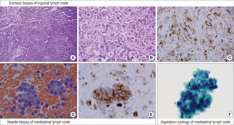

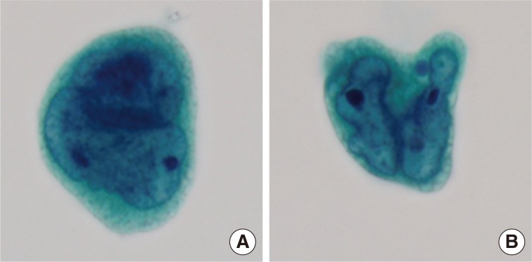
Figure & Data
References
Citations
Citations to this article as recorded by 
- Cytological Features of a Metastatic Angiosarcoma in the Lymph Node Diagnosed via Liquid-Based Cytology
Jie-Yang Jhuang, Chih-Yi Liu, Min-Hui Tseng, Shih-Sung Chuang
Diagnostics.2023; 13(12): 2124. CrossRef - Radiation-associated Angiosarcoma Presenting as Massive Pleural Effusion
Hirokazu Ogino, Makoto Tobiume, Kozo Kagawa, Hiroshi Kawano, Satoshi Sakaguchi, Atsuro Saijo, Daisuke Matsumoto, Hiromitsu Takizawa, Yuriko Morikawa, Yoshimi Bando, Hisatsugu Goto, Hiroshi Nokihara, Yasuhiko Nishioka
Internal Medicine.2022; 61(9): 1393. CrossRef - Delayed diagnosis of angiosarcoma of the spleen: clinically presenting as recurrent haemoperitoneum following embolisation
Verena Kornmann, Philip van Rijn, Dries Mulder, Koen Reijnders
BMJ Case Reports.2015; 2015: bcr2014208956. CrossRef - A Case of Angiosarcoma of the Scalp with Invasion to the Pleural Effusion
Yusuke Amano, Yukari Obana, Yoko Nakanishi, Ryusuke Tsujimura, Kayomi Wakamatsu, Fumiko Uemura, Yoshihisa Katsura, Masahiko Sugitani, Norimichi Nemoto
Journal of Nihon University Medical Association.2015; 74(3): 113. CrossRef
 PubReader
 Cite this Article

A Case of Metastatic Angiosarcoma Diagnosed by Liquid-Based Preparation: Peculiar Cytoplasmic Changes


Fig. 1 Overall histopathologic and cytopathologic findings in excision of inguinal lymph node, needle biopsy of mediastinal lymph node, and aspiration cytology by liquid-based preparation (LBP) of mediastinal lymph nodes. (A) Epithelioid tumor cells are arranged in a solid sheet and nest with focal vascular channel formation. (B) The major tumor cells are large and epithelioid with round nuclei, prominent nucleoli and sparse to plump cytoplasm. (C) Tumor cells are positive for CD31. (D) A few loose clusters or microacini with a central lumen are identified. (E) Tumor cells are positive for CD31. (F) Vague microacini formation is also identified in aspiration cytology by LBP (Papanicolaou stain).
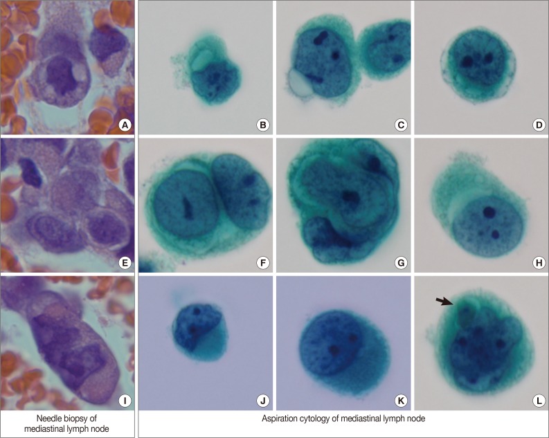
Fig. 2 Variable cytoplasmic features in needle biopsy of mediastinal lymph nodes and aspiration cytology by liquid-based preparation of mediastinal lymph nodes. Needle biopsy demonstrates intracytoplasmic vacuoles (A), perinuclear clearing (E), and juxtanuclear condensation (I). These histopathologic findings correspond to the cytopathologic findings in aspiration cytology. Intracytoplasmic vacuoles exhibit variable shapes: prominent intracytoplasmic vacuoles (B), a protruding vacuole beyond the cytoplasmic border (C), and several small vacuoles encircling the nuclei to form a band-like appearance (D). Perinuclear clearing is identified circumferentially (F, G) or sectionally (H). Condensed juxtanuclear cytoplasm with nuclear indentation makes a rhabdoid appearance (J, K). Intracytoplasmic degenerative red blood cells (L, arrow) are present (B-D, F-H, J-L, Papanicolaou stain).
Fig. 3 Variable nuclear features in aspiration cytology by liquid-based preparation (LBP) of mediastinal lymph nodes. (A) Tumor cells show binucleated, large nuclei with irregular borders. Many small vacuoles are present in the cytoplasm. (B) Tumor cells show markedly irregular nuclei with prominent nucleoli, resembling Reed-Sternberg cells (A, B, Papanicolaou stain).
Fig. 1
Fig. 2
Fig. 3
A Case of Metastatic Angiosarcoma Diagnosed by Liquid-Based Preparation: Peculiar Cytoplasmic Changes

