Warning: mkdir(): Permission denied in /home/virtual/lib/view_data.php on line 81

Warning: fopen(upload/ip_log/ip_log_2024-11.txt): failed to open stream: No such file or directory in /home/virtual/lib/view_data.php on line 83

Warning: fwrite() expects parameter 1 to be resource, boolean given in /home/virtual/lib/view_data.php on line 84
Bilateral Stafne Bone Cavity in the Anterior Mandible with Heterotopic Salivary Gland Tissue: A Case Report
Skip Navigation
Skip to contents

J Pathol Transl Med : Journal of Pathology and Translational Medicine

OPEN ACCESS
SEARCH
Search

Articles

Page Path
HOME > J Pathol Transl Med > Volume 48(3); 2014 > Article
Brief Case Report
Bilateral Stafne Bone Cavity in the Anterior Mandible with Heterotopic Salivary Gland Tissue: A Case Report
Hyunchul Kim, Jae Yeon Seok, Sangho Lee, Jungsuk An, Na Rae Kim, Dong Hae Chung, Hyun Yee Cho, Seung Yeon Ha
Korean Journal of Pathology 2014;48(3):248-249.
DOI: https://doi.org/10.4132/KoreanJPathol.2014.48.3.248
Published online: June 26, 2014

Department of Pathology, Gachon University Gil Medical Center, Incheon, Korea.

Corresponding Author: Seung Yeon Ha, M.D. Department of Pathology, Gachon University Gil Medical Center, 21 Namdong-daero 774beon-gil, Namdong-gu, Incheon 405-760, Korea. Tel: +82-32-460-3078, Fax: +82-32-460-2394, syha@gilhospital.com
• Received: June 7, 2013   • Revised: July 10, 2013   • Accepted: July 23, 2013

© 2014 The Korean Society of Pathologists/The Korean Society for Cytopathology

This is an Open Access article distributed under the terms of the Creative Commons Attribution Non-Commercial License (http://creativecommons.org/licenses/by-nc/3.0/) which permits unrestricted non-commercial use, distribution, and reproduction in any medium, provided the original work is properly cited.

prev next
  • 12,596 Views
  • 86 Download
  • 10 Crossref
  • 14 Scopus
Stafne bone cavity is a well demarcated defect of the mandible, usually asymptomatic and located in the posterior portion of the bone.1 Most cases have been reported in male patients between the age of 50 and 70 years.2 This lesion has been labeled with various terms, including ectopic salivary gland, idiopathic defect, mandibular salivary gland inclusion, Stafne bone cavity and cyst.2 The cavities are often filled with normal salivary gland tissue, but occasional cases showed cavity contents that included skeletal muscle, fibrous connective tissue and adipose tissue.1,2 Bilateral Stafne bone cavity of the anterior mandible is extremely rare and only seven such cases have been reported previously.2 Herein, we report a case of bilateral Stafne bone cavity of the anterior mandible.
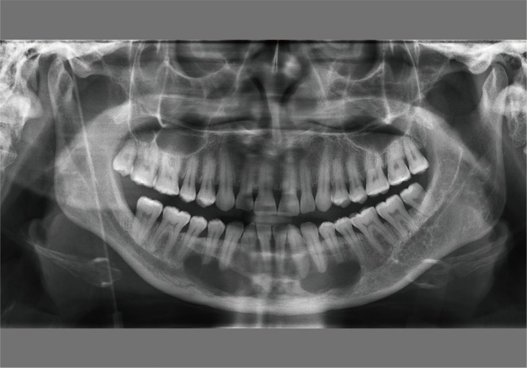
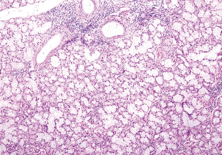
Two well demarcated ovoid radiolucent lesions (right, 3.9×1.5 cm and left, 2.9×1.1 cm) were found incidentally in the anterior mandible of a 44-year-old female patient on her panoramic dental X-ray (Fig. 1). The patient did not show any symptom of radiolucencies. The radiological findings were interpreted as radicular cysts. Two fragments of tissue from the cavities were submitted for histological diagnosis. The biopsied tissue fragments showed normal salivary gland tissue with mixed serous and mucinous cells. These findings were otherwise unremarkable, except for the small amount of lymphoid cell infiltration that was discovered (Fig. 2). The clinical, radiological, and pathological findings were consistent with Stafne bone cavity.
Since Stafne bone cavities were first reported in 1942, most cases have been found in the posterior mandible.3 However, these cavities were found also in the anterior portion of mandible in 1957 by Richard and Ziskind.4 Until now, only approximately fifty cases of anterior Stafne bone cavities have been reported.2
Similar to Stafne bone cavities located elsewhere in the mandible, anterior lesions show a wide age range (18 to 68 years) and higher prevalence in males (3:1, males:females).2 Most Stafne bone cavities are diagnosed on radiology, and only clinical follow-up with additional radiographic examination is sufficient unless there are symptoms or progression. The rarity of anterior Stafne bone cavities causes confusion and can hinder accurate and prompt diagnosis of this condition. Because the cavities resemble other periapical radiolucencies, they can appear similar to other cysts or tumors.2,5,6 The differential diagnosis of bilateral lesions includes, salivary gland tumors, fibro-osseous lesions, traumatic bone cysts, central giant cell lesions, hyperparathyroidism, ameloblastoma, eosinophilic granuloma, hemangioma, myxoma aneurysmal bone cyst, multiple myeloma, benign neurogenic tumors and arteriovenous fistula. Unlike tumors or the cysts, most Stafne bone cavities are asymptomatic and are therefore found accidentally.
In most cases, the content of the cavities was salivary gland tissue, and these lesions were sometimes referred to as heterotopic salivary glands. The salivary gland tissue contents suggeseted congenital malformation theory for the pathogenesis of the cavities,1 which states that the salivary gland tissue is congenitally entrapped within the mandible. Pressure from adjacent structures, including the hypertrophied salivary glands and facial arteries, has also been suggested as a possible pathogenic cause of Stafne bone cavities.7 A large-scale study of mandibular marrow tissue revealed the presence of intraosseous salivary gland tissue.8 As the authors of the article pointed out, such intraosseous heterotopias can serve as a pathogenic explanation and this finding can be interpreted as evidence supporting the former pathogenic theory of Stafne bone cavity formation. Our case lacks previous history and therefore, we were not able to determine the cavity's pathogenic cause. However, lymphocytic infiltration, which has been suggested as one of the causes of hypertrophied salivary glands in the pressure-induced acquired pathogenesis theory, is present in our case and therefore might be interpreted as a backup evidence for the theory.3
Although most Stafne bone cavities generally do not present any complications, there is one report of a Stafne bone cavity harboring pleomorphic adenoma.9 If a surgical specimen of the Stafne bone cavity is acquired, histological examination of the entire specimen is preferable for the detection of a possible salivary gland tumor within the specimen.
Because anterior Stafne bone cavity is very rare, accurate diagnosis of the lesion is challenging for clinicians.10 Pathologists should be aware of this lesion, because biopsies are performed in some cases and the cavity can harbor a salivary gland tumor.
  • 1. Apruzzese D, Longoni S. Stafne cyst in an anterior location. J Oral Maxillofac Surg 1999; 57: 333-338. ArticlePubMed
  • 2. Turkoglu K, Orhan K. Stafne bone cavity in the anterior mandible. J Craniofac Surg 2010; 21: 1769-1775. ArticlePubMed
  • 3. Sisman Y, Miloglu O, Sekerci AE, Yilmaz AB, Demirtas O, Tokmak TT. Radiographic evaluation on prevalence of Stafne bone defect: a study from two centres in Turkey. Dentomaxillofac Radiol 2012; 41: 152-158. ArticlePubMedPMC
  • 4. Richard EL, Ziskind J. Aberrant salivary gland tissue in mandible. Oral Surg Oral Med Oral Pathol 1957; 10: 1086-1090. ArticlePubMed
  • 5. Dorman M, Pierse D. Ectopic salivary gland tissue in the anterior mandible: a case report. Br Dent J 2002; 193: 571-572. ArticlePubMed
  • 6. Sisman Y, Etoz OA, Mavili E, Sahman H, Tarim Ertas E. Anterior Stafne bone defect mimicking a residual cyst: a case report. Dentomaxillofac Radiol 2010; 39: 124-126. ArticlePubMedPMC
  • 7. Buchner A, Carpenter WM, Merrell PW, Leider AS. Anterior lingual mandibular salivary gland defect: evaluation of twenty-four cases. Oral Surg Oral Med Oral Pathol 1991; 71: 131-136. ArticlePubMed
  • 8. Bouquot JE, Gnepp DR, Dardick I, Hietanen JH. Intraosseous salivary tissue: jawbone examples of choristomas, hamartomas, embryonic rests, and inflammatory entrapment: another histogenetic source for intraosseous adenocarcinoma. Oral Surg Oral Med Oral Pathol Oral Radiol Endod 2000; 90: 205-217. ArticlePubMed
  • 9. Simpson W. A Stafne's mandibular defect containing a pleomorphic adenoma: report of case. J Oral Surg 1965; 23: 553-556. ArticlePubMed
  • 10. Katz J, Chaushu G, Rotstein I. Stafne's bone cavity in the anterior mandible: a possible diagnostic challenge. J Endod 2001; 27: 304-307. ArticlePubMed
Fig. 1
Panoramic dental X-ray shows two radiolucencies beneath the apices of the canine and premolars in the right and left anterior mandible.
kjpathol-48-248-g001.jpg
Fig. 2
Biopsied cavity contents are composed of mixed seromucinous salivary gland tissue with lymphocytic infiltration.
kjpathol-48-248-g002.jpg

Figure & Data

References

    Citations

    Citations to this article as recorded by  
    • Diagnostic approach for the rare anterior variant of mandibular bone depression often misdiagnosed as tumorous lesions
      Hak-Sun Kim
      Journal of Dental Sciences.2024;[Epub]     CrossRef
    • Bilateral Stafne Bone Cavity in the Body of the Mandible: An Unusual Case Report and Literature Review
      Mayank Pahadia, Rutvi Vyas
      Cureus.2023;[Epub]     CrossRef
    • Effect of Stafne bone defect on the adjacent tooth: A review of the literature
      Mahdi Niknami, Azin Parsa, Zahra Khodadadi
      Imaging Science in Dentistry.2022; 52(2): 165.     CrossRef
    • Assessment of prevalence and volumetric estimation of possible Stafne bone concavities on cone beam computed tomography images
      Alaettin Koç, Cennet Neslihan Eroğlu, Ersen Bilgili
      Oral Radiology.2020; 36(3): 254.     CrossRef
    • Stafne’s bone cyst revisited and renamed: the benign mandibular concavity
      Johan K.M. Aps, Natasha Koelmeyer, Cina Yaqub
      Dentomaxillofacial Radiology.2020; 49(4): 20190475.     CrossRef
    • Cone‐beam computed tomography analysis of lingual mandibular bone depression in the premolar region: A case report
      Saeed Asgary, Naghmeh Emadi
      Clinical Case Reports.2020; 8(3): 523.     CrossRef
    • Letters to the Editor
      Ariyan S Araghi, Richard M Graham
      Dental Update.2019; 46(8): 792.     CrossRef
    • Radiographic features of lingual mandibular bone depression using dental cone beam computed tomography
      Liu Liu, Byung Cheol Kang, Suk Ja Yoon, Jae Seo Lee, Sel Ae Hwang
      Dentomaxillofacial Radiology.2018; 47(6): 20170383.     CrossRef
    • Stafne's bone cavity – unusual presentation in the anterior mandible
      Ioan Davies, Holly Boyes, James Wykes, Graham Smith
      Dental Update.2018; 45(4): 340.     CrossRef
    • Anterior stafne bone cyst mimicking periapical cyst: a case report
      Ji-Young Song
      Journal of Dental Rehabilitation and Applied Science.2016; 32(3): 209.     CrossRef

    • PubReader PubReader
    • Cite this Article
      Cite this Article
      export Copy Download
      Close
      Download Citation
      Download a citation file in RIS format that can be imported by all major citation management software, including EndNote, ProCite, RefWorks, and Reference Manager.

      Format:
      • RIS — For EndNote, ProCite, RefWorks, and most other reference management software
      • BibTeX — For JabRef, BibDesk, and other BibTeX-specific software
      Include:
      • Citation for the content below
      Bilateral Stafne Bone Cavity in the Anterior Mandible with Heterotopic Salivary Gland Tissue: A Case Report
      Korean J Pathol. 2014;48(3):248-249.   Published online June 26, 2014
      Close
    • XML DownloadXML Download
    Figure
    • 0
    • 1
    Bilateral Stafne Bone Cavity in the Anterior Mandible with Heterotopic Salivary Gland Tissue: A Case Report
    Image Image
    Fig. 1 Panoramic dental X-ray shows two radiolucencies beneath the apices of the canine and premolars in the right and left anterior mandible.
    Fig. 2 Biopsied cavity contents are composed of mixed seromucinous salivary gland tissue with lymphocytic infiltration.
    Bilateral Stafne Bone Cavity in the Anterior Mandible with Heterotopic Salivary Gland Tissue: A Case Report

    J Pathol Transl Med : Journal of Pathology and Translational Medicine
    TOP