 E-submission
Search
- Page Path
- HOME > Search
- HER2-low and ultralow breast cancer: interobserver challenges and lessons from a consensus study
- Jiwon Koh, Yoon Jin Cha, Eun Yoon Cho, Ahwon Lee, Ja Seung Koo, So Yeon Park, Min Hwan Kim, Jae Ho Jeong, Gyungyub Gong
- J Pathol Transl Med. 2026;60(3):331-337. Published online March 20, 2026
- DOI: https://doi.org/10.4132/jptm.2026.01.08
- 2,671 View
- 227 Download
-
 Abstract
 PDF
PDF - Background
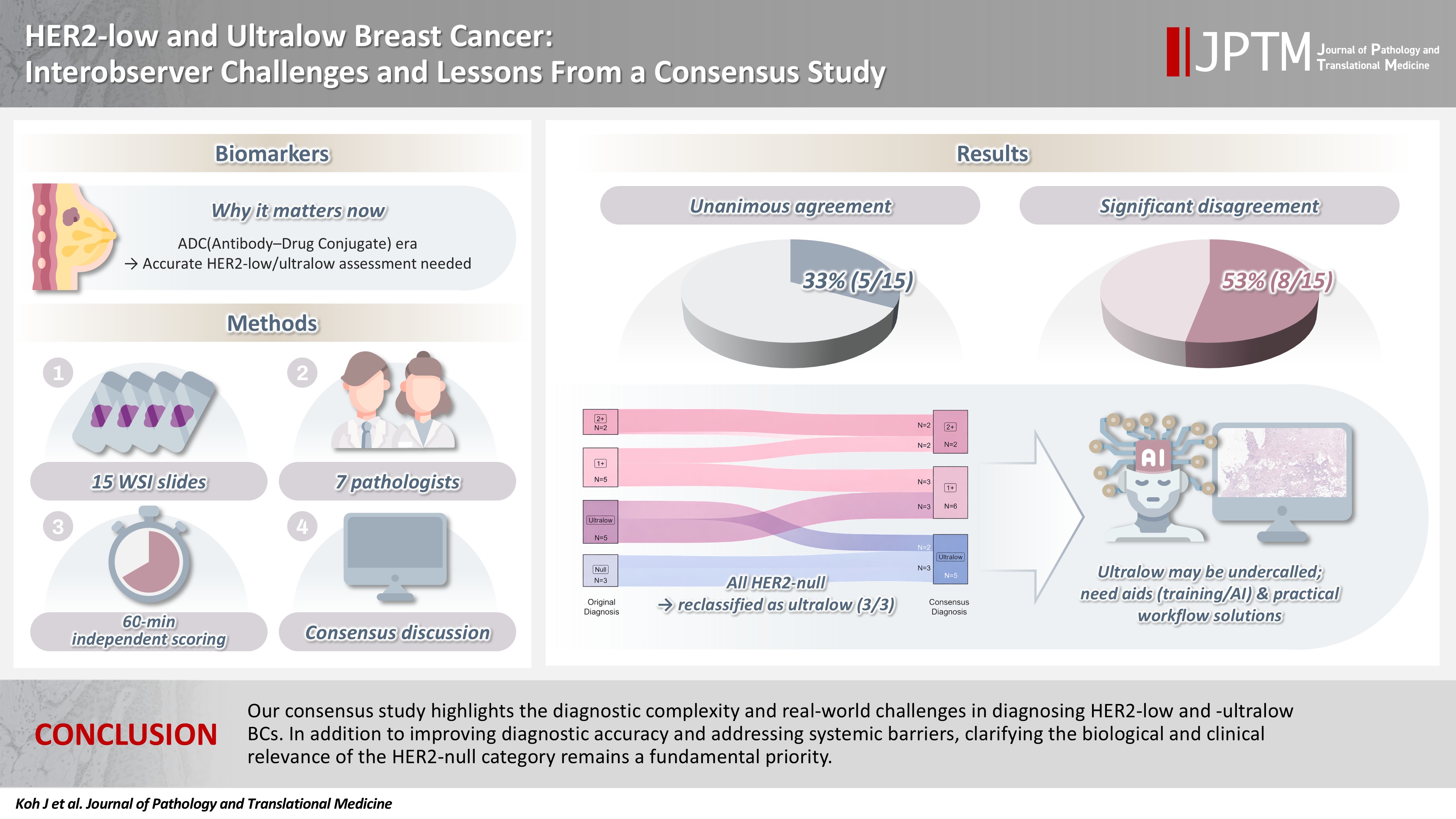
The recent approval of trastuzumab deruxtecan for human epidermal growth factor receptor 2 (HER2)–low and HER2-ultralow breast cancer mandates an adequate assessment of these categories. Methods: Seven breast pathologists from the Breast Pathology Study Group of the Korean Society of Pathologists held an on-site expert consensus meeting. Fifteen sets of virtual whole slide images (WSI) of hematoxylin and eosin stain and HER2 immunohistochemistry were provided. The pathologists were given 60 minutes to submit their diagnosis of HER2 expression into null, ultralow, 1+, 2+, or 3+. Afterwards, in-depth discussion and consensus diagnoses were made by real-time visualization of the WSI. Results: After the consensus meeting, unanimous 100% agreements were seen only in five (33.3%) of the examined cases, which consisted of three 1+ cases and two 2+ cases. Two cases (13.3%) had mild disagreement, with only one pathologist’s disagreement. Of note, eight cases (53.3%) showed significant disagreement, defined by more than two pathologists’ disagreement. All HER2-null cases were reclassified as ultralow after consensus review, suggesting potential widespread underclassification of ultralow cases in clinical practice. Conclusions: Experts had significant discrepancies in interpreting HER2-low/ultralow status. It is important to assess if the distinction between HER2-low and ultralow is strictly required and if HER2-null breast cancer exists in reality.
- Prevalence of HER2-ultralow breast cancer in South Korea: a multicenter study by reassessment of HER2-zero cases
- Min Chong Kim, Eun Yoon Cho, Hee Jin Lee, Ji Shin Lee, Jee Yeon Kim, Wan Seop Kim, Chungyeul Kim, Sun-Young Jun, Hye Jeong Choi, So Mang Lee, Ahrong Kim, Ji-Young Kim, Jeong Yun Shim, Gyungyub Gong, Young Kyung Bae
- J Pathol Transl Med. 2026;60(2):184-192. Published online February 23, 2026
- DOI: https://doi.org/10.4132/jptm.2025.10.22

- 2,421 View
- 190 Download
-
Abstract
PDF
 Supplementary Material
 - Background
This study aimed to determine the prevalence of human epidermal growth factor receptor 2 (HER2)–ultralow breast cancer among cases initially classified as HER2 immunohistochemistry (IHC) 0 and assess interobserver variability in interpreting low-level HER2 expression. Methods: In this multicenter retrospective study, all invasive breast cancer cases diagnosed between January and December 2022 across 10 Korean institutions were retrieved. Institutional pathologists reexamined HER2 IHC slides originally reported as IHC 0 according to the 2018 American Society of Clinical Oncology/College of American Pathologists guidelines and reclassified them as HER2-null (0), HER2-ultralow (0+), or HER2-low (1+). Slides from 10% of HER2-null and HER2-ultralow cases were digitized for central review and independently assessed by two pathologists, with discrepancies resolved by consensus. Results: Among 8,026 cases, 2,836 cases (35.5%) were initially reported as IHC 0. Upon re-review, 1,673 (59.0%), 1,139 (40.2%), and 24 (0.8%) cases were reclassified as HER2-null, HER2-ultralow, and HER2-low, respectively. The prevalence of HER2-ultralow breast cancer varied considerably across institutions (23.7%–78.1%). Central review of 268 digitized cases showed concordance in 193 cases (72.0%). Among the 75 discordant cases, 54 tumors (72.0%) were upgraded from HER2-null to HER2-ultralow, and 18 (24.0%) tumors were upgraded from HER2-ultralow to HER2-low. Furthermore, two tumors (2.7%) were downgraded from HER2-ultralow to HER2-null. Conclusions: Approximately 40% of cases initially categorized as IHC 0 were reclassified as HER2-ultralow. The substantial inter-institutional variability observed in interpreting low-level HER2 expression highlights the need for standardized training and quality assurance to ensure accurate identification of patients eligible for HER2-targeted antibody–drug conjugates.
- Development of CytoAcademy: a new web- and mobile-based E-learning platform for cytopathologists and cytotechnologists by the Korean Society for Cytopathology in the post-pandemic era
- Ran Hong, Yosep Chong, Seung Wan Chae, Seung-Sook Lee, Gyungyub Gong
- J Pathol Transl Med. 2024;58(6):261-264. Published online November 7, 2024
- DOI: https://doi.org/10.4132/jptm.2024.10.02

- 6,085 View
- 290 Download
- 3 Web of Science
- 4 Crossref
-
Abstract
PDF
- Since the late 1990s, online e-learning has offered unparalleled convenience and affordability, becoming increasingly popular among pathologists. Traditional learning theories have been successfully applied to web/mobile-based learning systems, with mobile technologies even enhancing conventional offline education. In cytopathology, hands-on microscope training has traditionally been paramount, complemented by real-case presentations and lectures. However, the coronavirus disease 2019 (COVID-19) pandemic disrupted regular academic activities, making online e-learning platforms essential. We designed a web/mobile-based learning platform to enhance continued medical education in cytopathology at various levels, particularly during the era of COVID-19 and beyond. Since 2021, we have integrated curriculum materials, virtual education files, and whole-slide images (WSIs) of cytopathology, submitted from over 200 institutions across Korea, with the support of numerous instructors. We develop a new e-learning platform named “CytoAcademy” composed of a basic session for each organ and level across the range of morphologic findings; on-demand lectures to enhance cytopathologic knowledge; WSI archives that allow users to explore various histologically confirmed cases; and a self-assessment test to help organize diagnostic knowledge acquired through the web/mobile-friendly learning system. The platform provides not just an opportunity to achieve a correct diagnosis, but also a learning experience based on problem-solving point. Members interact, identify their deficiencies, and focus on specific educational materials. In this manner, all participants can actively engage in creating and maintaining knowledge and foster a proactive approach to learning.
-
Citations
Citations to this article as recorded by
- Practice of Cytopathology in Korea: A 40‐Year Evolution Through Standardization, Digital Transformation, and Global Partnership
Yosep Chong, Ran Hong, Hyeong Ju Kwon, Haeryoung Kim, Lucia Kim, Soon Jae Kim, Yoon Jung Choi
Diagnostic Cytopathology.2026; 54(2): 146. CrossRef - Developing a smart and scalable tool for histopathological education—PATe 2.0
Lina Winter, Annalena Artinger, Hendrik Böck, Vignesh Ramakrishnan, Bruno Reible, Jan Albin, Peter J. Schüffler, Georgios Raptis, Christoph Brochhausen
Journal of Pathology Informatics.2026; 20: 100535. CrossRef - National quality assurance program using digital cytopathology: a 5-year digital transformation experience by the Korean Society for Cytopathology
Yosep Chong, Hyeong Ju Kwon, Soon Auck Hong, Sung Soon Kim, Bo-Sung Kim, Younghee Choi, Yoon Jung Choi, Jung-Soo Pyo, Ji Yun Jeong, Soo Jin Jung, Hoon Kyu Oh, Seung-Sook Lee
Journal of Pathology and Translational Medicine.2025; 59(5): 320. CrossRef - Integration of Digital Cytology in Quality Assurance Programs for Cytopathology
Yosep Chong, Maria Jesús Fernández Aceñero, Zaibo Li, Andrey Bychkov
Acta Cytologica.2025; 70(1): 126. CrossRef
- Practice of Cytopathology in Korea: A 40‐Year Evolution Through Standardization, Digital Transformation, and Global Partnership
- Diagnostic proficiency test using digital cytopathology and comparative assessment of whole slide images of cytologic samples for quality assurance program in Korea
- Yosep Chong, Soon Auck Hong, Hoon Kyu Oh, Soo Jin Jung, Bo-Sung Kim, Ji Yun Jeong, Ho-Chang Lee, Gyungyub Gong
- J Pathol Transl Med. 2023;57(5):251-264. Published online August 24, 2023
- DOI: https://doi.org/10.4132/jptm.2023.07.17
- 9,640 View
- 349 Download
- 10 Web of Science
- 11 Crossref
-
Abstract
PDFSupplementary Material
- Background
The Korean Society for Cytopathology introduced a digital proficiency test (PT) in 2021. However, many doubtful opinions remain on whether digitally scanned images can satisfactorily present subtle differences in the nuclear features and chromatin patterns of cytological samples.
Methods
We prepared 30 whole-slide images (WSIs) from the conventional PT archive by a selection process for digital PT. Digital and conventional PT were performed in parallel for volunteer institutes, and the results were compared using feedback. To assess the quality of cytological assessment WSIs, 12 slides were collected and scanned using five different scanners, with four cytopathologists evaluating image quality through a questionnaire.
Results
Among the 215 institutes, 108 and 107 participated in glass and digital PT, respectively. No significant difference was noted in category C (major discordance), although the number of discordant cases was slightly higher in the digital PT group. Leica, 3DHistech Pannoramic 250 Flash, and Hamamatsu NanoZoomer 360 systems showed comparable results in terms of image quality, feature presentation, and error rates for most cytological samples. Overall satisfaction was observed with the general convenience and image quality of digital PT.
Conclusions
As three-dimensional clusters are common and nuclear/chromatin features are critical for cytological interpretation, careful selection of scanners and optimal conditions are mandatory for the successful establishment of digital quality assurance programs in cytology. -
Citations
Citations to this article as recorded by- Practice of Cytopathology in Korea: A 40‐Year Evolution Through Standardization, Digital Transformation, and Global Partnership
Yosep Chong, Ran Hong, Hyeong Ju Kwon, Haeryoung Kim, Lucia Kim, Soon Jae Kim, Yoon Jung Choi
Diagnostic Cytopathology.2026; 54(2): 146. CrossRef - ThinPrep® Whole-Slide Digital Images versus Conventional Microscopy in Negative for Intraepithelial Lesion or Malignancy, Atypical Squamous Cells of Undetermined Significance, and Low-Grade Squamous Intraepithelial Lesion Cervical Lesions: European Federa
Ester Puntonen, Sara Bazzon, Massimo Bongiovanni, Rosario Granados, Ines Krivak Bolanca, Maria Nasioutziki, Maurizio Pinamonti, Danijela Vrdoljak-Mozetic, Arrigo Capitanio, Beatrix Cochand-Priollet, Ambrogio Fassina, Giovanni Negri, Laura Ventura, Ivana K
Pathobiology.2026; 93(4): 221. CrossRef - Determination of an optimal quality control method for Pap test analysis using digital cytology and artificial intelligence
Lakshmi Harinath, Esther Elishaev, Jonee Matsko, Amy Colaizzi, Haley Brown, Rohit Bhargava, Liron Pantanowitz, Chengquan Zhao
Journal of the American Society of Cytopathology.2026;[Epub] CrossRef - Accurate focal‐plane selection is crucial for artificial intelligence assessment of three‐dimensional urine cytology specimens for bladder cancer screening and surveillance
Yoseph Sayegh, Brody McNutt, Minh‐Khang Le, I‐Chuang Liao, Keluo Yao, Camille Ng, Ahmad Kohsar, Daniel Shou, Michael Yu, YinXian Jin, Louis J. Vaickus, Xiaoying Liu, Christopher J. VandenBussche, Samuel E. Harvey, Joshua J. Levy
Cancer Cytopathology.2026;[Epub] CrossRef - Sensitivity, Specificity, and Cost–Benefit Effect Between Primary Human Papillomavirus Testing, Primary Liquid‐Based Cytology, and Co‐Testing Algorithms for Cervical Lesions
Chang Gok Woo, Seung‐Myoung Son, Hye‐Kyung Hwang, Jung‐Sil Bae, Ok‐Jun Lee, Ho‐Chang Lee
Diagnostic Cytopathology.2025; 53(1): 35. CrossRef - Integration of AI‐Assisted in Digital Cervical Cytology Training: A Comparative Study
Yihui Yang, Dongyi Xian, Lihua Yu, Yanqing Kong, Huaisheng Lv, Liujing Huang, Kai Liu, Hao Zhang, Weiwei Wei, Hongping Tang
Cytopathology.2025; 36(2): 156. CrossRef - National quality assurance program using digital cytopathology: a 5-year digital transformation experience by the Korean Society for Cytopathology
Yosep Chong, Hyeong Ju Kwon, Soon Auck Hong, Sung Soon Kim, Bo-Sung Kim, Younghee Choi, Yoon Jung Choi, Jung-Soo Pyo, Ji Yun Jeong, Soo Jin Jung, Hoon Kyu Oh, Seung-Sook Lee
Journal of Pathology and Translational Medicine.2025; 59(5): 320. CrossRef - Integration of Digital Cytology in Quality Assurance Programs for Cytopathology
Yosep Chong, Maria Jesús Fernández Aceñero, Zaibo Li, Andrey Bychkov
Acta Cytologica.2025; 70(1): 126. CrossRef - Quantitative Assessment of Focus Quality in Whole-Slide Imaging of Thyroid Liquid-Based Cytology Using Laplacian Variance
Chan Kwon Jung, Chankyung Kim, Sora Jeon, Andrey Bychkov
Endocrine Pathology.2025;[Epub] CrossRef - Validation of digital image slides for diagnosis in cervico-vaginal cytology
Francisco Tresserra, Gemma Fabra, Olga Luque, Miriam Castélla, Carla Gómez, Carmen Fernández-Cid, Ignacio Rodríguez
Revista Española de Patología.2024; 57(3): 182. CrossRef - Improved Diagnostic Accuracy of Thyroid Fine-Needle Aspiration Cytology with Artificial Intelligence Technology
Yujin Lee, Mohammad Rizwan Alam, Hongsik Park, Kwangil Yim, Kyung Jin Seo, Gisu Hwang, Dahyeon Kim, Yeonsoo Chung, Gyungyub Gong, Nam Hoon Cho, Chong Woo Yoo, Yosep Chong, Hyun Joo Choi
Thyroid®.2024; 34(6): 723. CrossRef
- Practice of Cytopathology in Korea: A 40‐Year Evolution Through Standardization, Digital Transformation, and Global Partnership
- Current status of cytopathology practice in Korea: impact of the coronavirus pandemic on cytopathology practice
- Soon Auck Hong, Haeyoen Jung, Sung Sun Kim, Min-Sun Jin, Jung-Soo Pyo, Ji Yun Jeong, Younghee Choi, Gyungyub Gong, Yosep Chong
- J Pathol Transl Med. 2022;56(6):361-369. Published online October 27, 2022
- DOI: https://doi.org/10.4132/jptm.2022.09.21

- 6,036 View
- 107 Download
- 7 Web of Science
- 8 Crossref
-
Abstract
PDFSupplementary Material
- Background
The Continuous Quality Improvement program for cytopathology in 2020 was completed during the coronavirus pandemic. In this study, we report the result of the quality improvement program.
Methods
Data related to cytopathology practice from each institute were collected and processed at the web-based portal. The proficiency test was conducted using glass slides and whole-slide images (WSIs). Evaluation of the adequacy of gynecology (GYN) slides from each institution and submission of case glass slides and WSIs for the next quality improvement program were performed.
Results
A total of 214 institutions participated in the annual cytopathology survey in 2020. The number of entire cytopathology specimens was 8,220,650, a reduction of 19.0% from the 10,111,755 specimens evaluated in 2019. Notably, the number of respiratory cytopathology specimens, including sputum and bronchial washing/ brushing significantly decreased by 86.9% from 2019, which could be attributed to the global pandemic of coronavirus disease. The ratio of cases with atypical squamous cells to squamous intraepithelial lesions was 4.10. All participating institutions passed the proficiency test and the evaluation of adequacy of GYN slides.
Conclusions
Through the Continuous Quality Improvement program, the effect of coronavirus disease 2019 pandemic, manifesting with a reduction in the number of cytologic examinations, especially in respiratory-related specimen has been identified. The Continuous Quality Improvement Program of the Korean Society for Cytopathology can serve as the gold standard to evaluate the current status of cytopathology practice in Korea. -
Citations
Citations to this article as recorded by- Commercially Available Artificial Intelligence Solutions for Gynaecologic Cytology Screening and Their Integration Into Clinical Workflow
Yosep Chong, Andrey Bychkov
Cytopathology.2026; 37(1): 24. CrossRef - Practice of Cytopathology in Korea: A 40‐Year Evolution Through Standardization, Digital Transformation, and Global Partnership
Yosep Chong, Ran Hong, Hyeong Ju Kwon, Haeryoung Kim, Lucia Kim, Soon Jae Kim, Yoon Jung Choi
Diagnostic Cytopathology.2026; 54(2): 146. CrossRef - Telecytology in head and neck cytopathology: current applications and practical considerations
Yeongjoon Kim
Kosin Medical Journal.2026; 41(2): 126. CrossRef - A Study on the Workload of Cytotechnologists: Focus on Commercial Laboratories
Eun-Suk PARK
Korean Journal of Clinical Laboratory Science.2025; 57(2): 228. CrossRef - Integration of Digital Cytology in Quality Assurance Programs for Cytopathology
Yosep Chong, Maria Jesús Fernández Aceñero, Zaibo Li, Andrey Bychkov
Acta Cytologica.2025; 70(1): 126. CrossRef - National quality assurance program using digital cytopathology: a 5-year digital transformation experience by the Korean Society for Cytopathology
Yosep Chong, Hyeong Ju Kwon, Soon Auck Hong, Sung Soon Kim, Bo-Sung Kim, Younghee Choi, Yoon Jung Choi, Jung-Soo Pyo, Ji Yun Jeong, Soo Jin Jung, Hoon Kyu Oh, Seung-Sook Lee
Journal of Pathology and Translational Medicine.2025; 59(5): 320. CrossRef - A stepwise approach to fine needle aspiration cytology of lymph nodes
Yosep Chong, Gyeongsin Park, Hee Jeong Cha, Hyun-Jung Kim, Chang Suk Kang, Jamshid Abdul-Ghafar, Seung-Sook Lee
Journal of Pathology and Translational Medicine.2023; 57(4): 196. CrossRef - Diagnostic proficiency test using digital cytopathology and comparative assessment of whole slide images of cytologic samples for quality assurance program in Korea
Yosep Chong, Soon Auck Hong, Hoon Kyu Oh, Soo Jin Jung, Bo-Sung Kim, Ji Yun Jeong, Ho-Chang Lee, Gyungyub Gong
Journal of Pathology and Translational Medicine.2023; 57(5): 251. CrossRef
- Commercially Available Artificial Intelligence Solutions for Gynaecologic Cytology Screening and Their Integration Into Clinical Workflow
- Standardized pathology report for breast cancer
- Soo Youn Cho, So Yeon Park, Young Kyung Bae, Jee Yeon Kim, Eun Kyung Kim, Woo Gyeong Kim, Youngmee Kwon, Ahwon Lee, Hee Jin Lee, Ji Shin Lee, Jee Young Park, Gyungyub Gong, Hye Kyoung Yoon
- J Pathol Transl Med. 2021;55(1):1-15. Published online January 11, 2021
- DOI: https://doi.org/10.4132/jptm.2020.11.20

- 19,681 View
- 757 Download
- 12 Web of Science
- 8 Crossref
-
Abstract
PDFSupplementary Material
- Given the recent advances in management and understanding of breast cancer, a standardized pathology report reflecting these changes is critical. To meet this need, the Breast Pathology Study Group of the Korean Society of Pathologists has developed a standardized pathology reporting format for breast cancer, consisting of ‘standard data elements,’ ‘conditional data elements,’ and a biomarker report form. The ‘standard data elements’ consist of the basic pathologic features used for prognostication, while other factors related to prognosis or diagnosis are described in the ‘conditional data elements.’ In addition to standard data elements, all recommended issues are also presented. We expect that this standardized pathology report for breast cancer will improve diagnostic concordance and communication between pathologists and clinicians, as well as between pathologists inter-institutionally.
-
Citations
Citations to this article as recorded by- Adenoid Cystic Carcinoma of Breast Associated With an Incidental Radial Scar: A Cyto‐Histopathology Correlation
Rallapalli Rajyalakshmi, Valasapalli Rajani, Tanuku Sreedhar, Kollabathula Arpitha
Diagnostic Cytopathology.2026;[Epub] CrossRef - Refined risk stratification in residual triple-negative breast cancer after neoadjuvant therapy using residual cancer burden class and lymphovascular invasion
Tae Hoon Lee, Hyunwoo Lee, Jeong Yun Jang, Won Park, Won Kyung Cho, Eun Yoon Cho, Jin Seok Ahn, Yeon Hee Park, Seok Jin Nam, Seok Won Kim, Jeong Eon Lee, Haeyoung Kim
Breast Cancer Research and Treatment.2026;[Epub] CrossRef - Artificial intelligence-assisted three-dimensional imaging of breast microinvasive carcinoma reveals larger invasive focus size in a substantial proportion of cases
Yichieh Chien, Cher-Wei Liang, Chih-Yi Hsu, Yu-Chieh Lin, Yen-Yu Lin
Pathology - Research and Practice.2026; 283: 156388. CrossRef - Evaluation of a virtual Ayurvedic whole-systems lifestyle intervention for quality of life in breast cancer survivors: An exploratory randomized controlled trial
Shraddha Ravani, Charles Elder, Sabita Sawhney, Rammanohar Puthiyedath, Robert Schneider
EXPLORE.2026; 22(5): 103468. CrossRef - Retrospective Cohort Study: Extracting Coexisting Background Breast-Lesion Features from Stage I–III Invasive Breast Cancer
Ryan Jak Yang Lim, Phyu Nitar, Kah Weng Lau, Lester Chee Hao Leong, Veronique Kiak Mien Tan, Benita Kiat Tee Tan, Ern Yu Tan, Serene Si Ning Goh, Mikael Hartman, Fuh Yong Wong, Geok Hoon Lim, Jingmei Li
Cancers.2026; 18(12): 1965. CrossRef - Navigating discrepancies: The assessment of residual lymphovascular invasion in breast carcinoma after neoadjuvant treatment
Anikó Kovács, Åsa Rundgren-Sellei, Gunilla Rask, Annette Bauer, Anna Bodén, Johannes van Brakel, Eugenia Colón-Cervantes, Anna Ehinger, Johan Hartman, Balazs Acs
The Breast.2025; 82: 104519. CrossRef - Residual pure intralymphatic carcinoma component only (lymphovascular tumor emboli without invasive carcinoma) after neoadjuvant chemotherapy is associated with poor outcome: Not pathologic complete response
Hyunwoo Lee, Yunjeong Jang, Yoon Ah Cho, Eun Yoon Cho
Human Pathology.2024; 145: 1. CrossRef - Sentinel lymph node biopsy in patients with ductal carcinomain situ: systematic review and meta-analysis
Matthew G. Davey, Colm O’Flaherty, Eoin F. Cleere, Aoife Nohilly, James Phelan, Evan Ronane, Aoife J. Lowery, Michael J. Kerin
BJS Open.2022;[Epub] CrossRef
- Adenoid Cystic Carcinoma of Breast Associated With an Incidental Radial Scar: A Cyto‐Histopathology Correlation
- Breast implant–associated anaplastic large cell lymphoma: the first South Korean case
- Jongwon Lee, Hyungwoo Cho, Dok Hyun Yoon, Eun Key Kim, Gyungyub Gong, Cheolwon Suh, Joo-ryung Huh
- J Pathol Transl Med. 2020;54(5):432-434. Published online August 18, 2020
- DOI: https://doi.org/10.4132/jptm.2020.07.01

- 6,880 View
- 142 Download
- 5 Web of Science
- 5 Crossref
-
PDF
-
Citations
Citations to this article as recorded by- Breast Reconstruction after Breast Implant-Associated Anaplastic Large Cell Lymphoma Treatment: A Case Report and Literature Review
Won-Seob Lee, Tae-Gon Kim, Jun-Ho Lee, Il-Kug Kim
Journal of Clinical Medicine.2023; 12(5): 1885. CrossRef - Breast filler granuloma mistaken for implant rupture: A case report
Yong Seon Hwang, Je Yeon Byeon, Jun Hyuk Kim, Hwan Jun Choi, Mee Hye Oh, Da Woon Lee
Medicine.2023; 102(22): e33785. CrossRef - Implant replacement and anaplastic large cell lymphoma associated with breast implants: a quantitative analysis
Martina Vittorietti, Sergio Mazzola, Claudio Costantino, Daniele Domenico De Bella, Santo Fruscione, Nicole Bonaccorso, Martina Sciortino, Davide Costanza, Miriam Belluzzo, Alessandra Savatteri, Fabio Tramuto, Paolo Contiero, Giovanna Tagliabue, Palmira I
Frontiers in Oncology.2023;[Epub] CrossRef - Breast implant-associated anaplastic large cell lymphoma: a case report with a history of spontaneously resolved late seroma
Do Yeon Kim, Joon Hur, Woo Yeon Han, Kyunghyun Min, Jong Won Lee, Jin Sup Eom, Hyun Ho Han, Eun Key Kim
Archives of Aesthetic Plastic Surgery.2021; 27(4): 143. CrossRef - Comment on “Breast implant-associated anaplastic large cell lymphoma: the first South Korean case”
Il-Kug Kim, Tae Gon Kim
Journal of Pathology and Translational Medicine.2021; 55(6): 419. CrossRef
- Breast Reconstruction after Breast Implant-Associated Anaplastic Large Cell Lymphoma Treatment: A Case Report and Literature Review
- Primary Rhabdomyosarcoma of the Breast: Study of Three Cases at One Institution with a Review of Primary Breast Sarcomas
- Junyoung Shin, Hee Jeong Kim, Dae-Yeon Kim, Gyungyub Gong, Kyung-Ja Cho
- J Pathol Transl Med. 2019;53(5):308-316. Published online August 2, 2019
- DOI: https://doi.org/10.4132/jptm.2019.07.22
- 8,863 View
- 141 Download
- 7 Web of Science
- 9 Crossref
-
Abstract
PDF
- Background
Primary breast sarcoma (PBS) is rare, comprising approximately 1% of breast malignancies. Rhabdomyosarcoma (RMS) accounts for an extremely small proportion of PBSs, often leading to delayed histologic confirmation.
Methods
Upon reviewing Asan Medical Center’s pathology database between 2000 and 2018, 41 PBS cases were retrieved, including three cases of primary RMS of the breast. Their clinicopathological features were analyzed, and the literature related to PBS and primary RMS of the breast was reviewed.
Results
We identified three primary breast RMS cases from our institution database, comprising 7.3% of PBS: one case each of spindle cell/sclerosing RMS (ssRMS), alveolar RMS (aRMS), and embryonal RMS (eRMS). All cases involved adolescents or young adults (14, 16, and 25 years, respectively) who underwent mastectomy or radiotherapy and were confirmed using immunohistochemical testing for myogenin, desmin, and myogenic differentiation. The ssRMS patient experienced recurrence at the operation site 4 months post-surgery despite undergoing concurrent chemoradiotherapy. The aRMS patient had multiple metastases at diagnosis and showed FAX3-FOXO1 fusion transcripts; she died 22 months after the diagnosis. The eRMS patient had enlarged axillary lymph nodes; post-radiotherapy, the lesion recurred as multiple metastases to the bone and lung. She died 18 months post-diagnosis.
Conclusions
Our experience on RMS cases suggests that spindle cell or small round cell malignancy in breasts of young female should raise suspicion for the possibility of primary or secondary RMS. To our knowledge, this is the second report of primary breast ssRMS and it may help clinicians who encounter this rare disease in the future. -
Citations
Citations to this article as recorded by- Primary Ewing sarcoma of the breast in a male adolescent: A case report
Joie Sheen A. Bastian, Willie T. Hao, Lawrence Faith A. Lucañas-Yap
Journal of Pediatric Surgery Case Reports.2026; 127: 103205. CrossRef - Clinicopathological and molecular features of breast metastases in alveolar rhabdomyosarcoma: A series of 3 cases
Wanni Xu, Li Yang, Yuxin Hui, Peizhuo Yao, Yiwei Jia, Xinyu Wei, Shuqun Zhang
Annals of Diagnostic Pathology.2026; 83: 152627. CrossRef - Management of Pediatric Breast Masses for the Pediatric Surgeon: Expert Consensus Recommendations From the APSA Cancer Committee
Dana Schwartz, Elisabeth T. Tracy, Bindi Naik-Mathuria, Richard D. Glick, Stephanie F. Polites, Peter Mattei, David Rodeberg, Andres F. Espinoza, Sara A. Mansfield, Dave R. Lal, Meera Kotagal, Timothy Lautz, Jennifer Aldrink, Barrie S. Rich
Journal of Pediatric Surgery.2025; 60(2): 161916. CrossRef - Differential diagnosis of primary mesenchymal neoplasms of the breast
Mine Ozsen, Seyit Ali Volkan Polatkan, Ulviye Yalcınkaya, Sahsine Tolunay, Mustafa Sehsuvar Gokgoz
Clinical and Translational Oncology.2024; 27(1): 223. CrossRef - Primary breast rhabdomyosarcoma in a 17-year-old girl
Laxmi Singotia, V.S. Haritha
Journal of Cancer Research and Therapeutics.2023; 19(7): 2070. CrossRef - High-Grade Spindle Cell Lesions of the Breast
Esther Yoon, Qingqing Ding, Kelly Hunt, Aysegul Sahin
Surgical Pathology Clinics.2022; 15(1): 77. CrossRef - Primary Small Cell Malignancies of the Breast: Are They Rare Malignancies?
Kemal Behzatoğlu, Fernando Schmitt
Acta Cytologica.2022; 66(4): 347. CrossRef - Recurrent malignant phyllodes tumor of the breast: An extremely rare case of recurrence with only rhabdomyosarcoma components
Jia Han, Shuice Liu, Akihoro Shioya, Motona Kumagai, Emi Morioka, Miki Noguchi, Masafumi Inokuchi, Sohsuke Yamada
SAGE Open Medical Case Reports.2022;[Epub] CrossRef - Primary rhabdomyosarcoma: An extremely rare and aggressive variant of male breast cancer
Cătălin Bogdan Satală, Ioan Jung, Tivadar Jr Bara, Patricia Simu, Iunius Simu, Madalina Vlad, Rita Szodorai, Simona Gurzu
World Journal of Clinical Cases.2020; 8(19): 4466. CrossRef
- Primary Ewing sarcoma of the breast in a male adolescent: A case report
- Association between p53 Expression and Amount of Tumor-Infiltrating Lymphocytes in Triple-Negative Breast Cancer
- Miseon Lee, In Ah Park, Sun-Hee Heo, Young-Ae Kim, Gyungyub Gong, Hee Jin Lee
- J Pathol Transl Med. 2019;53(3):180-187. Published online March 11, 2019
- DOI: https://doi.org/10.4132/jptm.2019.02.08
- 11,069 View
- 209 Download
- 20 Web of Science
- 20 Crossref
-
Abstract
PDF
- Background
Most triple-negative breast cancers (TNBCs) have a high histologic grade, are associated with high endoplasmic stress, and possess a high frequency of TP53 mutations. TP53 missense mutations lead to the production of mutant p53 protein and usually show high levels of p53 protein expression. Tumor-infiltrating lymphocytes (TILs) accumulate as part of the anti-tumor immune response and have a strong prognostic and predictive significance in TNBC. We aimed to elucidate the association between p53 expression and the amount of TILs in TNBC.
Methods
In 678 TNBC patients, we evaluated TIL levels and expression of endoplasmic stress molecules. Immunohistochemical examination of p53 protein expression was categorized into three groups: no, low, and high expression.
Results
No, low, and high p53 expression was identified in 44.1% (n = 299), 20.1% (n = 136), and 35.8% (n = 243) of patients, respectively. Patients with high p53 expression showed high histologic grade (p < .001), high TIL levels (p = .009), and high expression of endoplasmic reticulum stress-associated molecules (p-eIF2a, p = .013; XBP1, p = .007), compared to patients with low p53 expression. There was no significant difference in disease-free (p = .406) or overall survival rates (p = .444) among the three p53 expression groups.
Conclusions
High p53 expression is associated with increased expression of endoplasmic reticulum stress molecules and TIL influx. -
Citations
Citations to this article as recorded by- A comparative analysis of mutational profiles between triple-negative breast cancer and non-triple-negative breast cancer
Wanlin Li, Chenchen Feng, Shunheng Zhou
Discover Oncology.2026;[Epub] CrossRef - The search for a TNBC vaccine: the guardian vaccine
Cory Fines, Helen McCarthy, Niamh Buckley
Cancer Biology & Therapy.2025;[Epub] CrossRef - Correlating p53 immunostaining patterns with somatic TP53 mutation and functional properties of mutant p53 in triple‐negative breast cancer
Meejeong Kim, Miseon Lee, Ahwon Lee, Byung‐Ock Choi, Woo‐Chan Park, Sung Hun Kim, Jieun Lee, Jun Kang
Histopathology.2025; 87(2): 299. CrossRef - GD3 synthase drives resistance to p53-induced apoptosis in breast cancer by modulating mitochondrial function
Vivek Anand, Fouad El-Dana, Natalia Baran, Jenny Borgman, Zheng Yin, Hong Zhao, Stephen T. Wong, Michael Andreeff, V. Lokesh Battula
Oncogene.2025; 44(30): 2646. CrossRef - Topoisomerase Inhibitor Resistance in Breast Cancer: Exploring Alterations in Cellular Metabolism and Properties Driving Drug Insensitivity
Juhong Lee, Youngjoo Kwon
Drug Targets and Therapeutics.2025; 4(2): 185. CrossRef - Updated Austrian treatment algorithm for metastatic triple-negative breast cancer
Rupert Bartsch, Gabriel Rinnerthaler, Edgar Petru, Daniel Egle, Michael Gnant, Marija Balic, Thamer Sliwa, Christian Singer
Wiener klinische Wochenschrift.2024; 136(11-12): 347. CrossRef - Triple negative breast cancer: Immunogenicity, tumor microenvironment, and immunotherapy
Sotiris Loizides, Anastasia Constantinidou
Frontiers in Genetics.2023;[Epub] CrossRef - Prognostic benefit of TILs independent of clinicopathological and molecular factors
Koen Brummel, Anneke L. Eerkens, Marco de Bruyn, Hans W. Nijman
British Journal of Cancer.2023; 129(5): 737. CrossRef - Dihydroartemisinin-Transferrin Adducts Enhance TRAIL-Induced Apoptosis in Triple-Negative Breast Cancer in a P53-Independent and ROS-Dependent Manner
Xinyu Zhou, Abel Soto-Gamez, Fleur Nijdam, Rita Setroikromo, Wim J. Quax
Frontiers in Oncology.2022;[Epub] CrossRef - New Challenges in the Differential Diagnosis of High-Grade Triple-Negative Breast Cancer and Serous Carcinoma
Andrii Puzyrenko, Chandler S Cortina, Julie M Jorns
International Journal of Surgical Pathology.2022; 30(7): 728. CrossRef - Prognostic analysis of cuproptosis-related gene in triple-negative breast cancer
Shengnan Sha, Luyi Si, Xinrui Wu, Yuanbiao Chen, Hui Xiong, Ying Xu, Wangrui Liu, Haijun Mei, Tao Wang, Mei Li
Frontiers in Immunology.2022;[Epub] CrossRef - p53 Missense Mutation is Associated with Immune Cell PD-L1 Expression in Triple-Negative Breast Cancer
Ai-Yan Xing, Long Liu, Ke Liang, Bin Wang
Cancer Investigation.2022; 40(10): 879. CrossRef - Crosstalk between Immune Checkpoint Modulators, Metabolic Reprogramming and Cellular Plasticity in Triple-Negative Breast Cancer
Arpita Poddar, Sushma R. Rao, Prashanth Prithviraj, George Kannourakis, Aparna Jayachandran
Current Oncology.2022; 29(10): 6847. CrossRef - The tumor microenvironment and triple-negative breast cancer aggressiveness: shedding light on mechanisms and targeting
Natsuki Furukawa, Vered Stearns, Cesar A. Santa-Maria, Aleksander S. Popel
Expert Opinion on Therapeutic Targets.2022; 26(12): 1041. CrossRef - Intracellular partners of fibroblast growth factors 1 and 2 - implications for functions
Katarzyna Dominika Sluzalska, Jakub Slawski, Martyna Sochacka, Agata Lampart, Jacek Otlewski, Malgorzata Zakrzewska
Cytokine & Growth Factor Reviews.2021; 57: 93. CrossRef - Targeted Chinese Medicine Delivery by A New Family of Biodegradable Pseudo-Protein Nanoparticles for Treating Triple-Negative Breast Cancer: In Vitro and In Vivo Study
Hiu Yee Kwan, Qinghua Xu, Ruihong Gong, Zhaoxiang Bian, Chih-Chang Chu
Frontiers in Oncology.2021;[Epub] CrossRef - Transcriptomic Properties of HER2+ Ductal Carcinoma In Situ of the Breast Associate with Absence of Immune Cells
Marie Colombe Agahozo, Marcel Smid, Ronald van Marion, Dora Hammerl, Thierry P. P. van den Bosch, Mieke A. M. Timmermans, Chayenne J. Heijerman, Pieter J. Westenend, Reno Debets, John W. M. Martens, Carolien H. M. van Deurzen
Biology.2021; 10(8): 768. CrossRef - With Our Powers Combined
Lawrence Kasherman, Katherine Karakasis, Amit M. Oza
The Cancer Journal.2021; 27(6): 511. CrossRef The Research Progress on the Prognostic Value of the Common Hematological Parameters in Peripheral Venous Blood in Breast Cancer
Li Chen, Xiangyi Kong, Chengrui Yan, Yi Fang, Jing Wang
OncoTargets and Therapy.2020; Volume 13: 1397. CrossRefBiomolecular Factors Represented by Bcl-2, p53, and Tumor-Infiltrating Lymphocytes Predict Response for Adjuvant Anthracycline Chemotherapy in Patients with Early Triple-Negative Breast Cancer
Xenia Elena Bacinschi, Anca Zgura, Inga Safta, Rodica Anghel
Cancer Management and Research.2020; Volume 12: 11965. CrossRef
- A comparative analysis of mutational profiles between triple-negative breast cancer and non-triple-negative breast cancer
- WITHDRAWN:Primary Rhabdomyosarcoma of the Breast: A Report of Two Cases and Literature Review
- Junyoung Shin, Hee Jeong Kim, Dae-Yeon Kim, Gyungyub Gong, Kyung-Ja Cho
- Received August 6, 2018 Accepted September 13, 2018 Published online October 4, 2018
- DOI: https://doi.org/10.4132/jptm.2018.09.14
- 4,656 View
- 62 Download
- 1 Crossref
-
Citations
Citations to this article as recorded by- Primary Alveolar Rhabdomyosarcoma of the Breast in an Adult: An Extremely Rare Case
Helen J. Trihia, Natasa Novkovic, Ioannis Provatas, Anastasios Mavrogiorgis, Evangelos Lianos
Case Reports in Pathology.2019; 2019: 1. CrossRef
- Primary Alveolar Rhabdomyosarcoma of the Breast in an Adult: An Extremely Rare Case
- Interobserver Variability of Ki-67 Measurement in Breast Cancer
- Yul Ri Chung, Min Hye Jang, So Yeon Park, Gyungyub Gong, Woo-Hee Jung, The Korean Breast Pathology Ki- Study Group
- J Pathol Transl Med. 2016;50(2):129-137. Published online February 15, 2016
- DOI: https://doi.org/10.4132/jptm.2015.12.24
- 15,499 View
- 134 Download
- 34 Web of Science
- 35 Crossref
-
Abstract
PDF
- Background
As measurement of Ki-67 proliferation index is an important part of breast cancer diagnostics, we conducted a multicenter study to examine the degree of concordance in Ki-67 counting and to find factors that lead to its variability. Methods: Thirty observers from thirty different institutions reviewed Ki-67–stained slides of 20 different breast cancers on whole sections and tissue microarray (TMA) by online system. Ten of the 20 breast cancers had hot spots of Ki-67 expression. Each observer scored Ki-67 in two different ways: direct counting (average vs. hot spot method) and categorical estimation. Intraclass correlation coefficient (ICC) of Ki-67 index was calculated for comparative analysis. Results: For direct counting, ICC of TMA was slightly higher than that of whole sections using average method (0.895 vs 0.858). The ICC of tumors with hot spots was lower than that of tumors without (0.736 vs 0.874). In tumors with hot spots, observers took an additional counting from the hot spot; the ICC of whole sections using hot spot method was still lower than that of TMA (0.737 vs 0.895). In categorical estimation, Ki-67 index showed a wide distribution in some cases. Nevertheless, in tumors with hot spots, the range of distribution in Ki-67 categories was decreased with hot spot method and in TMA platform. Conclusions: Interobserver variability of Ki-67 index for direct counting and categorical estimation was relatively high. Tumors with hot spots showed greater interobserver variability as opposed to those without, and restricting the measurement area yielded lower interobserver variability. -
Citations
Citations to this article as recorded by- Dynamic biomarkers in hormone receptor-positive/HER2-negative breast cancer trials: a new hope for precision oncology
Giuseppe Di Grazia, Rodrigo Sánchez-Bayona, Climent Casals-Pascual, Tomás Pascual, Daniele Generali, Alessandra Gennari, Paolo Vigneri, Nadia Harbeck, Javier Cortés, Aleix Prat, Francesco Schettini
npj Breast Cancer.2026;[Epub] CrossRef - Low-Proliferative Invasive Mucinous Carcinoma of the Breast in an Octogenarian: A Case Report Highlighting Pathology-Guided De-escalation
Rae-Anne Kastle, Christopher M Ahmad, Rachana Tadakamalla, Hima Patel, Amer Abboud
Cureus.2026;[Epub] CrossRef - Low E-cadherin expression is associated with poor prognosis in pulmonal adenocarcinoma
Fiete Gehrisch, Kiara A. Schmid, Martina Kluth, Georgia Makrypidi-Fraune, Katharina Möller, Maximilian Lennartz, Veit Bertram, Florian Lutz, Stefan Steurer, Philipp Busch, Birgit Hantzsch-Kuhn, Martin Reck, Till Olchers, David Benjamin Ellebrecht, Christo
Scientific Reports.2026;[Epub] CrossRef - Evaluating the Association of Ki-67 with Oncotype DX Recurrence Score in Early-Stage ER-Positive/HER2-Negative Breast Cancer
Dimitrios Dragoumis, George Kapetsis, Konstantinos Louis, Dimitrios Maniatis, Eleni Mpalampou, Konstantinos Bouloukos, Xenophon Xenakis, Nikolaos Papaioannou, Styliani Parpoudi, Grigorios Pesmatzoglou, Anna Sachoulidou, Eleftherios Sfakianakis, Sofia Tria
Cancers.2026; 18(11): 1731. CrossRef - Ki-67 as a treatment decision modifier in breast cancer
Amanda Dy, Jochen K Lennerz
The Oncologist.2026;[Epub] CrossRef - Comparison of three counting methods for Ki-67 labeling index in breast carcinoma: A study on core biopsies and resection specimens
Satyajit Singh Gill, Nutan Jumle, Gourav Agrawal, Shrikanth Muralidharan
International Journal of Molecular and Immuno Oncology.2026; 0: 1. CrossRef - Comparative analysis of Ki-67 labeling index morphometry using deep learning, conventional image analysis, and manual counting
Mohammad Rizwan Alam, Kyung Jin Seo, Kwangil Yim, Phoebe Liang, Joe Yeh, Chifu Chang, Yosep Chong
Translational Oncology.2025; 51: 102159. CrossRef - Machine Learning-Based Approaches for Breast Density Estimation from Mammograms: A Comprehensive Review
Khaldoon Alhusari, Salam Dhou
Journal of Imaging.2025; 11(2): 38. CrossRef - Letter re: A critical appraisal of the DATA trial analysis on the prognostic and predictive value of the luminal-like subtype
M. Rizk, K. Mokbel
ESMO Open.2025; 10(5): 105067. CrossRef - Clinico-Histomorphological and Mib-1 Analysis of Recurrent Meningiomas: A Retrospective Study
Sujata Sarangi, Asha Shenoy, Ashvini Kolhe, Kanchan Kothari
Asian Journal of Neurosurgery.2025; 20(04): 785. CrossRef - Emerging Diagnostics and Therapies in Neuroendocrine Neoplasms: A Critical Review
Jorge H. Hernandez-Felix, Monica Isabel Meneses-Medina, Rachel Riechelmann, Jonathan Strosberg, Rocio Garcia-Carbonero, Jaydira del Rivero
Cancers.2025; 17(22): 3632. CrossRef - Ki-67 Testing in Breast Cancer: Assessing Variability With Scoring Methods and Specimen Types and the Potential Subsequent Impact on Therapy Eligibility
Therese Bocklage, Virgilius Cornea, Caylin Hickey, Justin Miller, Jessica Moss, Mara Chambers, S. Emily Bachert
Applied Immunohistochemistry & Molecular Morphology.2024; 32(3): 119. CrossRef - Interobserver agreement and diagnostic challenges of Congo red staining for amyloid detection on fat pad aspiration biopsies
Levent Trabzonlu, T. Leif Helland, Melanie C. Kwan, Nathalie Kumiega, M. Lisa Zhang, Ivan Chebib, Vanda F. Torous
Journal of the American Society of Cytopathology.2024; 13(5): 359. CrossRef - Assessment of the Predictive Role of Ki-67 in Breast Cancer Patients’ Responses to Neoadjuvant Chemotherapy
Ghizlane Rais, Rania Mokfi, Farah Boutaggount, Meryem Maskrout, Soundouss Bennour, Chaymae Senoussi, Fadoua Rais
European Journal of Breast Health.2024; : 199. CrossRef - Improving the accuracy of reporting Ki-67 IHC by using an AI tool
Sahil Ajit Saraf, Aahan Singh, Wai Po Kevin Teng, Sencer Karakaya, M. Logaswari, Kaveh Taghipour, Rajasa Jialdasani, Li Yan Khor, Kiat Hon Lim, Sathiyamoorthy Selvarajan, Vani Ravikumar, Md Ali Osama, Priti Chatterjee, Santosh KV
Heliyon.2024; 10(22): e40193. CrossRef - Predictive Value of Ki-67 Index in Evaluating Sporadic Vestibular Schwannoma Recurrence: Systematic Review and Meta-analysis
Kunal Vakharia, Hirotaka Hasegawa, Christopher Graffeo, Mohammad H. A. Noureldine, Salomon Cohen-Cohen, Avital Perry, Matthew L. Carlson, Colin L. W. Driscoll, Maria Peris-Celda, Jamie J. Van Gompel, Michael J. Link
Journal of Neurological Surgery Part B: Skull Base.2023; 84(02): 119. CrossRef - Venous invasion and lymphatic invasion are correlated with the postoperative prognosis of pancreatic neuroendocrine neoplasm
Sho Kiritani, Junichi Arita, Yuichiro Mihara, Rihito Nagata, Akihiko Ichida, Yoshikuni Kawaguchi, Takeaki Ishizawa, Nobuhisa Akamatsu, Junichi Kaneko, Kiyoshi Hasegawa
Surgery.2023; 173(2): 365. CrossRef - Automated Molecular Subtyping of Breast Carcinoma Using Deep Learning Techniques
S. Niyas, Ramya Bygari, Rachita Naik, Bhavishya Viswanath, Dhananjay Ugwekar, Tojo Mathew, J Kavya, Jyoti R Kini, Jeny Rajan
IEEE Journal of Translational Engineering in Health and Medicine.2023; 11: 161. CrossRef - Grade Progression and Intrapatient Tumor Heterogeneity as Potential Contributors to Resistance in Gastroenteropancreatic Neuroendocrine Tumors
Diana Grace Varghese, Jaydira Del Rivero, Emily Bergsland
Cancers.2023; 15(14): 3712. CrossRef - Diagnostic Role and Prognostic Impact of PSAP Immunohistochemistry: A Tissue Microarray Study on 31,358 Cancer Tissues
Laura Sophie Tribian, Maximilian Lennartz, Doris Höflmayer, Noémi de Wispelaere, Sebastian Dwertmann Rico, Clara von Bargen, Simon Kind, Viktor Reiswich, Florian Viehweger, Florian Lutz, Veit Bertram, Christoph Fraune, Natalia Gorbokon, Sören Weidemann, C
Diagnostics.2023; 13(20): 3242. CrossRef - AI-Powered Segmentation of Invasive Carcinoma Regions in Breast Cancer Immunohistochemical Whole-Slide Images
Yiqing Liu, Tiantian Zhen, Yuqiu Fu, Yizhi Wang, Yonghong He, Anjia Han, Huijuan Shi
Cancers.2023; 16(1): 167. CrossRef - Expression of estrogen and progesterone receptors, HER2 protein and Ki-67 proliferation index in breast carcinoma in both tumor tissue and tissue microarray
UP Hacısalihoğlu, MA Dogan
Biotechnic & Histochemistry.2022; 97(4): 298. CrossRef - Diffusive Ki67 and vimentin are associated with worse recurrence-free survival of upper tract urothelial carcinoma: A retrospective cohort study from bench to bedside
Che Hsueh Yang, Wei Chun Weng, Yen Chuan Ou, Yi Sheng Lin, Li Hua Huang, Chin Heng Lu, Tang Yi Tsao, Chao Yu Hsu, Min Che Tung
Urologic Oncology: Seminars and Original Investigations.2022; 40(3): 109.e21. CrossRef - Should Ki-67 be adopted to select breast cancer patients for treatment with adjuvant abemaciclib?
P. Tarantino, H.J. Burstein, N.U. Lin, I.E. Krop, E.P. Winer, S.J. Schnitt, E.P. Hamilton, S.A. Hurvitz, H.S. Rugo, G. Curigliano, S.M. Tolaney
Annals of Oncology.2022; 33(3): 234. CrossRef - A novel deep classifier framework for automated molecular subtyping of breast carcinoma using immunohistochemistry image analysis
Tojo Mathew, S. Niyas, C.I. Johnpaul, Jyoti R. Kini, Jeny Rajan
Biomedical Signal Processing and Control.2022; 76: 103657. CrossRef - Deep learning for the standardized classification of Ki-67 in vulva carcinoma: A feasibility study
Matthias Choschzick, Mariam Alyahiaoui, Alexander Ciritsis, Cristina Rossi, André Gut, Patryk Hejduk, Andreas Boss
Heliyon.2021; 7(7): e07577. CrossRef - Oncotype DX Predictive Nomogram for Recurrence Score Output: The Novel System ADAPTED01 Based on Quantitative Immunochemistry Analysis
Fabio Marazzi, Roberto Barone, Valeria Masiello, Valentina Magri, Antonino Mulè, Angela Santoro, Federica Cacciatori, Luca Boldrini, Gianluca Franceschini, Francesca Moschella, Giuseppe Naso, Silverio Tomao, Maria Antonietta Gambacorta, Giovanna Mantini,
Clinical Breast Cancer.2020; 20(5): e600. CrossRef - Study of Ki-67 index in the molecular subtypes of breast cancer: Inter-observer variability and automated scoring
Divya Meermira, Meenakshi Swain, Swarnalata Gowrishankar
Indian Journal of Cancer.2020; 57(3): 289. CrossRef - Improving the accuracy of gastrointestinal neuroendocrine tumor grading with deep learning
Darshana Govind, Kuang-Yu Jen, Karen Matsukuma, Guofeng Gao, Kristin A. Olson, Dorina Gui, Gregory. E. Wilding, Samuel P. Border, Pinaki Sarder
Scientific Reports.2020;[Epub] CrossRef - Practical approaches to automated digital image analysis of Ki-67 labeling index in 997 breast carcinomas and causes of discordance with visual assessment
Ah-Young Kwon, Ha Young Park, Jiyeon Hyeon, Seok Jin Nam, Seok Won Kim, Jeong Eon Lee, Jong-Han Yu, Se Kyung Lee, Soo Youn Cho, Eun Yoon Cho, Irina V. Lebedeva
PLOS ONE.2019; 14(2): e0212309. CrossRef - Evaluation of Ki-67 Index in Core Needle Biopsies and Matched Breast Cancer Surgical Specimens
Soomin Ahn, Junghye Lee, Min-Sun Cho, Sanghui Park, Sun Hee Sung
Archives of Pathology & Laboratory Medicine.2018; 142(3): 364. CrossRef - Assessment of Ki-67 for Predicting Effective Prognosis in Breast Cancer Subtypes
Sangjung Park, Sunyoung Park, Jungho Kim, Sungwoo Ahn, Kwang Hwa Park, Hyeyoung Lee
Biomedical Science Letters.2018; 24(1): 9. CrossRef - Quantitative tumor heterogeneity assessment on a nuclear population basis
Anne‐Sofie Wessel Lindberg, Knut Conradsen, Rasmus Larsen, Michael Friis Lippert, Rasmus Røge, Mogens Vyberg
Cytometry Part A.2017; 91(6): 574. CrossRef - A comparison of Ki-67 counting methods in luminal Breast Cancer: The Average Method vs. the Hot Spot Method
Min Hye Jang, Hyun Jung Kim, Yul Ri Chung, Yangkyu Lee, So Yeon Park, William B. Coleman
PLOS ONE.2017; 12(2): e0172031. CrossRef - A Novel Breast Cancer Index for Prediction of Distant Recurrence in HR+ Early-Stage Breast Cancer with One to Three Positive Nodes
Yi Zhang, Brock E. Schroeder, Piiha-Lotta Jerevall, Amy Ly, Hannah Nolan, Catherine A. Schnabel, Dennis C. Sgroi
Clinical Cancer Research.2017; 23(23): 7217. CrossRef
- Dynamic biomarkers in hormone receptor-positive/HER2-negative breast cancer trials: a new hope for precision oncology
- Cytological Evaluation and REBA HPV-ID HPV Testing of Newly Developed Liquid-Based Cytology, EASYPREP: Comparison with SurePath
- Youn Soo Lee, Gyungyub Gong, Jin Hee Sohn, Ki Sung Ryu, Jung Hun Lee, Shin Kwang Khang, Kyung-Ja Cho, Yong-Man Kim, Chang Suk Kang
- Korean J Pathol. 2013;47(3):265-274. Published online June 25, 2013
- DOI: https://doi.org/10.4132/KoreanJPathol.2013.47.3.265
- 12,941 View
- 99 Download
- 4 Crossref
-
Abstract
PDF
Background The objective of this study was to evaluate a newly-developed EASYPREP liquid-based cytology method in cervicovaginal specimens and compare it with SurePath.
Methods Cervicovaginal specimens were prospectively collected from 1,000 patients with EASYPREP and SurePath. The specimens were first collected by brushing for SurePath and second for EASYPREP. The specimens of both methods were diagnosed according to the Bethesda System. Additionally, we performed to REBA HPV-ID genotyping and sequencing analysis for human papillomavirus (HPV) on 249 specimens.
Results EASYPREP and SurePath showed even distribution of cells and were equal in cellularity and staining quality. The diagnostic agreement between the two methods was 96.5%. Based on the standard of SurePath, the sensitivity, specificity, positive predictive value, and negative predictive value of EASYPREP were 90.7%, 99.2%, 94.8%, and 98.5%, respectively. The positivity of REBA HPV-ID was 49.4% and 95.1% in normal and abnormal cytological samples, respectively. The result of REBA HPV-ID had high concordance with sequencing analysis.
Conclusions EASYPREP provided comparable results to SurePath in the diagnosis and staining quality of cytology examinations and in HPV testing with REBA HPV-ID. EASYPREP could be another LBC method choice for the cervicovaginal specimens. Additionally, REBA HPV-ID may be a useful method for HPV genotyping.
-
Citations
Citations to this article as recorded by- Virome capture sequencing for comprehensive HPV genotyping in cervical samples
Thanayod Sasivimolrattana, Sasiprapa Liewchalermwong, Wasun Chantratita, Insee Sensorn, Arkom Chaiwongkot, Parvapan Bhattarakosol
Science Progress.2025;[Epub] CrossRef - High-Risk Human Papillomavirus Detection via Cobas® 4800 and REBA HPV-ID® Assays
Sasiprapa Liewchalermwong, Shina Oranratanaphan, Wichai Termrungruanglert, Surang Triratanachat, Patou Tantbirojn, Nakarin Kitkumthorn, Parvapan Bhattarakosol, Arkom Chaiwongkot
Viruses.2022; 14(12): 2713. CrossRef - Evaluation of nuclear chromatin using grayscale intensity and thresholded percentage area in liquid‐based cervical cytology
Hyekyung Lee, Myungein Han, Taejo Yoo, Chanho Jung, Hyun‐Jin Son, Migyung Cho
Diagnostic Cytopathology.2018; 46(5): 384. CrossRef - Comparison of EASYPREP® and SurePath® in thyroid fine‐needle aspiration
Yosep Chong, Ki Hyun Baek, Jee Young Kim, Tae‐Jung Kim, Eun Jung Lee, Chang Suk Kang
Diagnostic Cytopathology.2016; 44(4): 283. CrossRef
- Virome capture sequencing for comprehensive HPV genotyping in cervical samples
- Outcome of "Atypical Squamous Cells" in Cervical Cytology: Follow-up Assessment by Loop Electrical Excision Procedure
- Joon Seon Song, Ilseon Hwang, Gyungyub Gong
- Korean J Pathol. 2012;46(4):359-364. Published online August 23, 2012
- DOI: https://doi.org/10.4132/KoreanJPathol.2012.46.4.359
- 11,420 View
- 68 Download
- 1 Crossref
-
Abstract
PDF
Background We have retrospectively assessed the incidence and outcome of women diagnosed during a hospital-based cytology screening program with "atypical squamous cells (ASC)" and followed-up with loop electrical excision procedure (LEEP).
Methods We analyzed 173,947 cases of cervical smears' follow-up cytology and histology findings. Previous or archival cytology with LEEP results were retrieved for 390 women with ASC of undetermined significance (ASC-US) and 112 with ASC, cannot exclude high-grade squamous intraepithelial lesion (ASC-H).
Results On the follow-up cytology, of the 390 women initially diagnosed with ASC-US, 130 (33.3%) had no follow-up records of smears before LEEP; smears of 18 (4.6%) were negative for cytologic abnormalities, 193 (49.5%) were ASC-US, 24 (6.2%) were ASC-H, 111 (28.5%) were low grade squamous intraepithelial lesion (SIL), and 44 (11.4%) were high grade SIL. LEEP findings in these 390 women showed that 183 (46.9%) were negative, 73 (18.7%) were graded as cervical intraepithelial neoplasia (CIN) 1, 25 (6.4%) as CIN 2, 102 (26.2%) as CIN 3, and 7 (1.8%) had carcinoma. LEEP was performed in 112 women initially diagnosed with ASC-H; 36 (32.1%) were negative, 4 (3.6%) were graded as CIN 1, 7 (6.3%) as CIN 2, 60 (53.6%) as CIN 3, and 5 (4.5%) with carcinoma.
Conclusions Patients with ASC-H smears were at increased risk of SIL or carcnoma compared with patients with ASC-US. Careful follow-up is required in ASC patients.
-
Citations
Citations to this article as recorded by- Incisal margin condition after LEEP for cervical intraepithelial neoplasia patients and prognosis
Hong Chen, Xiufeang Liu, Lina Xu
Experimental and Therapeutic Medicine.2016; 12(2): 1019. CrossRef
- Incisal margin condition after LEEP for cervical intraepithelial neoplasia patients and prognosis
- Proposal for Creating a Guideline for Cancer Registration of Microinvasive Tumors of the Breast and Ovary (II)
- Jin Hee Sohn, Gyungyub Gong, Kyu Rae Kim, Chang Suk Kang, Youn Soo Lee, Jin Man Kim, Woo Hee Jung, Kwang Sun Suh
- Korean J Pathol. 2012;46(3):226-232. Published online June 22, 2012
- DOI: https://doi.org/10.4132/KoreanJPathol.2012.46.3.226
- 12,599 View
- 63 Download
- 2 Crossref
-
Abstract
PDF
Background Cancer registration in Korea has a longer than 30-years of history, during which time cancer registration has improved and become well-organized. Cancer registries are fundamental for cancer control and multi-center collaborative research. However, there have been discrepancies in assigning behavior codes. Thus, we intend to propose appropriate behavior codes for the International Classification of Disease Oncology, 3rd edition (ICD-O-3) for microinvasive tumors of the ovary and breast not only to improve the quality of the cancer registry but also to prevent conflicts.
Methods As in series I, two pathology study groups and the Cancer Registration Committee of the Korean Society of Pathologists (KSP) participated. To prepare a questionnaire on provisional behavior code, the relevant subjects were discussed in the workshop, and consensus was obtained by convergence of opinion from members of KSP.
Results Microinvasive tumor of the breast should be designated as a microinvasive carcinoma which was proposed as malignant tumor (/3). Serous borderline tumor with microinvasion of the ovary was proposed as borderline tumor (/1), and mucinous borderline tumor with microinvasion of the ovary as either borderline (/1) or carcinoma (/3) according to the tumor cell nature.
Conclusions Some issues should be elucidated with the accumulation of more experience and knowledge. Here, however, we present our second proposal.
-
Citations
Citations to this article as recorded by- Update on the Proposal for Creating a Guideline for Cancer Registration of the Gastrointestinal Tumors (I-2)
Eun Sun Jung, Yun Kyung Kang, Mee-Yon Cho, Joon Mee Kim, Won Ae Lee, Hee Eun Lee, Sunhoo Park, Jin Hee Sohn, So-Young Jin
Korean Journal of Pathology.2012; 46(5): 443. CrossRef - A Proposal for Creating a Guideline for Cancer Registration of the Fibromatosis, PEComa Group, Malignant LymphomaIn Situand Dendritic Cell Tumors (III)
Changyoung Yoo, Chang Suk Kang, Yoon La Choi, Hye Yoon Kang, Jin Man Kim, Young Hye Koh, Joo Hee Lee, Seung Sook Lee, In Sun Kim, Dong Hoon Kim, Yong Ku Park, Jin Hee Sohn
Korean Journal of Pathology.2012; 46(5): 436. CrossRef
- Update on the Proposal for Creating a Guideline for Cancer Registration of the Gastrointestinal Tumors (I-2)
- The Ratio of Atypical Ductal Hyperplasia Foci to Core Numbers in Needle Biopsy: A Practical Index Predicting Breast Cancer in Subsequent Excision
- Jeong-Ju Lee, Hee Jin Lee, Jun Kang, Jeong-Hyeon Jo, Gyungyub Gong
- Korean J Pathol. 2012;46(1):15-21. Published online February 23, 2012
- DOI: https://doi.org/10.4132/KoreanJPathol.2012.46.1.15
- 14,327 View
- 50 Download
- 1 Crossref
-
Abstract
PDF
Background Although core needle biopsy (CNB) is considered to be the standard technique for histological diagnosis of breast lesions, it is less reliable for diagnosing atypical ductal hyperplasia (ADH). We therefore assessed the characteristics of CNB-diagnosed ADH that are more likely to be associated with more advanced lesions on subsequent surgical excision.
Methods We retrospectively examined 239 consecutive CNBs, 127 of which were diagnosed as ADH following surgical excision, performed at Asan Medical Center between 1995 and 2010. Archival slides were analyzed for the number of cores per specimen, the number of ADH foci, and the ratio of ADH foci to number of cores (FC ratio).
Results We found that ADH foci in 3 or more cores (p=0.003) and the presence of ADH in 3 or more foci (p=0.002) were correlated with malignancy following excision lesion. Moreover, an FC>1.1 was significantly associated with malignancy in the subsequent excision (p=0.000).
Conclusions Including the number of ADH foci, the number of cores involved according to ADH, FC ratio, and histologic type in a pathology report of CNB may help in making clinical decisions about surgical excision.
-
Citations
Citations to this article as recorded by- Active Surveillance for Atypical Ductal Hyperplasia and Ductal Carcinoma In Situ
Rachel Miceli, Cecilia L Mercado, Osvaldo Hernandez, Chloe Chhor
Journal of Breast Imaging.2023; 5(4): 396. CrossRef
- Active Surveillance for Atypical Ductal Hyperplasia and Ductal Carcinoma In Situ
- Adrenal Cortical Adenoma Developed in Adrenohepatic Fusion, a Mimicry of Hepatocellular Carcinoma: A Case Report.
- Sun A Kim, Young Joo Lee, Kyoung Won Kim, Gyungyub Gong
- Korean J Pathol. 2011;45(2):196-200.
- DOI: https://doi.org/10.4132/KoreanJPathol.2011.45.2.196
- 5,802 View
- 44 Download
- 5 Crossref
-
Abstract
PDF
- Adrenohepatic fusion is the union of the liver and adrenal gland with close intermingling of their respective parenchymal cells. Adrenal cortical adenoma arising in adrenohepatic fusion tissue is extremely rare, although adrenohepatic fusion itself is relatively common. Here we report a case of a 59-year-old man with a mass in the right lobe of his liver. The mass showed slight hyperattenuation during arterial phase and hypoattenuation during portal phase on dynamic computed tomography with contrast enhancement. On pathology, the mass consisted of round to polygonal cells with clear microvesicular or eosinophilic cytoplasm, arranged in nests or in a trabecular pattern. The tumor cells were positive for inhibin and melan-A, but negative for Hep Par-1. In the periphery of the mass, adrenohepatic fusion was identified between the liver and adrenal gland, and was simultaneously resected with the mass. We report this rare case, and discuss its clinical implications, especially the differential diagnosis with hepatocellular carcinoma.
-
Citations
Citations to this article as recorded by- Adrenohepatic Adhesion/Fusion Mimicking Hepatic Invasion: Two Cases of Benign Right Adrenal Lesions and a Review of the Literature
Rastko Zivic, Milos Petrovic, Nemanja Pijanovic, Miljan Milanovic, Stefan Mitic, Vladan Zivaljevic
Diagnostics.2026; 16(14): 2297. CrossRef - An intrahepatic adrenal adenoma mimicking hepatocellular carcinoma: A case report and literature review
Cheng-Ju Yang, Cheng-Ming Peng
Formosan Journal of Surgery.2025; 58(5): 222. CrossRef - Adrenal cortical adenoma arising in an adreno-hepatic fusion: Case report and literature review of a potential diagnostic pitfall
Adam Stenman, Ivan Shabo, Jan Zedenius, C. Christofer Juhlin
Human Pathology Reports.2022; 29: 300656. CrossRef - Intrahepatic adrenocortical adenoma arising from adrenohepatic fusion mimicking hepatic malignancy
Yong Soo Cho, Jin Woong Kim, Hyun Ju Seon, Ju-Yeon Cho, Jun-Hee Park, Hyung Joong Kim, Yoo Duk Choi, Young Hoe Hur
Medicine.2019; 98(23): e15901. CrossRef - Direct and indirect imaging features of adrenohepatic fusion
Jung Jae Park, Byung Kwan Park, Chan Kyo Kim
Abdominal Radiology.2016; 41(2): 377. CrossRef
- Adrenohepatic Adhesion/Fusion Mimicking Hepatic Invasion: Two Cases of Benign Right Adrenal Lesions and a Review of the Literature
- Metastatic Tumors to the Breast from Extramammary Malignancies.
- Bong Hee Park, Yonghee Lee, Sei Hyun Ahn, Hak Hee Kim, Sung Bae Kim, Gyungyub Gong
- Korean J Pathol. 2010;44(1):70-76.
- DOI: https://doi.org/10.4132/KoreanJPathol.2010.44.1.70
- 4,454 View
- 50 Download
- 1 Crossref
-
Abstract
PDF
- BACKGROUND
Metastases to the breast from extramammary malignancies are very rare. We describe here the clinicopathologic features of the metastatic breast tumors that were identified in Korean patients at a single institute.
METHODS
We analyzed the clinicopathologic data of the patients who were diagnosed between January 1989 and April 2009 at Asan Medical Center.
RESULTS
Only 31 (0.21%) patients with metastases to the breast from extramammary malignancies were diagnosed over a 20-year period, and 29 of them had available data. The mean time to the diagnosis of metastasis after the diagnosis of the primary malignancy was 21 months (range, 0 to 102 months). The most common primary site was the stomach, followed by the uterus and lung. The most common histologic type was adenocarcinoma. A common clinical presentation was a unilateral palpable mass. Most metastatic tumors had morphological features that were similar to those of their respective primary tumors. However, in situ carcinoma, microcalcification and desmoplastic reactions were rarely observed.
CONCLUSIONS
Metastatic breast lesions from extramammary sites are extremely rare, and the stomach, uterus and lung could be considered as the common primary sites in Korean patients. The clinical history and comparing the morphology of the primary tumor with the morphology of the metastatic tumor are important for achieving the proper diagnosis. -
Citations
Citations to this article as recorded by- Histological clues to the diagnosis of metastasis to the breast from extramammary malignancies
Andrew H S Lee, Zsolt Hodi, Irshad Soomro, Vishakha Sovani, Areeg Abbas, Emad Rakha, Ian O Ellis
Histopathology.2020; 77(2): 303. CrossRef
- Histological clues to the diagnosis of metastasis to the breast from extramammary malignancies
- Analysis of Leptomeningeal Metastasis in Cerebrospinal Fluid Cytology.
- Ilseon Hwang, Joon Seon Song, Gyungyub Gong
- Korean J Pathol. 2009;43(1):63-67.
- DOI: https://doi.org/10.4132/KoreanJPathol.2009.43.1.63
- 4,652 View
- 64 Download
-
Abstract
PDF
- The confirmative diagnosis of leptomeningeal metastasis depends on detecting malignant cells on the cytologic examination of cerebrospinal fluid (CSF). The presence of leptomeningeal metastasis is a very important factor to determine the aggressiveness of treatment. We analyzed 273 cases that were diagnosed as malignancies on the CSF cytology. The most common metastatic carcinoma was lung cancer (76 cases, 27.8%). There were 49 cases (17.9%) and 40 cases (14.7%) of breast and stomach cancers, respectively. There were 49 cases (17.9%) of lymphoma and 40 cases (14.7%) of leukemia. There were 19 cases of other types of cancer (6.9%). For the cases of primary lung cancer, there were 60 cases (78.9%) of adenocarcinoma and 7 cases (9.2%) of squamous cell carcinoma, but only 4 cases (5.3%) of small cell carcinoma. For the case of breast cancer, all of those were invasive ductal carcinoma and 25 cases (51.0%) were grade 3. Diffuse large B-cell lymphoma was the most common type of all metastatic lymphomas (22 cases, 44.9%) and lymphoblastic lymphoma was the second most common (8 cases, 16.3%). In our hospital, the most common leptomeningeal metastasis was adenocarcinoma of the lung, and breast cancer and lymphoma were the second most common. On CSF cytology, malignancies that have a tendency towards CSF metastasis should be carefully examined for to select the proper treatment.
- Second Opinion Diagnoses of Cytologic Specimens on Consultation : Asan Medical Center Experience.
- Sohyung Park, Jae Y Ro, Kyung Ja Cho, Gyungyub Gong, Yong Mee Cho, Shin Kwang Khang
- J Pathol Transl Med. 2008;19(2):99-106.
- DOI: https://doi.org/10.3338/kjc.2008.19.2.99
- 2,978 View
- 13 Download
-
Abstract
PDF
- BACKGROUND
Second opinion diagnosis of outside pathology slides is a common practice for efficient and proper patient management. We analyzed cytology slides from outside hospitals submitted for a second opinion diagnosis to determine whether the second opinion diagnosis had any influence on patient care.
METHODS
We reviewed 1,153 outside cytology slides referred to Asan Medical Center for second opinions from January, 2007, to December, 2007. All cases were categorized into three groups; no diagnostic discrepancy, minor diagnostic discrepancies (no impact on the management), and major diagnostic discrepancies (significant impact on the management and subsequent follow-up).
RESULTS
The thyroid was the most common organ system (933 cases, 80.9%). Forty cases (3.6%) belonged to the major diagnostic discrepancy group and 149 cases (12.8%) to the minor discrepancy group. For validation of second opinion diagnoses in major discrepancy cases, subsequent biopsy or surgical resection specimens and clinical information were reviewed, which were available in 29 cases. The second opinion diagnoses resulted in alteration of clinical management in 21 of 29 cases.
CONCLUSION
For all referred patients, second opinion diagnosis is important and mandatory for appropriate patient care.
- A Cytomorphologic Study of Benign and Malignant Papillary Neoplasms of the Breast.
- Ho Jung Lee, Gyungyub Gong, Bohng Hee Kim, Sei Hyun Ahn, Jeong Mi Park, Jooryung Huh, Shin Kwang Khang, Jae Y Ro
- J Pathol Transl Med. 1999;10(1):27-34.
- 2,386 View
- 12 Download
-
Abstract
PDF
- Benign and malignant papillary neoplasms of the breast may be difficult to distinguish in both cytologic and histologic preparations. To define the cytologic features of benign and malignant papillary lesions, we retrospectively reviewed 18 cases of fine needle aspirates from histologically confirmed cases of papilloma or papillary carcinoma of the breast. This study included 3 intraductal papillary carci nomas, 3 invasive papillary carcinomas, and 12 intraductal papillomas. All cases were evaluated for presence or absence of papillary fragments, bloody background, apocrine metaplasia, macrophages, and degree of cellularity, atypia, and single isolated columnar epithelial cells. Papillary fragments were present in all cases. The background of the smear was bloody in all 6 carcinomas, but in only 7 out of 12 papillomas. Markedly increased cellularity was present in 4 carcinomas(67%) and 7 papillomas(58%). Single cells were present in 5 carcinomas(83%) and 8 papil lomas(67%). The majority of papillomas and papillary carcinomas had mild to moderate atypia, and severe atypia was noted in one case of intraductal papillary carcinoma and one case of invasive papillary carcinoma. Apocrine metaplasia was absent in all cases of papillary carcinomas, but present in 8 papillomas(67%). Macrophages were noted in 4 carcinomas and were present in all cases of papillomas. The constellation of severe atypia, bloody background, absence of apocrine metaplasia and/or macrophages were features to favor carcinoma. Malignant lesions tended to show higher cellularity and more single isolated cells. The cytologic features mentioned above would be helpful to distinguish benign from malignant papillary lesions of the breast. However, because of overlapping of cytologic features, surgical excision should be warranted in all cases of papillary lesions of the breast to further characterize the tumor.
- Pathological Analysis of Post-Transplantation Endomyocardial Biopsies.
- Jaegul Chung, Soonae Oak, Gheeyoung Choe, Gyungyub Gong, Jooryung Huh, Eunsil Yu, Inchul Lee, Meong Gun Song, Kwang Hyun Sohn, Jae Joong Kim, Jong Goo Lee
- Korean J Pathol. 1995;29(4):431-441.
- 2,472 View
- 17 Download
-
Abstract
PDF
- Heart transplantation was first performed in 1967. It is now regarded as a well-established treatment modality for end-stage cardiac diseases. Once the transplantation is performed, endomyocardial biopsy(EMB) is the examination of choice in monitoring the transplanted heart. We analyzed the pathological findings of follow-up EMB of 6 heart transplant patients. All patients have been suffered from severe heart failure. Four patients were adult male and two were adult females. All the hearts, except for one, displayed characteristic features of dilated cardiomyopathy. The remaining heart was diagnosed as having giant cell myocarditis. Post-transplantion EMBs were performed according to the protocol and standard cardiac biopsy grading of ISHT (1990). The standards were applied for grading of cellular rejection. In five patients, there were one or two episodes of biopsy proven acute rejection, grade II or IIIA without any clinical symptoms of rejection. Immediate "pulse therapy" was performed and follow-up biopsies were done. All episodes of rejection were cleared in subsequent biopsies. All patients are doing well without evidence of cardiac problem. The postoperative monitoring of acute rejection is critical since clinical signs of rejection are usually absent. At present, EMB is regarded as the most reliable method for diagnosis and grading of acute rejection and is an efficient guide to the monitoring of the cardiac recipients. Our experience of post-transplantation EMB corresponds with previously published reports.
- DNA Copy Number Changes in Thyroid Medullary Carcinomas Determined by Comparative Genomic Hybridization.
- Hyun Jung Kim, Kowan Ja Jee, Young Khee Shong, Suck Joon Hong, Gyungyub Gong
- Korean J Pathol. 2008;42(1):27-32.
- 2,280 View
- 17 Download
-
Abstract
PDF
- BACKGROUND
A point mutation in the RET proto-oncogene, in medullary thyroid carcinoma (MTC) is well known, but no other genetic causes of MTC have been found. This study was performed to identify the most common DNA copy number changes in MTC by comparative genomic hybridization (CGH).
METHODS
Twenty-nine surgically resected MTC specimens were retrospectively selected from patients operated on between 1996 and 2004 at the Asan Medical Center. A review of the clinical data and pathological findings was performed. Congored staining and immunohistochemical stains (calcitonin, chromogranin A and CEA) were processed by tissue microarray. CGH analysis was performed.
RESULTS
The Congo-red stain was positive in only 12 cases. The immunohistochemical results were positive in 29 cases for chromogranin A, 26 cases for CEA and 25 cases for calcitonin. DNA copy number changes were found in 23 cases (79.3%). The most frequent change was a gain of 19q (65.5%); less frequent changes were gain of 22 (55.2%), 19p (51.7%), 16p (27.58%), 17q (17.24%), and loss of 4q (27.6%) and 3p (17.24%).
CONCLUSIONS
DNA copy number changes of MTC were more common (79.3%) than reported in previous studies. The most frequent changes were gains in 19q, 22 and 19p.
- A Comparision of AutoCyte PREP with Matched Conventional Smear in Cervicovaginal Cytology.
- Jaejung Jang, Jungsun Kim, Kyung Ja Cho, Shin Kwang Khang, Joo Hyun Nam, Gyungyub Gong
- J Pathol Transl Med. 2002;13(1):8-13.
- 2,998 View
- 14 Download
-
Abstract
PDF
- This study was designed to compare the performance of liquid-based preparation from the AutoCyte PREP with the conventional cervicovaginal smear in masked split-samples. In randomly selected 840 cases, the conventional smear was always prepared first, and the AutoCyte PREP used the residual cells on the collecting device. Parallel AutoCyte PREP slides and matched conventional smears were screened in a blind fashion. All abnormals and 10% random normal cases were reviewed by two pathologists in a blind fashion. The Bethesda System was used for reporting the diagnosis and specimen adequacy. The diagnoses from the two methods were agreed exactly in 767(91.3%) of 840 cases. The AutoCyte PREP demonstrated a 25% overall improvement in the detection of squamous intraepithelial lesion(SIL). The ratio of ASCUS to SIL was decreased as 0.45 compared with 1.00 of conventional smear. The AutoCyte PREP produced excellent cellular preservation and superior sensitivity for detection of atypical cells as compared to the conventional smear. It makes us to be able to subclassify ASCUS into from WNL to HSIL. We thought that the AutoCyte PREP method might contribute to increase the detection rate of abnormal cells than conventional methods.
- Cytologic Features of Primary Tumors in Central Nervous System.
- Soonae Oak, Jaegul Chung, Gyungyub Gong, Gheeyoung Choe, Eunsil Yu, Inchul Lee
- J Pathol Transl Med. 1994;5(2):90-98.
- 2,127 View
- 14 Download
-
Abstract
PDF
- There has been a marked increase in the utility of aspiration cytology for pathologic diagnosis. It may be applied to any kinds of organs and substitutes surgical biopsy. Because of the high risk of complication and difficulties in localization, aspiration cytology in the central nervous system(CNS) has been used with less frequency compared to other sites. However, with the advent of sophisticated imaging instruments, aspiration cytology of lesions in the CNS is being used increasingly. Cytologic features of the CNS neoplasms were quite similar to those of histology except one spindle cell tumor. Reviewing various CNS neoplasms, it appears that cytology may be a useful diagnostic method.
- Fine Needle Aspiration Cytology of the Plasmablastic Lymphoma in Human Immunodeficiency Virus(HIV) Negative Patient: A Case Report.
- Hyang Im Lee, Hyun Ryung Koo, Eun Mee Han, Gyungyub Gong, Chulwon Suh, Min Hee Ryu, Yoon Goo Kang, Chan Jeong Park, Jooryung Huh
- J Pathol Transl Med. 2005;16(1):47-51.
- 2,444 View
- 31 Download
-
Abstract
PDF
- Plasmablastic lymphoma(PBL) is a recently described aggressive B-cell neoplasm, which usually manifests as a localized disease of the oral mucosa in individuals infected with human immunodeficiency virus(HIV). Recently, we encountered a case of plasmablastic lymphoma manifesting in the left maxillary sinus and cervical lymph node of a previously healthy HIV-negative man, 48 years of age. we conducted a fine-needle aspiration smear of the cervical lymph node, and this was found to be highly cellular with numerous large cells exhibiting eccentrically positioned nuclei, prominent nucleoli, and moderate quantities of basophilic cytoplasm. A biopsy of the mass in the maxillary sinus evidenced diffuse growth of similar plasmablastic cells. These tumor cells were negative for the leukocyte common antigens, CD20, CD3, CD30, and EMA. However, the cells tested positive for CD79a and CD138/syndecan-1. The tumor cells also exhibited L-light-chain restriction. The Ki-67 proliferation index was measured at almost 100%. The patient was diagnosed with plasmablastic lymphoma. After three cycles of combination chemotherapy and radiotherapy, the patient went into complete remission, and currently remains in this state.
- New Techniques for the Detection of the Malignant Cells in Urine Cytology.
- Gyungyub Gong
- J Pathol Transl Med. 2006;17(1):18-26.
- 2,636 View
- 38 Download
-
Abstract
PDF
- Transitional cell carcinoma of the urinary bladder is common in the genitourinary tract. The gold standard for the diagnosis of bladder cancer has been cystoscopy, along with urine cytology. Cystoscopy is an invasive and relatively expensive technique. By comparison, urine cytology is easy to perform and specific for a diagnosis of bladder cancer, although less sensitive, especially in low-grade tumors. For this reason, there has been a need for superior noninvasive technology to increase our confidence in being able to detect bladder cancer. There are many reports of the various urinary tests that are available to facilitate the diagnosis. In this article, I reviewed the literature on urinary markers and tests that may be clinically useful, including fluorescence in situ hybridization, uCyt+/Immunocyte, the BTA(R) test, the NMP 22TM, the FDP(R) test, the telomerase activity test, the HA and HAse tests, and flow cytometry. Most of these tests have a higher sensitivity and specificity than cytology. However, urine cytology has the highest specificity, especially in individuals with a high-grade tumor. We conclude that no urinary markers or tests can replace the role of cystoscopy along with cytology in the diagnosis of transitional cell carcinoma of the bladder. However, some markers could be used adjunctively to increase the diagnostic accuracy during screening or during the postoperative follow-up examination of patients with bladder cancer.
- Diagnostic Approach to Fine Needle Aspiration in a Breast Lesion.
- Gyungyub Gong
- J Pathol Transl Med. 2007;18(2):93-99.
- 2,046 View
- 13 Download
-
Abstract
PDF
- Fine needle aspiration has been widely used to diagnose of breast lesions whether they are malignant or not. When applied by experienced and well-trained practitioners, its accuracy can approach that of histopathology, In order to make optimal use of FNAB in breast lesions, this article has reviewed the criteria for sample adequacy, the diagnostic terminology and the cytomorphologic approach to making a diagnosis and avoiding diagnostic pitfalls.
- Pediatric Adrenal Cortical Neoplasm with Histologic Malignancy: A Case Report with Review of Literature.
- So Hyung Park, Daeyeon Kim, Gyungyub Gong
- Korean J Pathol. 2006;40(5):370-372.
- 2,264 View
- 18 Download
-
Abstract
PDF
- Adrenal cortical neoplasm, especially carcinoma, is extremely rare in pediatric patients. We describe here a rare pediatric case of adrenal cortical neoplasm. A 2-year-old girl presented with an enlarged clitoris. The other physical findings and laboratory tests were nonspecific. The magnetic resonance imaging showed a 4 cm-sized heterogeneously enhancing soft tissue mass with calcification in the left adrenal gland. The mass was removed by laparoscopic operation. Grossly, several fragments of reddish tan soft tissue were present, and they weighed 19 gm in total. Microscopically, there were capsular invasion, diffuse/solid growth pattern with focal necrosis, high cellularity, cytoplasmic eosinophilia, marked nuclear pleomorphism, high N/C ratio, prominent nucleoli, atypical mitotic figures and calcifications, which all suggested adrenal cortical neoplasm of histologic malignancy. On immunohistochemistrical staining, there were positive reactivities to pancytokeratin, cytokeratin 7/20, CEA, inhibin and p53. The Ki-67 labeling index was about 6%. All these findings were indicative of adrenal cortical neoplasm of histologic malignancy.
- Cytologic Analysis of Malignant Tumor Cells in Cerebrospinal Fluid.
- Jae Hee Suh, Gyungyub Gong, Shin Kwang Khang, On Ja Kim
- J Pathol Transl Med. 1998;9(1):21-28.
- 3,170 View
- 59 Download
-
Abstract
PDF
- Cytologic evaluation of cerebrospinal fluid(CSF) is an effective tool in diagnosing many disorders involving the central nervous system(CNS). CSF examination has been found to be of particular value in the diagnosis of metastatic carcinoma, lymphomatous or leukemic involvement of CNS and certain primary CNS tumors. As a survey of metastatic tumors to CSF and an evaluation of the preparation techniques increasing cellular yield in our laboratory, 713 CSF specimens examined between July 1995 and April 1997(1 year 10 months), were reviewed. There were 75 positive and 5 suspicious cases, the latter have had no evidence of tumors clinically. Primary tumors of 75 positive cases were classified as follows; 4(5.3%) as primary brain tumors, 40(53.3%) as secondary carcinomas, 13(17.3%) as leukemias, and 18 (24.0%) as lymphomas. The most common primary site of metastatic carcinomas was the lung in 17 cases(42.5%) followed by the stomach in 13(32.5%), breast in 8 (20.0%), and unknown primary in 2(5.0%). Four primary brain tumors were 3 cerebellar medulloblastomas and a supratentorial primitive neuroectodermal tumor (PNET). All 40 metastatic carcinomas were adenocarcinoma presented as single cells or cell clusters. Although signet ring cells were frequent in the cases of gastric primary cancers, no significant cytologic differences according to the primary site were observed. The cytologic features of leukemia and lymphoma were characterized by hypercellular smears presenting as individual atypical cells with increased N/C ratio, presence of nucleoli, and nuclear protrusions. In medulloblastomas and PNET, the principal cytologic findings were small undifferentiated cells arranged singly or in loose clusters with occasional rosettoid features. This study suggests that the CSF cytology is useful in the diagnosis of malignancy, especially metastatic extracranial tumors and the diagnostic accuracy can be improved by increasing cellular yield using cytocentrifuge.
- Quality Improvement Methods in Cervico-vaginal Cytology: Cytologic/Histologic Correlation vs. 10% Random Rescreening .
- Ghil Suk Yoon, Jooryung Huh, Kyung Hee Son, On Ja Kim, Gyungyub Gong
- J Pathol Transl Med. 1998;9(2):129-138.
- 2,180 View
- 14 Download
-
Abstract
PDF
- Although the success of the Papanicolaou test as a screening tool of cervical cancer is evident, there still exists 2-5% of discrepancy rate by both human and machine. To improve the qualilty of cervico-vaginal cytology, the authors compared cervicovaginal smear with cervical biopsy diagnoses, and analysed the causes of discrepancies. Among 30,922 cervicovaginal smears from June 1996 to April 1997 at our hospital, there were 271 cases of cervicovaginal smear with subsequent cervical punch or LEEP cone biopsies within several months. The biopsies and smears from a total of 98 discordant cases were reviewed. The discrepancy was attributed to sampling errors in 43 cases(43.9%), and to cytologic diagnosis in 49 cases(50.0%). Among these, 43 cases were interpretative errors(categories A;19, B;16 and C;8), whereas six cases were screening errors(categories B;2 and C;4). Among cervical biopsy cases, errors were present in four. As for 10% random rescreening, cytote chnologists reviewed 3,196 of 30,922 smears during the same period. There were 43 cases of screening error(categories A;27, B;16). Cytologic/histologic correlation was superior to 10% random rescreening of negative cases. The most effective method for quality improvement in cervicovaginal cytology was to implement both quality control(rescreening) and quality assurance(cytologic/histologic correlation) programs.
- Fine Needle Aspiration Cytology of Intraoral Lesions .
- Ho Jung Lee, Gyungyub Gong, Dong Hae Chung, Jooryung Huh, On Ja Kim
- J Pathol Transl Med. 1998;9(2):155-160.
- 2,496 View
- 21 Download
-
Abstract
PDF
- Fine needle aspiration cytology(FNAC) has become a popular method for the diagnosis of a wide variety of both superficial and deep-seated lesions. However, there are few reports about the potential of FNAC for the diagnosis of intraoral lesions. We experienced 44 FNACs from intraoral lesions at Asan Medical Center: 22 from the palate, 6 from the tongue, 5 from the parapharyngeal space, 5 from the lip, 2 from the floor of mouth, 1 from the buccal mucosa, and 3 from other intraoral sites. Histological confirmation was obtained in 32 cases and we analyzed 27 cases excluding 5 cases of inadequate aspirates. Diagnosis was specifically made in 19 of 27 cases(70%) including 1 mucoepidermoid carcinoma, 1 undifferentiated carcinoma, 1 chordoma, 9 pleomorphic adenomas, 1 neurofibroma, and 6 benign lesions. There were three false negative cases(sensitivity, 62.5%) and no false positive cases(specificity, 100%): Two cases diagnosed as "cystic lesion" were confirmed to be mucoepidermoid carcinomas and a case diagnosed as pleomorphic adenoma was proved to be adenoid cystic carcinoma. The results of our study suggest that FNAC is a useful technique in the diagnosis of intraoral lesion.
 First
First Prev
Prev


