 E-submission
Search
- Page Path
- HOME > Search
Original Articles
- PSMA expression in hepatic colorectal cancer metastasis
- Eundong Park, Michel Kmeid, Xin Wang, Haiyan Qiu, Clifton G. Fulmer, Marcello P. Toscano, Nusret Bekir Subasi, Maciej Gracz, Hwajeong Lee
- J Pathol Transl Med. 2026;60(1):107-123. Published online January 14, 2026
- DOI: https://doi.org/10.4132/jptm.2025.10.20
- 3,294 View
- 160 Download
- 1 Web of Science
- 1 Crossref
-
 Abstract
 PDF
PDF Supplementary Material
 - Background
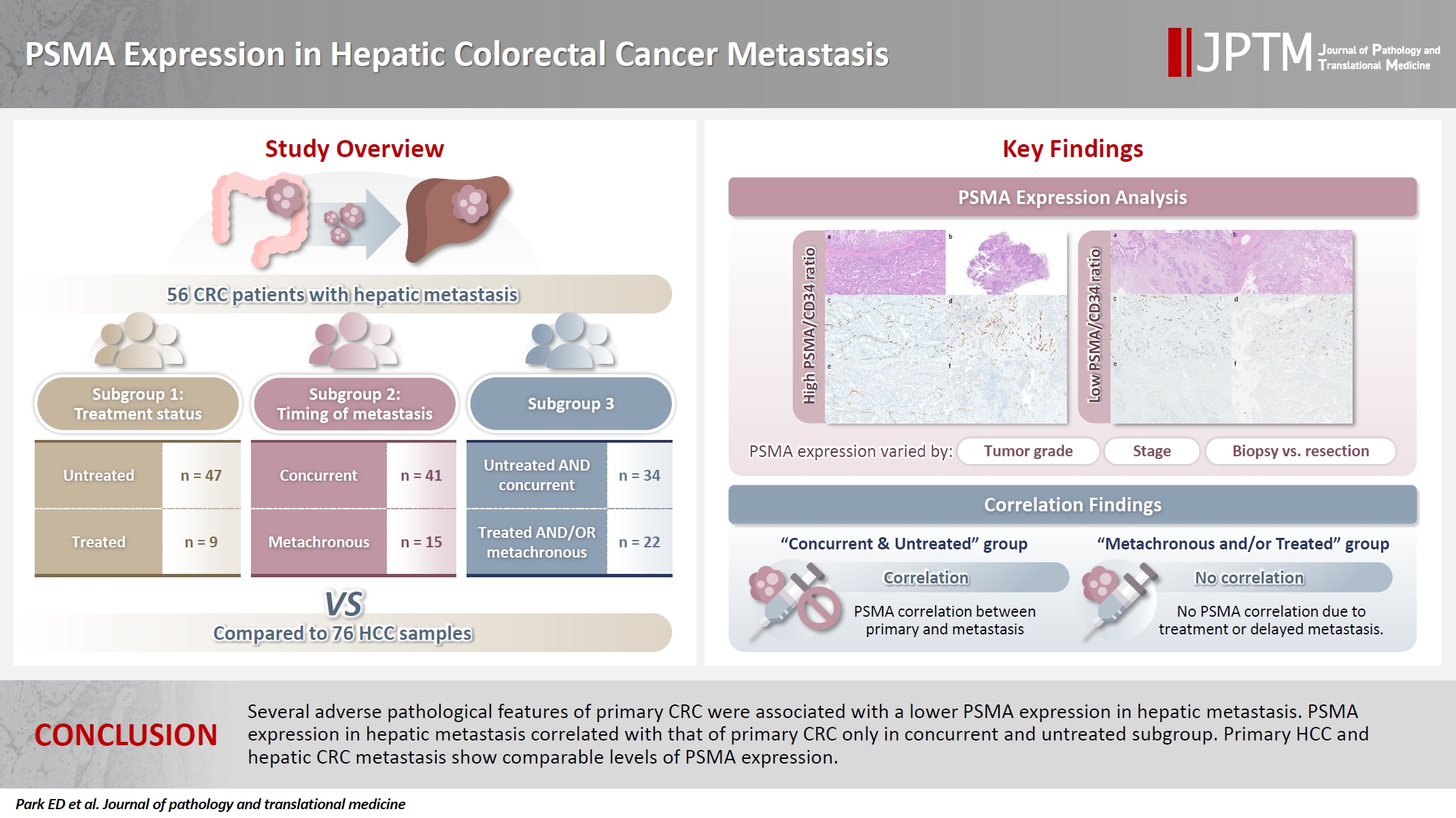
Prostate-specific membrane antigen (PSMA) is expressed in the neovasculature of various malignancies, such as colorectal cancer (CRC) and hepatocellular carcinoma (HCC). However, PSMA expression in hepatic CRC metastasis has not been studied in detail. Methods: The PSMA expression in primary CRC and corresponding hepatic metastasis was evaluated by immunohistochemistry in a metastatic CRC cohort (n = 56), which was divided into subgroups according to treatment history and timing of metastasis. Demographic and histological characteristics of primary CRC were collected and their relationships with PSMA expression were examined. Additionally, the PSMA expression in resected HCC (n = 76) was compared with that of hepatic CRC metastasis. Results: In primary CRC, PSMA level showed a positive association with tumor size. Lower PSMA expression in hepatic metastasis was associated with higher primary CRC grade, advanced pTNM stage at the time of CRC resection, presence of tumor deposit, and unresectability of metastatic lesion. PSMA expression in primary CRC correlated with that in hepatic metastasis only in concurrent and untreated metastasis subgroup. PSMA expression in primary CRC and hepatic metastasis, regardless of treatment history and timing of metastasis, was not significantly different from that of HCC. Conclusions: Several adverse pathological features of primary CRC were associated with a lower PSMA expression in hepatic metastasis. PSMA expression in hepatic metastasis correlated with that of primary CRC only in concurrent and untreated subgroup. Primary HCC and hepatic CRC metastasis show comparable levels of PSMA expression. -
Citations
Citations to this article as recorded by
- Incidental detection of PDAC via 18F-PSMA PET/CT in a patient with recurrent prostate cancer. A case report
Giordano Savelli, Mattia Bonacina, Alberto Soffientini, Elvira Archiati, Claudio Bnà, Alberto Zaniboni
Frontiers in Nuclear Medicine.2026;[Epub] CrossRef
- Incidental detection of PDAC via 18F-PSMA PET/CT in a patient with recurrent prostate cancer. A case report
- Prognostic significance of viable tumor size measurement in hepatocellular carcinomas after preoperative locoregional treatment
- Yoon Jung Hwang, Youngeun Lee, Hyunjin Park, Yangkyu Lee, Kyoungbun Lee, Haeryoung Kim
- J Pathol Transl Med. 2021;55(5):338-348. Published online September 2, 2021
- DOI: https://doi.org/10.4132/jptm.2021.07.26

- 8,030 View
- 124 Download
- 5 Web of Science
- 5 Crossref
-
Abstract
PDFSupplementary Material
- Background
Preoperative locoregional treatment (LRT) for hepatocellular carcinoma (HCC) often induces intratumoral necrosis without affecting the overall tumor size, and residual viable tumor size (VTS) on imaging is an important clinical parameter for assessing post-treatment response. However, for surgical specimens, it is unclear whether the VTS would be more relevant to prognosis compared to total tumor size (TTS).
Methods
A total of 142 surgically resected solitary HCC cases were retrospectively reviewed. The TTS and VTS were assessed by applying the modified Response Evaluation Criteria in Solid Tumors method to the resected specimens, and correlated with the clinicopathological features and survival.
Results
As applying VTS, 13/142 cases (9.2%) were down-staged to ypT1a. Although the survival analysis results for overall survival according to TTS or VTS were similar, VTS was superior to predict disease-free survival (DFS; p = .023) compared to TTS (p = .08). In addition, multivariate analysis demonstrated VTS > 2 cm to be an independent predictive factor for decreased DFS (p = .001). In the subpopulation of patients with LRT (n = 54), DFS in HCCs with TTS or VTS > 2 cm were significantly shorter than those with TTS or VTS ≤ 2 cm (p = .047 and p = .001, respectively). Interestingly, HCCs with TTS > 2 cm but down-staged to VTS ≤ 2 cm after preoperative LRT had similar survival to those with TTS ≤ 2 cm.
Conclusions
Although the prognostic impact of tumor size was similar regardless of whether TTS or VTS was applied, reporting VTS may help to increase the number of candidates for surgery in HCC patients with preoperative LRT. -
Citations
Citations to this article as recorded by- PET-Assessed Metabolic Tumor Volume Across the Spectrum of Solid-Organ Malignancies: A Review of the Literature
Anusha Agarwal, Chase J. Wehrle, Sangeeta Satish, Paresh Mahajan, Suneel Kamath, Shlomo Koyfman, Wen Wee Ma, Maureen Linganna, Jamak Modaresi Esfeh, Charles Miller, David C. H. Kwon, Andrea Schlegel, Federico Aucejo
Biomedicines.2025; 13(1): 123. CrossRef - Measures for response assessment in HCC treatment
Fereshteh Yazdanpanah, Omar Al-Daoud, Moein Moradpour, Stephen Hunt
Hepatoma Research.2024;[Epub] CrossRef - Machine Learning for Dynamic Prognostication of Patients With Hepatocellular Carcinoma Using Time-Series Data: Survival Path Versus Dynamic-DeepHit HCC Model
Lujun Shen, Yiquan Jiang, Tao Zhang, Fei Cao, Liangru Ke, Chen Li, Gulijiayina Nuerhashi, Wang Li, Peihong Wu, Chaofeng Li, Qi Zeng, Weijun Fan
Cancer Informatics.2024;[Epub] CrossRef - Construction and validation of a novel signature based on epithelial-mesenchymal transition–related genes to predict prognosis and immunotherapy response in hepatocellular carcinoma by comprehensive analysis of the tumor microenvironment
Biao Gao, Yafei Wang, Shichun Lu
Functional & Integrative Genomics.2023;[Epub] CrossRef - Cellular senescence affects energy metabolism, immune infiltration and immunotherapeutic response in hepatocellular carcinoma
Biao Gao, Yafei Wang, Shichun Lu
Scientific Reports.2023;[Epub] CrossRef
- PET-Assessed Metabolic Tumor Volume Across the Spectrum of Solid-Organ Malignancies: A Review of the Literature
- Guanabenz Acetate Induces Endoplasmic Reticulum Stress–Related Cell Death in Hepatocellular Carcinoma Cells
- Hyo Jeong Kang, Hyang Sook Seol, Sang Eun Lee, Young-Ah Suh, Jihun Kim, Se Jin Jang, Eunsil Yu
- J Pathol Transl Med. 2019;53(2):94-103. Published online January 16, 2019
- DOI: https://doi.org/10.4132/jptm.2019.01.14
- 10,731 View
- 201 Download
- 14 Web of Science
- 14 Crossref
-
Abstract
PDFSupplementary Material
- Background
Development of chemotherapeutics for the treatment of advanced hepatocellular carcinoma (HCC) has been lagging. Screening of candidate therapeutic agents by using patient-derived preclinical models may facilitate drug discovery for HCC patients.
Methods
Four primary cultured HCC cells from surgically resected tumor tissues and six HCC cell lines were used for high-throughput screening of 252 drugs from the Prestwick Chemical Library. The efficacy and mechanisms of action of the candidate anti-cancer drug were analyzed via cell viability, cell cycle assays, and western blotting.
Results
Guanabenz acetate, which has been used as an antihypertensive drug, was screened as a candidate anti-cancer agent for HCC through a drug sensitivity assay by using the primary cultured HCC cells and HCC cell lines. Guanabenz acetate reduced HCC cell viability through apoptosis and autophagy. This occurred via inhibition of growth arrest and DNA damage-inducible protein 34, increased phosphorylation of eukaryotic initiation factor 2α, increased activating transcription factor 4, and cell cycle arrest.
Conclusions
Guanabenz acetate induces endoplasmic reticulum stress–related cell death in HCC and may be repositioned as an anti-cancer therapeutic agent for HCC patients. -
Citations
Citations to this article as recorded by- ER stress signaling at the interphase between MASH and HCC
Younis Hazari, Eric Chevet, Béatrice Bailly-Maitre, Claudio Hetz
Hepatology.2026; 83(2): 387. CrossRef - The Integrated Stress Response in Cancer: Paradox and Therapeutic Promise
Chenliang Zhang, Ting Zhang, Qiulin Tang, Yu Zeng, Dan Cao
Comprehensive Physiology.2026;[Epub] CrossRef - Optogenetic Control of the Integrated Stress Response Limits Glioblastoma Invasion
Lisa K. Månsson, Ethan Dickson, Lun Hao, Angela A. Pitenis, Maxwell Z. Wilson
Cell Biochemistry and Function.2026;[Epub] CrossRef - The Construction of ceRNA Regulatory Network Unraveled Prognostic Biomarkers and Repositioned Drug Candidates for the Management of Pancreatic Ductal Adenocarcinoma
Busra Aydin, Keziban Okutan, Ozge Onluturk Aydogan, Raghu Sinha, Beste Turanli
Current Issues in Molecular Biology.2025; 47(7): 496. CrossRef - Current trends and future prospects of drug repositioning in gastrointestinal oncology
Nayeralsadat Fatemi, Mina Karimpour, Hoda Bahrami, Mohammad Reza Zali, Vahid Chaleshi, Andrea Riccio, Ehsan Nazemalhosseini-Mojarad, Mehdi Totonchi
Frontiers in Pharmacology.2024;[Epub] CrossRef - Small molecules for impairing endoplasmic reticulum in cancer
Tripti Mishra, Navneet Dubey, Sudipta Basu
Organic & Biomolecular Chemistry.2024; 22(44): 8689. CrossRef -
Guanabenz acetate, an antihypertensive drug repurposed as an inhibitor of
Escherichia coli
biofilm
Arakkaveettil Kabeer Farha, Olivier Habimana, Harold Corke, Olaya Rendueles
Microbiology Spectrum.2024;[Epub] CrossRef - The integrated stress response in cancer progression: a force for plasticity and resistance
Caleb L. Lines, Morgan J. McGrath, Tanis Dorwart, Crystal S. Conn
Frontiers in Oncology.2023;[Epub] CrossRef - Endoplasmic reticulum stress: Multiple regulatory roles in hepatocellular carcinoma
Jiacheng Wu, Shan Qiao, Yien Xiang, Menying Cui, Xiaoxiao Yao, Ruixin Lin, Xuewen Zhang
Biomedicine & Pharmacotherapy.2021; 142: 112005. CrossRef - The two faces of the Integrated Stress Response in cancer progression and therapeutic strategies
Eugenia Licari, Luis Sánchez-del-Campo, Paola Falletta
The International Journal of Biochemistry & Cell Biology.2021; 139: 106059. CrossRef - Repurposing of Guanabenz acetate by encapsulation into long-circulating nanopolymersomes for treatment of triple-negative breast cancer
Yusuf A. Haggag, Mohamed Yasser, Murtaza M. Tambuwala, Suleiman S. El Tokhy, Mohammad Isreb, Ahmed A. Donia
International Journal of Pharmaceutics.2021; 600: 120532. CrossRef - Endoplasmic reticulum stress: New insights into the pathogenesis and treatment of retinal degenerative diseases
Marina S. Gorbatyuk, Christopher R. Starr, Oleg S. Gorbatyuk
Progress in Retinal and Eye Research.2020; 79: 100860. CrossRef - Delineating the role of eIF2α in retinal degeneration
Christopher R. Starr, Marina S. Gorbatyuk
Cell Death & Disease.2019;[Epub] CrossRef - Repositioning of Guanabenz in Conjugation with Gold and Silver Nanoparticles against Pathogenic Amoebae Acanthamoeba castellanii and Naegleria fowleri
Areeba Anwar, Mohammad Ridwane Mungroo, Ayaz Anwar, William J. Sullivan, Naveed Ahmed Khan, Ruqaiyyah Siddiqui
ACS Infectious Diseases.2019; 5(12): 2039. CrossRef
- ER stress signaling at the interphase between MASH and HCC
- Prognosis of Hepatocellular Carcinoma after Liver Transplantation: Comparative Analysis with Partial Hepatectomy
- Kyuho Lee, Kyoung-Bun Lee, Nam-Joon Yi, Kyung-Suk Suh, Ja-June Jang
- J Pathol Transl Med. 2017;51(1):79-86. Published online December 25, 2016
- DOI: https://doi.org/10.4132/jptm.2016.10.13
- 10,376 View
- 152 Download
- 6 Web of Science
- 5 Crossref
-
Abstract
PDF
- Background
Liver transplantation (LT) is the treatment of choice for hepatocellular carcinoma (HCC). The aim of this study was to investigate the recurrence rate of HCC after LT and prognostic factors for recurrence by comparing LT with non-transplanted resection. Methods: The participants were 338 patients who underwent LT between 1996 and 2012 at Seoul National University Hospital (LT group) and 520 HCC patients who underwent partial hepatectomy between 1995 and 2006 (control group, non-LT group). Results: In the LT group, 68 of 338 patients (19.8%) showed relapse, and the recurrence rate was lower than that in the non-LT group (64.9%, 357/520, p < .001). Stratification analysis by American Joint Committee on Cancer (AJCC) stage showed that the stage I-II LT group had a lower recurrence rate than the non-LT group. Univariate comparative analysis demonstrated that multiplicity of tumor, tumor size, gross type, Edmondson- Steiner (ES) nuclear grade, extent of tumor, angioinvasion, AJCC stage, Milan criteria, University of California at San Francisco criteria on explant pathology (all p < .001), positive expression of cytokeratin 19 (p = .002), and preoperative α-fetoprotein (AFP) (p < .001) were predictors of tumor recurrence. In multivariate analysis, LT, preoperative AFP, multiplicity of tumor, extent of tumor, size of tumor, and ES nuclear grade were independent prognostic factors. Conclusions: LT might have a protective effect against the late recurrence of stage I-II HCC compared to non-LT, and the prognostic factors for recurrence were similar to previously well-known prognostic factors for HCC. -
Citations
Citations to this article as recorded by- Locoregional and Surgical Treatment of Single-Nodule Hepatocellular Carcinoma Recurrence After Liver Transplantation: A Systematic Review and a Meta-Analysis
Marco Maria Pascale, Camilla Marandola, Francesco Frongillo, Erida Nure, Salvatore Agnes
Cancers.2025; 17(9): 1501. CrossRef - Risk Factor Analysis of Death in Patients With Hepatic Cellular Carcinoma After Radical Operation: A Consecutive Cohort of 433 Patients
Zhengyang He, Wenfeng Lu, Dongze Qiu, Weimin She
Health Science Reports.2025;[Epub] CrossRef - Related Factors of Hepatocellular Carcinoma Recurrence Associated With Hyperglycemia After Liver Transplantation
Yujian Zheng, Qing Cai, Lishan Peng, Shibo Sun, Shaoping Wang, Jie Zhou
Transplantation Proceedings.2021; 53(1): 177. CrossRef - Oncological Outcomes of Hepatic Resection vs Transplantation for Localized Hepatocellular Carcinoma
A.T. Akcam, A.G. Saritas, A. Ulku, A. Rencuzogullari
Transplantation Proceedings.2019; 51(4): 1147. CrossRef - Clustering Asian Countries According to the Trend of liver cancer Mortality Rates: an Application of Growth Mixture Models
Maryam Salari, Anoshirvan Kazemnejad, Farid Zayeri
Iranian Red Crescent Medical Journal.2017;[Epub] CrossRef
- Locoregional and Surgical Treatment of Single-Nodule Hepatocellular Carcinoma Recurrence After Liver Transplantation: A Systematic Review and a Meta-Analysis
Brief Case Reports
- Periductal Stromal Tumor of Breast: A Case Report and A Review of Literature
- Salma L. Abbasi, Kate McNamara, Mohammed S. Absar, Alison Darlington, Francene Clucas, Sami Titi
- Korean J Pathol. 2014;48(6):442-444. Published online December 31, 2014
- DOI: https://doi.org/10.4132/KoreanJPathol.2014.48.6.442
- 14,526 View
- 89 Download
- 5 Crossref
-
PDF
-
Citations
Citations to this article as recorded by- Genetic Profiling of Mammary Periductal Stromal Tumors With Histologic Correlation Highlights High-Grade and Low-Grade Groups and Similarities to Phyllodes Tumors
Gregor Krings, Gregory R. Bean, Elizabeth M. Hosfield, J Jordi Rowe, Joseph Geradts, Yunn-Yi Chen
Modern Pathology.2026; 39(3): 100961. CrossRef - Survey of recurrent diagnostic challenges in breast phyllodes tumours
Benjamin Yongcheng Tan, Stephen B Fox, Sunil R Lakhani, Puay Hoon Tan
Histopathology.2023; 82(1): 95. CrossRef - Management of a periductal stromal tumor in a young woman: Our breast unit experience
Irene Valente, Adela Ristani, Cristina Mancini, Eugenia Martella, Leonardo Quartieri, Cecilia D'Aloia
The Breast Journal.2020; 26(7): 1375. CrossRef - A Diagnostic Approach to Fibroepithelial Breast Lesions
Benjamin Yongcheng Tan, Puay Hoon Tan
Surgical Pathology Clinics.2018; 11(1): 17. CrossRef - A case of local recurrence of periductal stromal sarcoma of the breast
Kana TERAMOTO, Yasuro DOI, Kayo YAMAMOTO, Kaname MATSUKAWA, Hisaka IWAIHARA, Rumi MOTOSHIMA, Noboru TAKATA, Ichiro YOSHINAKA, Kazunori HARADA
Choonpa Igaku.2018; 45(1): 61. CrossRef
- Genetic Profiling of Mammary Periductal Stromal Tumors With Histologic Correlation Highlights High-Grade and Low-Grade Groups and Similarities to Phyllodes Tumors
- Solid Form of Epithelioid Hemangioma: A Case Report
- Jin Roh, Min Jeong Song, Mi Woo Lee, Chan-Sik Park
- Korean J Pathol. 2014;48(5):394-397. Published online October 27, 2014
- DOI: https://doi.org/10.4132/KoreanJPathol.2014.48.5.394
- 12,916 View
- 93 Download
- 4 Crossref
-
PDF
-
Citations
Citations to this article as recorded by- Clinicopathologic and radio-genomic insights into hepatic epithelioid hemangioma: An illustrated review
Elijah Brown, Karen Schoedel, Adarsh Barwad, Alessandro Furlan, Paul J.L. Zhang, Nigar Anjuman Khurram
Human Pathology.2026; 170: 105955. CrossRef - Epithelioid Haemangioma of Bone: A Case Series and Comprehensive Literature Review Reappraising the Diagnostic Classification of All Epithelioid Vascular Neoplasms of Bone
Subramaniam Ramkumar
Cureus.2021;[Epub] CrossRef - Update on cutaneous epithelioid vascular tumours
Boštjan Luzar, Eduardo Calonje
Diagnostic Histopathology.2018; 24(8): 273. CrossRef - Epithelioid Hemangioma (Angiolymphoid Hyperplasia With Eosinophilia) of the Heart With Peripheral Eosinophilia and Nephrotic Syndrome
Isidro Machado, Agustín Chong, Anisia Serrano, Alfredo Mario Naranjo Ugalde, Damian Pineda, Laynes Savón, Ever Olivera, Antonio Llombart-Bosch
International Journal of Surgical Pathology.2016; 24(1): 59. CrossRef
- Clinicopathologic and radio-genomic insights into hepatic epithelioid hemangioma: An illustrated review
Original Articles
- Histologic Disorderliness in the Arrangement of Tumor Cells as an Objective Measure of Tumor Differentiation
- Sungwook Suh, Gyeongsin Park, Young Sub Lee, Yosep Chong, Youn Soo Lee, Yeong Jin Choi
- Korean J Pathol. 2014;48(5):339-345. Published online October 27, 2014
- DOI: https://doi.org/10.4132/KoreanJPathol.2014.48.5.339
- 8,274 View
- 68 Download
-
Abstract
PDF
- Background: Inter-observer and intra-observer variation in histologic tumor grading are well documented. To determine whether histologic disorderliness in the arrangement of tumor cells may serve as an objective criterion for grading, we tested the hypothesis the degree of disorderliness is related to the degree of tumor differentiation on which tumor grading is primarily based. Methods: Borrowing from the statistical thermodynamic definition of entropy, we defined a novel mathematical formula to compute the relative degree of histologic disorderliness of tumor cells. We then analyzed a total of 51 photomicrographs of normal colorectal mucosa and colorectal adenocarcinoma with varying degrees of differentiation using our formula. Results: A one-way analysis of variance followed by post hoc pairwise comparisons using Bonferroni correction indicated that the mean disorderliness score was the lowest for the normal colorectal mucosa and increased with decreasing tumor differentiation. Conclusions: Disorderliness, a pathologic feature of malignant tumors that originate from highly organized structures is useful as an objective tumor grading proxy in the field of digital pathology.
- Fine Needle Aspiration Cytology of hepatocellular Carcinoma: A Study on 247 Cases.
- Kwang Gil Lee, Jong Tae Lee, Soo Im Choi, Chan II Park
- J Pathol Transl Med. 1990;1(1):1-17.
- 43,507 View
- 99 Download
-
Abstract
PDF
- Hepatocellular carcinoma(HCC) is malignant tumor frequently occurring in Koreans. There have been few reports regarding the cytologic findings of fine needle aspiration(FNA) of HCC. Most have suggested a diagnostic problem in the cytology distinguishing HCC from some benign hepatic lesion-for example, a regeneration nodule in cirrhosis and liver cell adenoma. In spite of its high frequency in Korea, no cytologic study has been reported, concerning the FNA of HCC. In an attempt to achieve cytologic criteria for the diagnosis of HCC, the authors studied retrospectively cytopathologic findings of 247 cases of HCC. These cases were confirmed either by histologic examination including lobectomy, biopsy, or cell block material, or, when tissue diagnosis was unavailable, by a high serum alpha-fetoprotein level(over 400 I. U.). All aspiration smears were stained by the Papainicolaou method. In each case, the smears were analyzed for cell patterns and various cytomorphology of the tumor cells. The smear background was assessed for the presence of tumor cell necrosis and inflammatory components and compared to that of metastatic carcinomas. The cell patterns were classified as trabecular, acinar, dispersed, and irregular. The cytologic parameters analyzed included the degree of nuclear atypia and the presence of mitoses, intranuclear cytoplasmic inclusions, nucleolar prominency, endothelial lining, multinucleated giant cells, eosinophilc, globules bile and Mallory body. Most of the FNA of HCC showed markedly cellular smears. The tumor cells were most frequently arranged in a trabecular pattern(80.3%). The irregular(12.6%), the acinar(5.5%), and the dispersed patterns(1.7%) followed in decreasing frequency. Individual hepatoma cells were larger than normal liver cells. However, they had morphologic features characteristic of the hepatic cells : the cells were round or polygonal, their cytoplasm was abundant and granular with eosinophilic or amphophilic stainability, and their nuclei were round to oval, located centrally, and tended to have prominent nucleoli. Anaplasia and pleomorphism of tumor cells were generally mild to moderate. These findings existed even in very well differentiated cases. Mitotic figures were present in about 85% of the cases. Prominent nucleoli were observed only in about half the cases. The frequency of other cytologic features was as follows : intranuclear cytoplasmic inclusion in 86.8% ; endothelial lining in 56.1% ; bile in 19.8% ; and giant cells in 60.1%. Clear cells were often present in 11.7%, Most aspiration smears of HCC displayed clean background without necrosis or inflammatory material in contrast to the dirty, necrotic background of metastatic cancers and cholangiocarcinomas. Based on the above mentioned features, it is suqqested that the cytologic critieria most important for the diagnosis of HCC include a markedly cellular smear, trabecular pattern. hepatocytoid appearance of tumor cells, endothelial lining, the presence of bile, giant cells, intranuclear cytoplasmic inclusions, and prominent nucleoli, Among these, trabecular pattern, endothelial lining, giant cells and clean smear background are points to be considered in differentiating HCC from metastatic and cholangiocellular carcinoma.
- Fine Needle Aspiration Cytology of Hepatoblastoma: Report of Two Cases.
- Young Nyun Park, Kwang Gil Lee, Chan II Park
- J Pathol Transl Med. 1990;1(1):98-102.
- 2,396 View
- 35 Download
-
Abstract
PDF
- Hepatoblastoma(HB) is a rare embryonic malignant tumor of the liver. Most morphological studies on HB have limited to the histological characteristics and only 3 cases of HB have been described in the cytology literature. We present 2 cases of HB occurring in children aged 1 year and 3 years, respectively. The distinctive cytologic features of fine needle aspiration of HB were clusters of tumor cells showing acinar and trabecular pattern, smaller tumor cells with a high nuclear-cytoplasmic ratio and hyperchromatic nuclei having prominent nucleoli, and the presence of extramedullary hematopoiesis and osteoid material. These features were also found in the cell block and the biopsy specimen, and appeared very useful in the differentiation of HB from hepatocellular carcinoma.
- Enhanced Protein Expression of Signal Transducer and Activator of Transcription 3 and Protein Kinase Substrate p36 in Hepatocellular Carcinoma.
- Hongxiu Han, Si Hyong Jang, Chan Kum Park
- Korean J Pathol. 2009;43(5):393-399.
- DOI: https://doi.org/10.4132/KoreanJPathol.2009.43.5.393
- 3,890 View
- 28 Download
-
Abstract
PDF
- BACKGROUND
Signal transducers and activators of transcription 3 (STAT3) and protein kinase substrate p36 may be involved in cell proliferation, differentiation and growth.
METHODS
Immunohistochemistry for STAT3 and p36 was performed in 46 patients with hepatocellular carcinoma (HCC).
RESULTS
STAT3 staining was present in the cytoplasm and/or nucleus, while p36 staining was present in the nucleus. STAT3 and p36 expression occurred in 78.3% (36/46) and 47.8% (22/46) of HCC patients, respectively. However, no correlation was found between STAT3 and p36 protein expression (p>0.05). Enhanced expression of STAT3 was negatively correlated with portal vein invasion (p=0.033). Expression of STAT3 in the nucleus was correlated with tumor grade (p=0.004). Enhanced expression of p36 was correlated with tumor grade (p=0.031). HCC was correlated with HBV infection (p=0.032). The patients'5-year survival was related to expression of p36 (p=0.044), but not to total STAT3 or nuclear STAT3 (p>0.05).
CONCLUSIONS
The enhanced expression of STAT3 in the nucleus and the enhanced expression of p36 are associated with the aggressive phenotype of HCC. Enhanced p36 expression may contribute to poor survival of patients with HCC.
Case Report
- Gastric Adenocarcinoma with Coexistent Hepatoid Adenocarcinoma and Neuroendocrine Carcinoma: A Case Report.
- Aeri Kim, Sang Woon Kim, Sun Kyo Song, Young Kyung Bae
- Korean J Pathol. 2009;43(1):79-82.
- DOI: https://doi.org/10.4132/KoreanJPathol.2009.43.1.79
- 4,717 View
- 36 Download
- 2 Crossref
-
Abstract
PDF
- This report represents a very rare case of a gastric adenocarcinoma that was coexistent with hepatoid adenocarcinoma and neuroendocrine carcinoma. A 77-year-old man was admitted to our hospital due to a huge ulcerofungating mass identified at the proximal body of the stomach. After a pathological diagnosis of the tumor as a poorly differentiated adenocarcinoma was made, the patient underwent a total gastrectomy with lymph node dissection. Microscopically, the tumor consisted of three morphologically distinct components-tubular adenocarcinoma, hepatoid adenocarcinoma and neuroendocrine carcinoma. The hepatoid adenocarcinoma component resembled a hepatocellular carcinoma and produced alpha-fetoprotein. The neuroendocrine carcinoma component was positive for chromogranin and synaptophysin immunostains. This is an example of the diverse morphological and immunophenotypical differentiation of gastric carcinomas.
-
Citations
Citations to this article as recorded by- An Intestinal Type Gastric Neuroendocrine Tumor: A Case Report
Mohammad Abu-Jeyyab, Renata Kakish, Malak Alkatib, Leen Alshawabkeh, Rawan Bani Hamad, Mary Almadani, Ma'wia Santarisi, Mohammad Al-Jafari, Abdulqadir J. Nashwan
Case Reports in Oncology.2023; 16(1): 1113. CrossRef - Gastric adenocarcinoma is concurrent with metastatic neuroendocrine cancer treated with nivolumab and chemotherapy: A case report
Bing Yan, Meiqi Cui, Junhao You, Fang Li, Hui Liu
Molecular and Clinical Oncology.2018;[Epub] CrossRef
- An Intestinal Type Gastric Neuroendocrine Tumor: A Case Report
Original Articles
- The Effects of Transforming Growth Factor beta1 on Apoptosis in Rat Hepatocellular Carcinoma.
- Young Euy Park, Young Hee Choi, Won Yo Lee, Jin Ja Park, Kyung Chan Choi, Hyung Shik Shin
- Korean J Pathol. 1999;33(2):71-79.
- 2,133 View
- 10 Download
-
Abstract
- Based upon the concept that carcinogenesis is associated with apoptosis, specific therapies designed to enhance the susceptibility of cancer cells to undergo apoptosis could be developed. Thus, in this paper, it was designed to investigate whether, using rat animal model with chemical-induced hepatocellular carcinoma, TGF-1 in vivo could induce apoptosis in cancer. The chemical hepatocarcinogenic procedure of Solt-Farber method was used on Sprague-Dawley rats. Experimental groups were divided into group A treated with the standard Solt-Farber regimen of diethylnitrosamine (DEN) and 2-Acetaminofluorene (AAF), group B TGF-, group C TGF-1, and group D adriamycin after hepatocellular carcinoma developed. For detection of apoptotic cells, apoptotic indices were examined by the in situ end DNA labelling method. The expression of proliferating cell nuclear antigen was examined by immunohistochemical staining. Apoptosis of rat hepatocellular carcinoma cells increased significantly to 4.92+/-2.32/HPF in the group C compared with the control group (A) (2.54+/-1.13/HPF; P<0.05). Two distinctly different populations of proliferating hepatocellular carcinoma cells were identified. The cells at G1/S boundary (weak granular staining) increased to 15.75+/-6.19/HPF and 6.45+/-2.93/HPF in the groups C and D, respectively, but decreased to 2.42+/-2.06/HPF in the group B compared with the control group (A) (6.38+/-2.18/HPF; p<0.05). The cells at S phase (strong granular staining) increased to 3.37+/-2.69/HPF in the group B but decreased to 0.32+/-0.47/HPF in the group D (p<0.05). In conclusion, these results indicate that the TGF-1 may be used as an effective anticancer agent.
- Kupffer Cells in Hepatocellular Carcinoma.
- Young Nyun Park, Soon Hee Jung, Chan Il Park
- Korean J Pathol. 1989;23(3):305-310.
- 3,440 View
- 97 Download
-
Abstract
PDF
- Kupffer cells are tissue macrophages (histiocytes) fixed in hepatie sinusoids. Since malignant hepatocytes are the only tumor parencymal cells of the hepatocellular carcinoma, theoretically there are no Kupffer cells within the hepatocellular carcinoma. To clarify whether it is true or not, 12 cases of hepatocellular carcinoma of the trabecular type with some extents of the non-neoplastic surrounding liver were subjected to immunoperoxidase staining for lysozyme and S-100 protein and the results are as follows. 1) Kupffer cells were stained positively by the immunoperoxidase staining for lysozyme but not for S-100 protein, indicating that they are monocyte derived macrophages. 2) Kupffer cells were also present within the hepatocellular carcinoma, but were 2-7 times fewer within the hepatocellular carcinoma than in the non-neoplastic areas (p<0.05). 3) The non-neoplastic hepatic tissue of patients with serum HBsAg shows a tendency to have more kupffer cells than those without HBsAg.
- Clinicopathologic Features of Early Hepatocellular Carcinoma.
- Chang Ohk Sung, Suk Jin Choi, Cheol Keun Park
- Korean J Pathol. 2004;38(3):138-144.
- 2,699 View
- 23 Download
-
Abstract
PDF
- BACKGROUND
Early hepatocellular carcinoma (HCC) is an early stage HCC, and it is sometimes difficult to identify the margins of the cancer nodules in the resected specimens.
METHODS
We studied 22 cases of early HCC to investigate the clinicopathologic features of early stage HCC.
RESULTS
Seven of 22 cases were single HCC, and 15 were multicentric HCC. The average tumor size was 1.34 cm (0.4-2.7 cm). Early HCCs didn't destroy the basic architecture of the liver lobules or pseudolobules and the lesions had an indistinct margin. Most tumors were uniformly composed of well-differentiated cancer tissue that was characterized by an increased cell density and an irregular thin-trabecular pattern. The tumor retained a varying number of portal tracts. There was a replacing growth pattern at the tumor-nontumor boundary without tumor capsule. Three of 22 cases had a "nodule-in-nodule" lesion, and the inner nodules consisted of moderately differentiated HCC without portal tracts. All 22 cases showed no vascular invasion. All 7 patients with single early HCC have survived for the past 11-54 months without any local recurrence. But in one patient with single early HCC, multicentric HCC developed 20 months after surgery.
CONCLUSION
The clinicopathologic features of early HCCs are quite different from those of advanced HCCs. The increased recognition of early HCC during routine clinical practice will contribute to improved patient survival.
- Cytologic Diagnosis of Metastatic Hepatocellular Carcinoma by Aspiration Cytology of Sacrum.
- Jungweon Shim, Illhyang Ko
- J Pathol Transl Med. 1990;1(2):179-184.
- 2,355 View
- 17 Download
-
Abstract
PDF
- Bone metastasis of hepatocellular carcinoma appears to be peculiar when clinical manifestation of liver disease is not apparent, and initial diagnosis of metastatic hepatocellular carcinoma by fine needle aspiration cytology is rarely obtained. We experienced a case of 45-year-old man with metastatic hepatocellular carcinoma in the sacrum, which was diagnosed by fine needle aspiration cytology. The intrahepatic mass, measuring 1.2 cm in diameter and kept unchanged in size for two years, was never proved to be hepatocellular carcinoma histopathologically. The aspirated neoplastic cells were mostly in sheets, showing abundant acidophilic cytoplasm and large, round. centrally located nuclei with single, prominent acidophilic mucleoti. In the cell block section, diagnosis of metastatic well-differentiated hepatocellular carcinoma was made without difficulty, and definite trabecular fashion with sinusoidal endothelial cell lining was found.
- Analysis of DNA Ploidy Patterns and Nuclear Morphometry in Diethylnitrosamine Induced Hepatocyte Nodules and Hepatocellular Carcinoma of Rats.
- Chan Choi, Myung Kwan Kim, Kwan Mook Chae, Eun Cheol Kim, Hyung Bae Moon
- Korean J Pathol. 1993;27(3):226-234.
- 2,502 View
- 24 Download
-
Abstract
PDF
- This study was designed to answer the question; (1) How does the DNA ploidy pattern change in hepatocarcinogenesis? (2) How does the nuclear morphology change in hepatocarcinogenesis? Diethylnitrosamine(DEN) (16.5 mg per kg) was subcutaneously injected to female Sprague-Dawley rats(150~200g) by weekly interval for 30 weeks. DNA ploidy and parameters of nuclear morphology were measured by image analyser(IBAS 200, Kontron, FRG). The DNA ploidy pattern was divided into three basic patterns(diploid, polyploid, and aneuploid modes). In 8 cases of saline-injected control rats, the DNA histograms showed all polyploid pattern. Inhepatocyte nodules(hyperplastic nodules), DNA diploidy was the most frequent pattern, being followed by polyploid and aneuploid DNA patterns, contrast to hepatocelular carcinomas in which polyploid DNA pattern was most frequently noted being followed by diploid and aneuploid DNA pattern. Although the nuclei of hepatocytes in hepatocyte nodules and hepatocellular carcinomas were larger and more pleomorphic than those of normal hepatocytes, they were as same as those of normal hepatocytes in regard to nuclear hyperchromasia. DNA content, which was increased in hepatocarcinogenesis, was significantly related to the nuclear area.
- Immunohistochemical Study of the Expression of p53, Pan-ras, c-erbB-2 and PCNA in N-Nitrosomorpholine(NNM)-Induced Hepatocellular Carcinoma of Rats.
- Ok Kyung Kim, Ryun Jo Shin
- Korean J Pathol. 1995;29(6):727-739.
- 1,993 View
- 14 Download
-
Abstract
PDF
- The focus of this study was o aialyze the morphologic expression of p53, Pan-ras, c-erbB-2, and PCNA in preneoplastic and neoplastic liver lesions induced with NNM of rats. The development of hepatocellular tumors was investigated by histology and electron microscope in 65 Splague-Dawley rats administered with NNM in drinking water at low dose(5 mg/100 ml) and high dose(20 mg/100 ml). Three types of hepatocytic degeneration glycogenotic, eosinophilic and basophilic changes were followed by the appearance of hepatocellullar carcinoma. Hepatocellular carcinoma was increased in number and size according to NNM dosage and to duration of exposure. The histological classifications of hepatocelular carcinoma wer trabecular type, which was which was the most common, large eosinophilic, small cell, adenocarcinomatous and clear cell type. The expression of p53, Pan-ras, c-erbB-2 PCNA was examined by immunohistochemical stains. Eosinophilic degeneration revealed mild positivity at 18-26 weeks for expression of all oncogenic proteins studied and PCNA, whereas precancerous lesions showed variable expression from negative to moderate positivity on PCNA. Hepatocellular carcinoma lesions showed strong positivity for all stains and increased intensity during experimental period. These may indicate that chemical carcinogen produce hepatic eosinophilic degeneration and preCancerOus lesions by genetic mutation, resulting in hepatocellular carcinoma.
- The Tissue Expression of HBsAg and HBcAg in Hepatocellular Carcinoma and Peritumoral Liver.
- Jee Young Han, Woo Hee Jung, Chae Yoon Chon, Chan Il Park
- Korean J Pathol. 1993;27(4):371-378.
- 2,638 View
- 27 Download
-
Abstract
PDF
- To evaluate the tissue expression rate and pattenr of HBsAg and HBcAg in tumors and peritumoral livers, an immunohistochemical study was undertaken on 47 surgically resected hepatocellular carcinomas(HCCs). The results are as follows. 1. Patient's sera were positive for HBsAg in 40 cases(85.1%). In the remaining 7 cases, the tumor and peritumoral liver expressed neither HBcAg nor HbSaG, suggesting that they were caused by other etiologies than hepatitis B virus. 2. The peritumoral liver had HBsAg and HBcAg in 95.0% and 27.5% among the 40 cases, respectively. But the tumor expressed HBsAg in 50.0% and HBcAg in none. 3. The expression of HBsAg within the tumor and both HBsAg and HBcAg in the peritumoral liver tended to be more frequent in the pretreated cases before surgery. 4. Edmondson-Steiner grade IV tumors revealed a lower expression rate of HBsAg than the low grade tumors(p<0.05). Incases with cirrhosis at peritumoral tissues, HBcAg was less frequently found than in those without cirrhosis. The majority of tissue HBsAg and HBcAg was represented as groups of positive cells. These results suggest that, during the development and progression of HCCs, the HBcAg containing cells are repeatedly removed and the HBcAg negative cells are selected, because cellular expression of HBcAg is the target of host immune response.
- Relationship between Proliferative Activity and Expression of HBcAg and p53 Protein in Hepatocellular Carcinoma and Surrounding Nontumorous Liver.
- So Ya Paik, Ho Guen Kim, Chan Il Park
- Korean J Pathol. 1997;31(8):773-781.
- 2,042 View
- 17 Download
-
Abstract
PDF
- In an attempt to discover the factors contributing to the increased proliferative activity in hepatocytes and subsequent development of HCC, the proliferative activity of hepatocytes was compared with the size of regenerative nodules and HBcAg expression status in the surrounding nontumorous liver of 45 surgically resected hepatocellular carcinomas, including 34 HBV related ones. In the tumor, the difference in proliferative activity and the histological grade was analyzed in terms of p53 gene alteration. The proliferative activity was assessed by immunohistochemical methods using Ki-67 monoclonal antibody. HBcAg expression in the surrounding nontumorous liver correlated with both the inflammatory and proliferative activity of hepatocytes (p<0.05). p53 overexpression was associated with high proliferative activity and aggressive phenotype of tumor. No correlation was observed between the proliferative activity of hepatocytes and the size of regenerative nodules in cirrhosis (p>0.05). p53 overexpression was not evident in surrounding nontumorous liver including cirrhosis. In conclusion, the above results are in line with the view that hepatic carcinogenesis is a mutistep, progressive process. In the initial stage, chronic cellular injury incurred by immumologic reaction against HBcAg seems to play a pivotal role in increased cellular regeneration. However, once transformation of hepatocytes occur the major contributor to tumor growth seems to be alteration in p53 tumor suppresor gene.
- Application of Argyrophilic Nucleolar Organizer Regions(AgNORs) in the Diagnosi of Hepatocellular Carcinoma.
- Cheol Hee Yun, Sang Sook Lee, Eun Sook Chang
- Korean J Pathol. 1993;27(6):553-560.
- 2,292 View
- 10 Download
-
Abstract
- Necleolar organizer regions(NORs) ARE LOOPS OF DNA which transcribe to ribosomal RNA by RNA polymerase I. Since NOR-associated proteins are argyrophilic, silver staining method has been used for demonstration of AgNORs. The numbers and/or configurations of NORs may reflect the activities of cells in hyperplastic and neoplastic conditions. To evaluated the applicability of AgNORs in the diagnosis of hepatocellular carcinoma, the author had performed silver staining on the routinely processed, formalin-fixed, paraffin-embedded sections of 14 cases of normal liver(control), 23 cirrhotic liver, and 21 hepatocellular carcinoma. The results are summarized as follows: 1) The mean number of AgNORs per nucleus(mAgNOR) of normal liver, cirrhotic liver and hepatocellular carcinoma was 1.45+/-0.07, 2.53+/-0.38 and 5.52+/-1.63, respectively. The difference of mAgNOR between normal and cirrhotic liver, and between cirrhotic liver and hepatocellular carcinoma was statistically significant, respectively(p<0.01). 2) The percentage of nuclei showing five or more AgNORs per nucleus(pAgNOR) was 0.07% in normal liver, 7.59% in cirrhotic liver, and 60.49% in hepatocellular carcinoma. 3) AgNORs in hepatocellular carcinoma were large, pleomorphic and irregularly clumped, in addition to increase of mAgNOR and high pAgNOR. In conclusion, the increase of mAgNOR, high pAgNOR and large, irregular AgNORs are regarded as an additional helpful finding for the histopathological diagnosis of hepatocellular carcinoma.
- Clonality of Large Regenerative Nodule Accompanied by Hepatocellular Carcinoma.
- Zhe Piao, Bong Kyun Chun, Woo Jung Lee, Young Nyun Park, Ho guen Kim, Chanil Park
- Korean J Pathol. 1997;31(9):884-890.
- 2,209 View
- 15 Download
-
Abstract
PDF
- In order to clarify the preneoplastic nature of large regenerative nodules without dysplastic change, we analysed the clonality of hepatocellular carcinomas (HCCs) and large nodules, diameter > or =0.5 cm, of cirrhotic liver by X-linked human androgen receptor (HUMARA) gene assay, using the principle of random X chromosome methylation and inactivation in female. Ten cases of HCC and 5 cases of large nodules without dysplasia from 9 female patients were selected. All the tumors, large nodules and paired normal control cells were selectively microdissected from deparaffinized hematoxylin and eosin stained slides. Genomic DNA was isolated and digested with HhaI. Polymerase chain reaction(PCR) amplication of the HUMARA locus was performed using 32P-a-dCTP containing PCR mixtures. The PCR amplified products were separated by gel electrophoresis and analysed by autoradiography. Nine HCCs from 8 patients were monoclonal and 1 case was polyclonal and the remaining 1 case was not polymorphic at the HUMARA locus. The HCC case which showed polyclonality contained many inflammatory cells. All the large nodules were polyclonal by HUMARA assay. These results suggest that all or most of the cells composing the large regenerative nodules without dysplasia are polyclonal. This assay may be informative for the differentiation between regenerative and preneoplastic nodules in cirrhotic liver and the size of nodule may be not important in hepatocarcinogenesis.
- Flow Cytometric DNA Analysis of Hepatocellular Carcinoma.
- Young Lyun Oh, Yong Il Kim
- Korean J Pathol. 1993;27(6):581-589.
- 2,151 View
- 15 Download
-
Abstract
PDF
- A flow cytometric analysis of the nuclear DNA content of solid tumors using paraffin-embedded tissues has become available since 1983, and its ploidy pattern has been designated as an important prognostic parameter in many human tumors. Hepatocellular carcinoma(HCC) is one of the most common malignant tumors among Koreans, but little information is consolidated about the significance of ploidy pattern. We measured the nuclear DNA content of 62 surgically resected HCCs and 45 non-neoplastic tissues from the surrounding parenchyma by flow cytometry. Aneuploid was detected in 18 cases(29.0%) in HCCs and 2 cases(4.4%) in nonneoplastic hepatic parenchyma(p<0.005). Correlations between the DNA ploidy pattern and various clinicopathologic findings of HCCs were analized. The mean tumor size was significantly different(p<0.05) between the aneuploid group(8.8 cm) and the diploid group(6.1 cm). Mean age of the aneuploid group was younger(47 year) than the diploid group(51 years), but the difference was not statistically significant(p=0.052). The DNA pattern did not show any meaningful correlation with the gross and microscopic features of HCC except for the presence of capsule. These results suggest that DNA ploidy correlates with growth rate of the tumor and it may be a possibly useful prognostic factor in HCCs.
- Gross Anatomical Typing of Hepatocellular Carcinoma: Classification of 49 lobectomized hepatocellular carcinomas.
- Young Nyun Park, Eun Kyung Han, Chan Il Park
- Korean J Pathol. 1991;25(2):83-92.
- 2,560 View
- 23 Download
-
Abstract
PDF
- Forty-nine lobectomized hepatocellular carcinomas(HCC) were classified according to the gross anatomical features. Because the presence of cirrhosis in the remaining liver has a good clinico-pathological implication, cases of HCC were divided into non-cirrhotic(non-LC) and cirrhotic(LC) groups. In both groups, the tumors themselves belonged to either expanding, focal spreading, spreading or mixed type. Another special type, which has been called a "diffuse type" is added in the LC group with the name of "cirrhotomimetic type" Among 49 cases, 21 belonged to the non-LC group and 28 to the LC group. Most common was expanding type(20 cases, 40.8%), which was followed by spreading(32.7%), focal spreading(16.3%), mixed(6.1%) and cirrhotomimetic(4.1%) types. Expanding type of the LC group was the single most common type(13 cases, 26.5%). The accordance rate of gross typing was 0.94. Tumor masses of the LC group showed a greater tendency of having a fibrous capsule(60.7%) and a lobulated cut surface(82.1%), in contrast to those of the non-LC group (28.6% and 42.9% respectively). The patient's age and the HBsAg seropositivity were not different between the groups and between the types. Increased serum level of AFP was particularly frequent in the spreading type(81.3%) of both groups and in the cirrhotomimetic type(100%).
Case Report
- A Case of Metastatic Hepatocellular Carcinoma of the Ovary: An Immunohistochemical Study and Literature Review.
- Mi Jin Kim
- Korean J Pathol. 2005;39(4):287-290.
- 2,282 View
- 16 Download
-
Abstract
PDF
- Hepatocellular carcinomas rarely metastasize to the ovaries. To our knowledge, only nine cases of metastatic hepatocellular carcinoma of the ovary have been reported in the literature. Here, we present an additional case in which an ovarian lesion was the initial presentation in a 43-year-old female patient. An exploratory laparotomy revealed a left ovarian solid mass measuring 6.5*4.0*3.5 cm, with a lobulated greenish brown sectioned surface. A subsequent ultrasonogram and CT scan revealed a concurrent hepatic mass, and laboratory tests showed high serum AFP and CA125 levels. Microscopically the tumor showed predominantly solid and trabecular patterns, and intercellular canaliculi containing bile pigments. A postoperative hepatic biopsy confirmed the hepatocellular carcinoma. The main differential diagnosis involved ovarian metastasis of the hepatocellular carcinoma, the hepatoid carcinoma of the ovary with liver metastasis, and a hepatoid yolk sac tumor. Diagnosis in such cases should be reached by careful clinical evaluation and a thorough pathologic examination accompanied by a histochemical and immunohistochemical work-up.
Original Articles
- Morphometric Analysis of Cirrhotic Nodules in Hepatocellular Carcinoma-bearing Livers.
- Gyeong Hoon Kang, Yong Il Kim
- Korean J Pathol. 1991;25(4):338-345.
- 2,526 View
- 14 Download
-
Abstract
PDF
- It has been well known that liver cirrhosis, regardless of its etiology, is an important predisposing factor in hepatocarcinogenesis. However, the type of cirrhosis in hepatocellular carcinoma(HCC)-bearing liver varies not only by geographic areas but also with the cirteria applied for morphological classification of cirrhosis. To elucidate the relationship between the nodule size of HCC-bearing cirrhotic liver and clinicopathologic features, we measured cirrhotic nodule areas of 49 surgically resected HCC cases using image analyzer. The morphological type of cirrhosis was predominantly macronodular(49%), and followed by mixed(37%) and micronodular(14%). Seventy percent of the cases showed seropositivity for HBsAg. The average area of cirrhotic nodules was significantly larger in HBsAg-positive cases(mean: 6.14 mm2) than that of HBsAg-negative cases(mean: 2.5 mm2)(p<0.05), and their size was bigger in cases with grossly expansile pattern of HCC than those cases with infiltrative ones(p<0.05). Based on the above findings, we assume that seropositivity of HBsAg may influence on the regenerative activity of cirrhotic nodules and also subsequent increase of risk for further development of HCC. The presence of cirrhohsis and nodule size seem to be the important contributing factors to determine the growing patterns of HCC.
- DNA Ploidy and S-Phase Fraction in Proliferative Hepatic Lesions of Rat Liver Induced by Dietylnitrosamine and Partial Hepatectomy.
- Chan Choi, Sung Hee Cho, Hyung Bae Moon, Ki Jung Yun, Hun Taeg Chung, Sang Woo Juhng
- Korean J Pathol. 1991;25(4):346-356.
- 2,199 View
- 13 Download
-
Abstract
PDF
- We have investigated the changes of DNA ploidy and S-phase fraction in proliferative lesions of rat liver. Proliferative lesions were induced by diethylnitrosamine and partial hepatectomy. DNA ploidy was measured by flow cytometer, and S-phase fraction was measured by in situ bromodeoxyuridine(BRdU)-anti BRdU monoclonal antibody techniques. Normal liver and initiated lesion revealed DNA diploidy or DNA tetraploidy. Hepatocyte nodule (NODULE) and hepatocelular carcinoma (HCC) revealed DNA diploidy, tetraploidy or aneuploidy. S-phase fraction was 1.0+/-0.9, 1.0+/-0.9m 3.7+/-2.3, 5.5+/-4.9, and 13.8+/-11.6 in normal liver, initiated lesion, NODULE not associated with HCC, NODULE associated with HCC, and HCC, respectively. In NODULE associated with HCC, it was widely distributed, ranging from 0.8 to 15.5%. In conclusion, S-phase fraction appeared to be increased as the hepatocarcinogenesis proceeded, but DNA ploidy did not. There was a heterogeneity of DNA ploidy and S-phase fraction in the proliferative hepatic lesions.
Case Report
- A Case Report of Pedunculated Hepatocellular Carcinomas.
- Jae Bok Park, Sae Kwang Moon
- Korean J Pathol. 1991;25(5):467-470.
- 2,354 View
- 11 Download
-
Abstract
- Pedunculated hepatocellular carcinoma was first described by Roux in 1987, and Goldberg and Wallerstein presented a case with pathological description in 1934. Since then 37 cases have been reported in the world literature. A pedunculated hepatocellular carcinoma, occurred in a 69-year-old woman, was encountered. It was attached to the Glission's capsule of the right lobe of liver inferiorly, and was connected to the porta hepatis bt a mesenteric pedicle containing arteries, veins, bile ducts and nerve bundles. The tumor mass was completely encapsulated, and measured 8.5x8x6 cm and 255 gm. It was composed of hepatocellular carcinoma of a well-differentiated, acinar type. Tumor cells were positive for HBsAg, but negative for alpha-fetoprotein on peroxidase-antiperoxidase staining. The liver exhibited macronodular cirrhosis on gross examination. This tumor is thought to have on in the supernumerary lobe of liver.
Original Articles
- Analysis of DNA Ploidy Pattern of Hepatocellular Carcinoma with Comparison between Flow Cytometric and Image Cytometric Study.
- Sung Sook Kim, Seok Gun Park, Dong Sun Han, Man Ha Huh
- Korean J Pathol. 1992;26(1):1-9.
- 2,029 View
- 16 Download
-
Abstract
PDF
- Fifty cases of hepatocellularcarcinoma were studied using retrospective flow cytometric(FCM) and image cytometric(ICM) DNA analysis to determine the prevalence of aneuploid cell population and whether they were associated with any particular clinico-pathologic findings. At the same time, we compared the difference between FCM and ICM. The materials were prepared from 50 micrometer cut of paraffin embedded blocks. The DNA modal values, which could be defined in 42 cases by FCM with 74% aneuploidy and in 50 cases by ICM with 76% aneuploidy. So 95% of the cases had concordant DNA ploidy results by both techniques of FCM and ICM. Abnormal DNA pattern was correlated with age(<50), presence of cirrhosis, pathologic grade and some pathologic types(p<0.05), but was not correlated with presence of HBsAg, sex, alphafeto protein, and alcohol history(p>0.05). Also we found that ICM technique was easier to perform and interprete.
- Small Hepatocellular Carcinoma: Pathologic Features of 39 Cases A Comparison with Large Hepatocellular Carcinoma.
- Yong Il Kim, Geon Kook Lee, Sang Yong Song
- Korean J Pathol. 1992;26(2):103-116.
- 2,506 View
- 18 Download
-
Abstract
PDF
- With advance of diagnostic imaging technics, the detection rate of small hepatocellular carcinoma (HCC) has become much increased, but the questions whether the growth pattern and histologic nature of the HCC keep maintain the original gross and microscopic features with its advancement of tumor size remain still unclear. We reviewed 39 surgically resected hepatocellular carcinomas(HCCs) with a tumor size less than or equal to 3 cm in diameter(s-HCC), and their gross and microscopic features were compared with the HCCs bigger than 3 cm (i-HCC, 199 cases). Single nodular type(SN) was the most common gross type(60%) in s-HCCs, and was followed by single nodular type with perinodular extension(SNPE; 15.4%), multinodular-discrete type(10.3%) and multinodular-confluent type(5.1%). These figures contrasted to SNPE(42.2%) and SN(20.6%) in the i-HCCs. Of the 39 s-HCCs, 25 cases(64.1%) were encapsulated, and 14 cases(36%) demonstrated intratumoral fibrous septations, being contrasted to the i-HCCs in which fibrous septa formation was mord prominent but complete capsule formation was found only in 40.2% of the larger ones. Microscopically, the trabecular type was the most frequent one(53.9%), and increased with their size while the compact type transformed into trabecular one. Thirty three cases(84.6%) were associated with macronodular cirrhosis. Seropositivity for HBsAg was found in 26 cases(66.6%), and high serum alpha-fetoprotein level over 500 IU/L was found in 15 s-HCC cases(38.4%), while 53.3% in i-HCC. The above results suggest that HCCs change their pathologic features by increase of their size, and a comparison of the details with regard to the possible mechanisms involved is discussed.
- The Effect of Preoperative Treatment on Cell Kinetics and Patients Survival in Hepatocellular Carcinoma.
- Yoon Jung Choi, Ho Guen Kim, Chan Il Park, Woo Hee Jung
- Korean J Pathol. 1994;28(6):605-611.
- 2,154 View
- 13 Download
-
Abstract
PDF
- To evaluate the effect of preoperative treatment on proliferative activity and prognosis of the hepatocellular carcinomas(HCCs), fifty-three surgically resected HCCs were studied. Twenty cases were treated preoperatively and thirty-three were not treated before surgery. The proliferation index(PI, % of proliferating cell nuclear antigen positive cells) of the remaining cancer cases(35.41). Although PI was similar among gross types and among histologic grades, tumors of the expanding type and of the histologic grade I revealed distinctly low PI in pretreated cases. Two-year survival rate was not significantly different between pretreated and not-pretreated cases(67.4 vs 52.7). But the differences between gross types(p<0.05) and between histologic grades(p<0.01) were significant. Total necrosis of tumor occurred in five pretreated patients, all of whom were alive during two-year follow-up. Smaller HCCs showed better prognosis(p<0.01). Although PI appeared not correlated well with the two tear survival rate, the pretreated HCCs preoperative modalities induce tumor necrosis, but do not reduce the proliferative activity of tumor cells significantly, and that pretreatment does not affect the long-term prognosis of HCCs except for the accasions of total necrosis of tumor.
- Immunohistochemical Profile of Sclerosing Hepatic Carcinoma.
- Chan Il Park, Young Nyun Park
- Korean J Pathol. 1994;28(6):636-642.
- 2,175 View
- 22 Download
-
Abstract
PDF
- Sclerosing hepatic carcinoma (SHC) is composed of slender cords or small nests of tumor cells with peripheral palisading, and abundant intervening sclerosis. The tumor seems to have the histologic features of both hepatocellular carcinoma (HCC) and cholangiocarcinoma. To evaluate the phenotypic expression of SHC and to investigate its cellular origin, immunohistochemical studies on three cases of SHC were performed. In all cases, the tumor cells showed positive staining for cytokeratins AE1, AE3 and 19, carcinoembryonic antigen (CEA) and epithelial membrane antigen (EMA). The expressions of cytokeratins AE1 and 19 were stronger in the palisading cells than the interior of the cords and nests. Conversely, CEA and EMA were expressed mainly in the inner portion. Alpha-fetoprotein was expressed in only one case, mainly in the palisading cells. In summary, SHC has the histological as well as the immunohistochemical profiles intermediate between HCC and cholangiocarcinoma, and the immunohistochemical profile suggests that SHC arises from primitive hepatoblast with a tendency of differentiation to the bile duct epithelium.
- Combined Hepatocellular-Cholangiocarcinoma: Analysis of 8 cases with special reference to their growth patterns.
- Yong Il Kim, Geun Kook Lee, Woo Ho Kim, Eun Sil Yu, Ghee Young Choe
- Korean J Pathol. 1992;26(3):229-241.
- 2,288 View
- 14 Download
-
Abstract
PDF
- Eight cases of combine hepatocellular-cholangiocarcinoma(HCC-CC) of the liver were analysed along with their growth patterns and histologic subtypes to draw a possible implication in understanding of their histogenesis. The relative incidence of combined HCC-CC among the surgically resected primary carcinomas of the liver(485 cases) was 1.6%. The combination patterns varied and were classified as follows; the multinodular tumor, each consisting of HCC or CC element(type 1) was found in 1 case, the single tumor mass with two distinct compartments of HCC and CC(type 2) in 3, and the single tumor with random mixture of two elements(type 3) comprised the remaining 4 cases. Of the 7 cases of single tumor mass(type 2 and 3) HCC and CC comprised the major component in 5 and 2 cases, respectively. The histologic features of both HCC and CC elements were not different from those of isolated HCC and CC, except for two of CC element in type 3 which disclosed the intermediated differentiation or socalled cholangiolocellular carcinoma in part. We conclude that there is no significant difference in the relative incidence of combined HCC-CC among primary carcinomas of the liver and their subtypes compared to that in other countries, regardless of high incidence of both HCC and CC in Korea. Also, we discussed a possible histogenisis along a hypothesis that some of the combined HCC-CC be the consequence of interposition of different cell type from a new subclone into the growth of an initial single cell type of primary carcinoma of the liver.
- Changes of the Kupffer Cell Number in the Course of Metastasis of Hepatocellular Carcinoma.
- Chan Il Park, Yee Jeong Kim, Young Nyun Park, Sun Hee Sung
- Korean J Pathol. 1992;26(3):247-252.
- 2,204 View
- 11 Download
-
Abstract
PDF
- The number of Kupffer cells was evaluated in hepatocellular carcinomas, including 18 primary lesions, 3 tumor emboli within the portal vein radicles and 4 metastatic lesions and in non-neoplastic liver adjacent to the primary lesions, to persue the origin of Kupffer cells dwelling in hepatocellular carcinoma. Hepatocellular carcinomas of the sinusoidal(trabecular) type were carefully selected, and excluded were those carcinomas which showed inflammation or other changes evoking inflammation. The immunohistochemical stains for CD 68 and lysozyme were done to identify Kupffer cells and to draw the mean Kupffer cell number per high power microscopic field of each lesion. Kupffer cell was most numerous in primary lesions followed by tumor emboli and still fewer in metastatic lesions. The Kupffer cell number in the primary lesions of hepatocellular carcinoma was in turn smaller than that of the adjacent non-neoplastic liver. The results suggest that, during the early neoplastic transformation, sinusoids of the non-neoplastic liver could creep into the carcinomatous tissue accompanying Kupffer cells.
- Liver Cell Dysplasia: Analysis of 141 cases with reference to histopathologic Characterization and proliferative activity.
- Sang Yong Song, Yong Il Kim
- Korean J Pathol. 1992;26(4):338-347.
- 2,053 View
- 12 Download
-
Abstract
PDF
- Liver cell dysplasia of Anthony(LCD) is a common association in hepatocellular carcinoma(HCC)-bearing liver and has been regarded as a premalignant condition with strong linkage to hepatitis B virus infection and cirrhosis. A total of 189 surgically resected livers [HCC(168 cases), cholangiocarcinoma(3 cases), metastatic carcinoma(3 cases), and non-neoplastic lesions(15 cases)] were reviewed to elucidate the nature of LCD by means of light microscopic examination, in situ hybridization method for HBV DNA and expression of proliferatin cell nuclear antigen(PCNA) using immunohistochemical technique. LCD was present in 141 cases(74.6%), and its prevalence rate was independently significant in HCCs with or without cirrhosis than other groups. There was no difference in mean age, although LCD-positive group was younger than its negative counterpart. Association rate of LCD in HCC-cirrhosis group was statistically significant than the non-cirrhotic group, and higher histological grading of LCD was correlated well with wider distribution pattern and clustering. Seropositivity of HBsAg was not correlated with presence of LCD or with histological grading. In situ hybridization techique using HBV DNA probe demonstrated fine granular stainable particles even in LCD cells. Immunohistochemical study for PCNA revealed that the proliferative activity of LCD was lower than that of the cirrhotic cell. With the above results it is concluded that LCD reflects neither a regenerating condition nor a premalignant lesion but suggest a reactive change.
- Atypical Nodule Arising in a Hepatocellular Adenoma.
- Kun Chang Song, Young Nyun Park, Chanil Park
- Korean J Pathol. 1995;29(2):251-255.
- 2,224 View
- 14 Download
-
Abstract
PDF
- This report presents a case of an atypical nodule arising in a hepatocellular adenoma(HCA) in a non-cirrhotic liver of a 42-year-old man. The patient had been relatively healthy until he developed right upper abdominal pain. Abdominal sonography and computerized tomogram revealed a 7.5x7cm sized mass in the right inferior segment of liver. The mass revealed the histologic features of HCA. At near center of the HCA, was found a I cm sized discrete nodule, a nodule within a nodule. The nodule revealed higher cellularity than the HCA and was composed of monotonous hepatocytes with an increased nuclear-cytoplasmic ratio, resembling atypical adenomatous hyperplasia. Interestingly, the atypical nodule showed a focal pseudoacinar arrangement of tumor cells. The histologic features of the atypical nodule arising in HCA may the morphological sequence of transformation from HCA to hepatocellular carcinoma
 First
First Prev
Prev


