 E-submission
Search
- Page Path
- HOME > Search
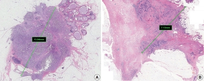
- Standardized Pathology Report for Colorectal Cancer, 2nd Edition
- Baek-hui Kim, Joon Mee Kim, Gyeong Hoon Kang, Hee Jin Chang, Dong Wook Kang, Jung Ho Kim, Jeong Mo Bae, An Na Seo, Ho Sung Park, Yun Kyung Kang, Kyung-Hwa Lee, Mee Yon Cho, In-Gu Do, Hye Seung Lee, Hee Kyung Chang, Do Youn Park, Hyo Jeong Kang, Jin Hee Sohn, Mee Soo Chang, Eun Sun Jung, So-Young Jin, Eunsil Yu, Hye Seung Han, Youn Wha Kim
- J Pathol Transl Med. 2020;54(1):1-19. Published online November 13, 2019
- DOI: https://doi.org/10.4132/jptm.2019.09.28
- 31,946 View
- 1,342 Download
- 49 Web of Science
- 41 Crossref
-
 Abstract
 PDF
PDF Supplementary Material
 - The first edition of the ‘Standardized Pathology Report for Colorectal Cancer,’ which was developed by the Gastrointestinal Pathology Study Group (GIP) of the Korean Society of Pathologists, was published 13 years ago. Meanwhile, there have been many changes in the pathologic diagnosis of colorectal cancer (CRC), pathologic findings included in the pathology report, and immunohistochemical and molecular pathology required for the diagnosis and treatment of colorectal cancer. In order to reflect these changes, we (GIP) decided to make the second edition of the report. The purpose of this standardized pathology report is to provide a practical protocol for Korean pathologists, which could help diagnose and treat CRC patients. This report consists of “standard data elements” and “conditional data elements.” Basic pathologic findings and parts necessary for prognostication of CRC patients are classified as “standard data elements,” while other prognostic factors and factors related to adjuvant therapy are classified as “conditional data elements” so that each institution could select the contents according to the characteristics of the institution. The Korean version is also provided separately so that Korean pathologists can easily understand and use this report. We hope that this report will be helpful in the daily practice of CRC diagnosis.
-
Citations
Citations to this article as recorded by
- Proteogenomic profiling predicts outcomes of adjuvant chemotherapy in extrahepatic cholangiocarcinoma
Hyehyun Jeong, Ji-Hye Oh, Hee-Sung Ahn, Baek-Yeol Ryoo, Kyu-pyo Kim, Jae Ho Jeong, Inkeun Park, Dae Wook Hwang, Jae Hoon Lee, Ki Byung Song, Woohyung Lee, Ki-Hun Kim, Deog-Bog Moon, Gi Won Song, Dong-Hwan Jung, Seung-Mo Hong, Chae Won Park, In-Pyo Baek, Y
Journal of Hepatology.2026; 84(1): 122. CrossRef - A study on the effect of CBSM-based psychological intervention on self-management ability and quality of life in colorectal cancer patients from the perspective of benefit finding: A quasi-experimental study
Linzhi Jiang, Zhouyuan Peng, Rongrong Liu, Lei Chen, Yang Wu, Xingqun Tan, Fan Wang, Liyuan Sun, Chong-Chi Chiu
PLOS One.2026; 21(1): e0339472. CrossRef - Impact of gross tumor morphology on the clinical outcomes of colon cancer: multicenter retrospective cohort study
So Jung Han, Hyun Seok Lee, Byung Ik Jang, Jae Hyun Kim, Hyun Gun Kim, Il Hyun Baek, Jun Lee, Bun Kim, Dae Bum Kim, Jae Jun Park
International Journal of Colorectal Disease.2026;[Epub] CrossRef - Vitrification-based tissue-to-organoid cryopreservation workflow for colorectal cancer
Juyoung Choi, Hye Kyung Hong, Mi-Sook Lee, Ye Jin Lim, Yurimi Lee, Yong Beom Cho, Yoon-La Choi
New Biotechnology.2026; 93: 341. CrossRef - Diagnostic accuracy and pitfalls of MRI for restaging locally advanced rectal cancer in patients following anti-PD1 therapy plus neoadjuvant chemoradiotherapy: a multicenter study
Lixue Xu, Liting Sun, Yuhuan Fu, Guangyong Chen, Hongwei Yao, Zhenchang Wang, Zhenghan Yang, Jie Zhang
Abdominal Radiology.2025; 51(1): 14. CrossRef - Unraveling the role of perineural invasion in cancer progression across multiple tumor types
Muqtada Shaikh, Sanket Shirodkar, Gaurav Doshi
Medical Oncology.2025;[Epub] CrossRef - MALT lymphoma affecting the oral cavity: a clinical, pathologic and genetic study of MALT1 gene translocation
Juan Manuel Arteaga Legarrea, Mauro Lima dos Santos, Nathalia Gomes Rodrigues, Ricardo Santiago Gomez, Ricardo Alves Mesquita, Silvia Ferreira de Sousa, Cinthia Verónica Bardález López de Cáceres, Hélder Antônio Rebelo Pontes, Pablo Agustin Vargas, Luiz A
Oral Surgery, Oral Medicine, Oral Pathology and Oral Radiology.2025; 140(6): 740. CrossRef - Additional staining for lymphovascular invasion is associated with increased estimation of lymph node metastasis in patients with T1 colorectal cancer: Systematic review and meta‐analysis
Jun Watanabe, Katsuro Ichimasa, Yuki Kataoka, Atsushi Miki, Hidehiro Someko, Munenori Honda, Makiko Tahara, Takeshi Yamashina, Khay Guan Yeoh, Shigeo Kawai, Kazuhiko Kotani, Naohiro Sata
Digestive Endoscopy.2024; 36(5): 533. CrossRef - The use of core descriptors from the ENiGMA code study in recent literature: a systematic review
Saher‐Zahra Khan, Andrea Arline, Kate M. Williams, Matthew J. Lee, Emily Steinhagen, Sharon L. Stein
Colorectal Disease.2024; 26(3): 428. CrossRef - Efficacy and safety of PD-1 blockade plus long-course chemoradiotherapy in locally advanced rectal cancer (NECTAR): a multi-center phase 2 study
Zhengyang Yang, Jiale Gao, Jianyong Zheng, Jiagang Han, Ang Li, Gang Liu, Yi Sun, Jie Zhang, Guangyong Chen, Rui Xu, Xiao Zhang, Yishan Liu, Zhigang Bai, Wei Deng, Wei He, Hongwei Yao, Zhongtao Zhang
Signal Transduction and Targeted Therapy.2024;[Epub] CrossRef - Diagnostic Accuracy of Highest-Grade or Predominant Histological Differentiation of T1 Colorectal Cancer in Predicting Lymph Node Metastasis: A Systematic Review and Meta-Analysis
Jun Watanabe, Katsuro Ichimasa, Yuki Kataoka, Shoko Miyahara, Atsushi Miki, Khay Guan Yeoh, Shigeo Kawai, Fernando Martínez de Juan, Isidro Machado, Kazuhiko Kotani, Naohiro Sata
Clinical and Translational Gastroenterology.2024; 15(3): e00673. CrossRef - Comparative evaluation of CT and MRI in the preoperative staging of colon cancer
Effrosyni Bompou, Aikaterini Vassiou, Ioannis Baloyiannis, Konstantinos Perivoliotis, Ioannis Fezoulidis, George Tzovaras
Scientific Reports.2024;[Epub] CrossRef - Pathologic Implications of Magnetic Resonance Imaging-detected Extramural Venous Invasion of Rectal Cancer
Hyun Gu Lee, Chan Wook Kim, Jong Keon Jang, Seong Ho Park, Young Il Kim, Jong Lyul Lee, Yong Sik Yoon, In Ja Park, Seok-Byung Lim, Chang Sik Yu, Jin Cheon Kim
Clinical Colorectal Cancer.2023; 22(1): 129. CrossRef - A standardized pathology report for gastric cancer: 2nd edition
Young Soo Park, Myeong-Cherl Kook, Baek-hui Kim, Hye Seung Lee, Dong-Wook Kang, Mi-Jin Gu, Ok Ran Shin, Younghee Choi, Wonae Lee, Hyunki Kim, In Hye Song, Kyoung-Mee Kim, Hee Sung Kim, Guhyun Kang, Do Youn Park, So-Young Jin, Joon Mee Kim, Yoon Jung Choi,
Journal of Pathology and Translational Medicine.2023; 57(1): 1. CrossRef - IGFL2-AS1, a Long Non-Coding RNA, Is Associated with Radioresistance in Colorectal Cancer
Jeeyong Lee, Da Yeon Kim, Younjoo Kim, Ui Sup Shin, Kwang Seok Kim, Eun Ju Kim
International Journal of Molecular Sciences.2023; 24(2): 978. CrossRef - A Standardized Pathology Report for Gastric Cancer: 2nd Edition
Young Soo Park, Myeong-Cherl Kook, Baek-hui Kim, Hye Seung Lee, Dong-Wook Kang, Mi-Jin Gu, Ok Ran Shin, Younghee Choi, Wonae Lee, Hyunki Kim, In Hye Song, Kyoung-Mee Kim, Hee Sung Kim, Guhyun Kang, Do Youn Park, So-Young Jin, Joon Mee Kim, Yoon Jung Choi,
Journal of Gastric Cancer.2023; 23(1): 107. CrossRef - Incremental Detection Rate of Dysplasia and Sessile Serrated Polyps/Adenomas Using Narrow-Band Imaging and Dye Spray Chromoendoscopy in Addition to High-Definition Endoscopy in Patients with Long-Standing Extensive Ulcerative Colitis: Segmental Tandem End
Ji Eun Kim, Chang Wan Choi, Sung Noh Hong, Joo Hye Song, Eun Ran Kim, Dong Kyung Chang, Young-Ho Kim
Diagnostics.2023; 13(3): 516. CrossRef - Prognostic Impact of Extramural Lymphatic, Vascular, and Perineural Invasion in Stage II Colon Cancer: A Comparison With Intramural Invasion
Sang Sik Cho, Ji Won Park, Gyeong Hoon Kang, Jung Ho Kim, Jeong Mo Bae, Sae-Won Han, Tae-You Kim, Min Jung Kim, Seung-Bum Ryoo, Seung-Yong Jeong, Kyu Joo Park
Diseases of the Colon & Rectum.2023; 66(3): 366. CrossRef - Towards targeted colorectal cancer biopsy based on tissue morphology assessment by compression optical coherence elastography
Anton A. Plekhanov, Marina A. Sirotkina, Ekaterina V. Gubarkova, Elena B. Kiseleva, Alexander A. Sovetsky, Maria M. Karabut, Vladimir E. Zagainov, Sergey S. Kuznetsov, Anna V. Maslennikova, Elena V. Zagaynova, Vladimir Y. Zaitsev, Natalia D. Gladkova
Frontiers in Oncology.2023;[Epub] CrossRef - Is High-Grade Tumor Budding an Independent Prognostic Factor in Stage II Colon Cancer?
Jung Kyong Shin, Yoon Ah Park, Jung Wook Huh, Seong Hyeon Yun, Hee Cheol Kim, Woo Yong Lee, Seok Hyung Kim, Sang Yun Ha, Yong Beom Cho
Diseases of the Colon & Rectum.2023; 66(8): e801. CrossRef - Detection of Human cytomegalovirus UL55 Gene and IE/E Protein Expression in Colorectal Cancer Patients in Egypt
May Raouf, Ahmed A. Sabry, Mahinour A. Ragab, Samar El Achy, Amira Amer
BMC Cancer.2023;[Epub] CrossRef - Polo-like kinase 4 as a potential predictive biomarker of chemoradioresistance in locally advanced rectal cancer
Hyunseung Oh, Soon Gu Kim, Sung Uk Bae, Sang Jun Byun, Shin Kim, Jae-Ho Lee, Ilseon Hwang, Sun Young Kwon, Hye Won Lee
Journal of Pathology and Translational Medicine.2022; 56(1): 40. CrossRef - A Prediction Model for Tumor Recurrence in Stage II–III Colorectal Cancer Patients: From a Machine Learning Model to Genomic Profiling
Po-Chuan Chen, Yu-Min Yeh, Bo-Wen Lin, Ren-Hao Chan, Pei-Fang Su, Yi-Chia Liu, Chung-Ta Lee, Shang-Hung Chen, Peng-Chan Lin
Biomedicines.2022; 10(2): 340. CrossRef - Rationale and design of a prospective, multicenter, phase II clinical trial of safety and efficacy evaluation of long course neoadjuvant chemoradiotherapy plus tislelizumab followed by total mesorectal excision for locally advanced rectal cancer (NCRT-PD1
Zhengyang Yang, Xiao Zhang, Jie Zhang, Jiale Gao, Zhigang Bai, Wei Deng, Guangyong Chen, Yongbo An, Yishan Liu, Qi Wei, Jiagang Han, Ang Li, Gang Liu, Yi Sun, Dalu Kong, Hongwei Yao, Zhongtao Zhang
BMC Cancer.2022;[Epub] CrossRef - Potential of DEK proto‑oncogene as a prognostic biomarker for colorectal cancer: An evidence‑based review
Muhammad Habiburrahman, Muhammad Wardoyo, Stefanus Sutopo, Nur Rahadiani
Molecular and Clinical Oncology.2022;[Epub] CrossRef - Reproducibility and Feasibility of Classification and National Guidelines for Histological Diagnosis of Canine Mammary Gland Tumours: A Multi-Institutional Ring Study
Serenella Papparella, Maria Crescio, Valeria Baldassarre, Barbara Brunetti, Giovanni Burrai, Cristiano Cocumelli, Valeria Grieco, Selina Iussich, Lorella Maniscalco, Francesca Mariotti, Francesca Millanta, Orlando Paciello, Roberta Rasotto, Mariarita Roma
Veterinary Sciences.2022; 9(7): 357. CrossRef - Composite scoring system and optimal tumor budding cut-off number for estimating lymph node metastasis in submucosal colorectal cancer
Jeong-ki Kim, Ye-Young Rhee, Jeong Mo Bae, Jung Ho Kim, Seong-Joon Koh, Hyun Jung Lee, Jong Pil Im, Min Jung Kim, Seung-Bum Ryoo, Seung-Yong Jeong, Kyu Joo Park, Ji Won Park, Gyeong Hoon Kang
BMC Cancer.2022;[Epub] CrossRef - Automated Hybrid Model for Detecting Perineural Invasion in the Histology of Colorectal Cancer
Jiyoon Jung, Eunsu Kim, Hyeseong Lee, Sung Hak Lee, Sangjeong Ahn
Applied Sciences.2022; 12(18): 9159. CrossRef - Clinical Implication of Perineural and Lymphovascular Invasion in Rectal Cancer Patients Who Underwent Surgery After Preoperative Chemoradiotherapy
Young Il Kim, Chan Wook Kim, Jong Hoon Kim, Jihun Kim, Jun-Soo Ro, Jong Lyul Lee, Yong Sik Yoon, In Ja Park, Seok-Byung Lim, Chang Sik Yu, Jin Cheon Kim
Diseases of the Colon & Rectum.2022; 65(11): 1325. CrossRef - Molecular Pathology of Gastric Cancer
Moonsik Kim, An Na Seo
Journal of Gastric Cancer.2022; 22(4): 264. CrossRef - Selective approach to arterial ligation in radical sigmoid colon cancer surgery with D3 lymph node dissection: A multicenter comparative study
Sergey Efetov, Albina Zubayraeva, Cüneyt Kayaalp, Alisa Minenkova, Yusuf Bağ, Aftandil Alekberzade, Petr Tsarkov
Turkish Journal of Surgery.2022; 38(4): 382. CrossRef - Evaluation of lncRNA FOXD2-AS1 Expression as a Diagnostic Biomarker in Colorectal Cancer
Hooman Shalmashi, Sahar Safaei, Dariush Shanehbandi, Milad Asadi, Soghra Bornehdeli, Abdolreza Mehdi Navaz
Reports of Biochemistry and Molecular Biology.2022; 11(3): 471. CrossRef - Improvement in the Assessment of Response to Preoperative Chemoradiotherapy for Rectal Cancer Using Magnetic Resonance Imaging and a Multigene Biomarker
Eunhae Cho, Sung Woo Jung, In Ja Park, Jong Keon Jang, Seong Ho Park, Seung-Mo Hong, Jong Lyul Lee, Chan Wook Kim, Yong Sik Yoon, Seok-Byung Lim, Chang Sik Yu, Jin Cheon Kim
Cancers.2021; 13(14): 3480. CrossRef - Addition of V-Stage to Conventional TNM Staging to Create the TNVM Staging System for Accurate Prediction of Prognosis in Colon Cancer: A Multi-Institutional Retrospective Cohort Study
Jung Hoon Bae, Ji Hoon Kim, Jaeim Lee, Bong-Hyeon Kye, Sang Chul Lee, In Kyu Lee, Won Kyung Kang, Hyeon-Min Cho, Yoon Suk Lee
Biomedicines.2021; 9(8): 888. CrossRef - Gene Expression Profiles Associated with Radio-Responsiveness in Locally Advanced Rectal Cancer
Jeeyong Lee, Junhye Kwon, DaYeon Kim, Misun Park, KwangSeok Kim, InHwa Bae, Hyunkyung Kim, JoonSeog Kong, Younjoo Kim, UiSup Shin, EunJu Kim
Biology.2021; 10(6): 500. CrossRef - A Patient-Derived Organoid-Based Radiosensitivity Model for the Prediction of Radiation Responses in Patients with Rectal Cancer
Misun Park, Junhye Kwon, Joonseog Kong, Sun Mi Moon, Sangsik Cho, Ki Young Yang, Won Il Jang, Mi Sook Kim, Younjoo Kim, Ui Sup Shin
Cancers.2021; 13(15): 3760. CrossRef - Comparison between Local Excision and Radical Resection for the Treatment of Rectal Cancer in ypT0-1 Patients: An Analysis of the Clinicopathological Factors and Survival Rates
Soo Young Oh, In Ja Park, Young IL Kim, Jong-Lyul Lee, Chan Wook Kim, Yong Sik Yoon, Seok-Byung Lim, Chang Sik Yu, Jin Cheon Kim
Cancers.2021; 13(19): 4823. CrossRef - Comparison of Vascular Invasion With Lymph Node Metastasis as a Prognostic Factor in Stage I-III Colon Cancer: An Observational Cohort Study
Jung Hoon Bae, Ji Hoon Kim, Bong-Hyeon Kye, Abdullah Al-Sawat, Chul Seung Lee, Seung-Rim Han, In Kyu Lee, Sung Hak Lee, Yoon Suk Lee
Frontiers in Surgery.2021;[Epub] CrossRef - Clinicopathological significance of Ki67 expression in colorectal cancer
Jing Li, Zhi-ye Liu, Hai-bo Yu, Qing Xue, Wen-jie He, Hai-tao Yu
Medicine.2020; 99(20): e20136. CrossRef - Lateral lymph node and its association with distant recurrence in rectal cancer: A clue of systemic disease
Young Il Kim, Jong Keon Jang, In Ja Park, Seong Ho Park, Jong Beom Kim, Jin-Hong Park, Tae Won Kim, Jun-Soo Ro, Seok-Byung Lim, Chang Sik Yu, Jin Cheon Kim
Surgical Oncology.2020; 35: 174. CrossRef - Transformation of Pathology Reports Into the Common Data Model With Oncology Module: Use Case for Colon Cancer
Borim Ryu, Eunsil Yoon, Seok Kim, Sejoon Lee, Hyunyoung Baek, Soyoung Yi, Hee Young Na, Ji-Won Kim, Rong-Min Baek, Hee Hwang, Sooyoung Yoo
Journal of Medical Internet Research.2020; 22(12): e18526. CrossRef
- Proteogenomic profiling predicts outcomes of adjuvant chemotherapy in extrahepatic cholangiocarcinoma
- Association between Expression of 8-OHdG and Cigarette Smoking in Non-small Cell Lung Cancer
- Ae Ri An, Kyoung Min Kim, Ho Sung Park, Kyu Yun Jang, Woo Sung Moon, Myoung Jae Kang, Yong Chul Lee, Jong Hun Kim, Han Jung Chae, Myoung Ja Chung
- J Pathol Transl Med. 2019;53(4):217-224. Published online March 11, 2019
- DOI: https://doi.org/10.4132/jptm.2019.02.20
- 10,662 View
- 251 Download
- 24 Web of Science
- 24 Crossref
-
Abstract
PDF
- Background
Exposure to cigarette smoking (CS) is a major risk factor for the development of lung cancer. CS is known to cause oxidative DNA damage and mutation of tumor-related genes, and these factors are involved in carcinogenesis. 8-Hydroxydeoxyguanosine (8-OHdG) is considered to be a reliable biomarker for oxidative DNA damage. Increased levels of 8-OHdG are associated with a number of pathological conditions, including cancer. There are no reports on the expression of 8-OHdG by immunohistochemistry in non-small cell lung cancer (NSCLC).
Methods
We investigated the expression of 8-OHdG and p53 in 203 NSCLC tissues using immunohistochemistry and correlated it with clinicopathological features including smoking.
Results
The expression of 8-OHdG was observed in 83.3% of NSCLC. It was significantly correlated with a low T category, negative lymph node status, never-smoker, and longer overall survival (p < .05) by univariate analysis. But multivariate analysis revealed that 8-OHdG was not an independent prognostic factor for overall survival in NSCLC patients. The aberrant expression of p53 significantly correlated with smoking, male, squamous cell carcinoma, and Ki-67 positivity (p < .05).
Conclusions
The expression of 8-OHdG was associated with good prognostic factors. It was positively correlated with never-smokers in NSCLC, suggesting that oxidative damage of DNA cannot be explained by smoking alone and may depend on complex control mechanisms. -
Citations
Citations to this article as recorded by- N-acetyl-cysteine alleviates nandrolone decanoate-induced hippocampal cell apoptosis in rats via reversing protein expressions of S1P1, Akt and FOXO3a signaling pathway
Alireza Shirpoor, Zahra Zarrini, Roya Naderi
Steroids.2026; 228: 109759. CrossRef - The distinct roles of ROS in tumor immunity: from mechanisms to immunotherapeutic applications
Jiayi Li, Chen Huang, Pan Tang, Ruiyan Wu, Quanyou Wu, Chenliang Zhang
Journal of Hematology & Oncology.2026;[Epub] CrossRef - Sustainable framework for automated segmentation and prediction of lung cancer in CT image using CapsNet with U-net segmentation
S.R. Vijayakumar, S. Aarthy, D. Deepa, P. Suresh
Biomedical Signal Processing and Control.2025; 99: 106873. CrossRef - Endolysosomal cation channel MCOLN as the novel regulator of redox homeostasis
Yahao Gao, Lei Xu, Ying Chen
Biochimica et Biophysica Acta (BBA) - Molecular Basis of Disease.2025; 1871(7): 167910. CrossRef - Catalase: The golden key to regulate oxidative stress in breast cancer
Jia-Wei Liu, Wen-Jia Chen, Yang-Zheng Lan, Jing Liu
World Journal of Clinical Oncology.2025;[Epub] CrossRef - Association of sirtuin 1 rs10997868 and rs730821 polymorphisms with sirtuin 1 and hydroxy-2′-deoxyguanosine levels in healthy smokers: A case–control study
Samar Sultan
Journal of International Medical Research.2025;[Epub] CrossRef - Increased pretreatment triglyceride glucose-body mass index associated with poor prognosis in patients with advanced non-small cell lung cancer
Shaoming Guo, Yi Zhao, Yue Jiang, Huaping Ye, Ying Wang
Clinical Nutrition ESPEN.2024; 59: 412. CrossRef - Oxidative Damage and Telomere Length as Markers of Lung Cancer Development among Chronic Obstructive Pulmonary Disease (COPD) Smokers
Elizabeth Córdoba-Lanús, Luis M. Montuenga, Angélica Domínguez-de-Barros, Alexis Oliva, Delia Mayato, Ana Remírez-Sanz, Francisca Gonzalvo, Bartolomé Celli, Javier J. Zulueta, Ciro Casanova
Antioxidants.2024; 13(2): 156. CrossRef - Automated determination of 8-OHdG in cells and tissue via immunofluorescence using a specially created antibody
Tobias Jung, Nicole Findik, Bianca Hartmann, Katja Hanack, Kai Grossmann, Dirk Roggenbuck, Marc Wegmann, René Mantke, Markus Deckert, Tilman Grune
Biotechnology Reports.2024; 42: e00833. CrossRef - Combination treatment of zinc and selenium intervention ameliorated BPA-exposed germ cell damage in SD rats: elucidation of molecular mechanisms
Chittaranjan Sahu, Gopabandhu Jena
Naunyn-Schmiedeberg's Archives of Pharmacology.2024; 397(9): 6685. CrossRef - Interplay of arsenic exposure and cigarette smoking on oxidative DNA damage in healthy males
Sepideh Nemati-Mansour, Mohammad Mosaferi, Javad Babaie, Asghar Mohammadpoorasl, Reza Dehghanzadeh, Leila Nikniaz, Mohammad Miri
Environmental Sciences Europe.2024;[Epub] CrossRef - The role of tissue persistent organic pollutants and genetic polymorphisms in patients with benign and malignant kidney tumors
Rasih Kocagöz, İlgen Onat, Merve Demirbügen Öz, Burak Turna, Banu Sarsık Kumbaracı, Mehmet Nurullah Orman, Halit Sinan Süzen, Hilmi Orhan
Environmental Toxicology and Pharmacology.2024; 110: 104495. CrossRef - Mitochondrial Plasticity and Glucose Metabolic Alterations in Human Cancer under Oxidative Stress—From Viewpoints of Chronic Inflammation and Neutrophil Extracellular Traps (NETs)
Hui-Ting Lee, Chen-Sung Lin, Chao-Yu Liu, Po Chen, Chang-Youh Tsai, Yau-Huei Wei
International Journal of Molecular Sciences.2024; 25(17): 9458. CrossRef - Oxidative DNA Damage and Arterial Hypertension in Light of Current ESC Guidelines
Radka Hazuková, Zdeněk Zadák, Miloslav Pleskot, Petr Zdráhal, Martin Pumprla, Miloš Táborský
International Journal of Molecular Sciences.2024; 25(23): 12557. CrossRef - Significance of 8-OHdG Expression as a Predictor of Survival in Colorectal Cancer
Myunghee Kang, Soyeon Jeong, Sungjin Park, Seungyoon Nam, Jun-Won Chung, Kyoung Oh Kim, Jungsuk An, Jung Ho Kim
Cancers.2023; 15(18): 4613. CrossRef - Serum 8-Hydroxy-2′-deoxyguanosine Predicts Severity and Prognosis of Patients with Acute Exacerbation of Chronic Obstructive Pulmonary Disease
Peng Cao, Chen Zhang, Dong-Xu Hua, Meng-Die Li, Bian-Bian Lv, Lin Fu, Hui Zhao
Lung.2022; 200(1): 31. CrossRef - Redox signaling at the crossroads of human health and disease
Jing Zuo, Zhe Zhang, Maochao Luo, Li Zhou, Edouard C. Nice, Wei Zhang, Chuang Wang, Canhua Huang
MedComm.2022;[Epub] CrossRef - Assessment of MDA and 8-OHdG expressions in ovine pulmonary adenocarcinomas by immunohistochemical and immunofluorescence methods
Emin Karakurt, Enver Beytut, Serpil Dağ, Hilmi Nuhoğlu, Ayfer Yıldız, Emre Kurtbaş
Acta Veterinaria Brno.2022; 91(3): 235. CrossRef - Dietary Antioxidants and Lung Cancer Risk in Smokers and Non-Smokers
Naser A. Alsharairi
Healthcare.2022; 10(12): 2501. CrossRef - Targeting oxidative stress in disease: promise and limitations of antioxidant therapy
Henry Jay Forman, Hongqiao Zhang
Nature Reviews Drug Discovery.2021; 20(9): 689. CrossRef - Association between tobacco substance usage and a missense mutation in the tumor suppressor gene P53 in the Saudi Arabian population
Mikhlid H. Almutairi, Bader O. Almutairi, Turki M. Alrubie, Sultan N. Alharbi, Narasimha R. Parine, Abdulwahed F. Alrefaei, Ibrahim Aldeailej, Abdullah Alamri, Abdelhabib Semlali, Alvaro Galli
PLOS ONE.2021; 16(1): e0245133. CrossRef - Measurement of uranium concentrations in urine samples of adult healthy groups in Najaf governorate with estimation of urine concentrations of 8-OHdG compound as biomarker for DNA damage
Samia K. Abbas, Dhuha S. Saleh, Hayder S. Hussain
Journal of Physics: Conference Series.2021; 1879(3): 032097. CrossRef - Common Data Model and Database System Development for the Korea Biobank Network
Soo-Jeong Ko, Wona Choi, Ki-Hoon Kim, Seo-Joon Lee, Haesook Min, Seol-Whan Oh, In Young Choi
Applied Sciences.2021; 11(24): 11825. CrossRef - EVALUATION OF OXIDATIVE STATUS IN PATIENTS WITH CHRONIC PERIODONTITIS AND ADDITIONAL TOBACCO ABUSE: A CROSS-SECTIONAL STUDY
Didem ÖZKAL EMİNOĞLU, Varol ÇANAKÇI
Atatürk Üniversitesi Diş Hekimliği Fakültesi Dergisi.2020; : 1. CrossRef
- N-acetyl-cysteine alleviates nandrolone decanoate-induced hippocampal cell apoptosis in rats via reversing protein expressions of S1P1, Akt and FOXO3a signaling pathway
- Immunohistochemical Expression and Clinical Significance of Suggested Stem Cell Markers in Hepatocellular Carcinoma
- Jong Jin Sung, Sang Jae Noh, Jun Sang Bae, Ho Sung Park, Kyu Yun Jang, Myoung Ja Chung, Woo Sung Moon
- J Pathol Transl Med. 2016;50(1):52-57. Published online November 18, 2015
- DOI: https://doi.org/10.4132/jptm.2015.10.09
- 12,634 View
- 78 Download
- 21 Web of Science
- 18 Crossref
-
Abstract
PDF
- Background
Increasing evidence has shown that tumor initiation and growth are nourished by a small subpopulation of cancer stem cells (CSCs) within the tumor mass. CSCs are posited to be responsible for tumor maintenance, growth, distant metastasis, and relapse after curative operation. We examined the expression of CSC markers in paraffin-embedded tissue sections of hepatocellular carcinoma (HCC) and correlated the results with clinicopathologic characteristics. Methods: Immunohistochemical staining for the markers believed to be expressed in the CSCs, including epithelial cell adhesion molecule (EpCAM), keratin 19 (K19), CD133, and CD56, was performed in 82 HCC specimens. Results: EpCAM expression was observed in 56% of the HCCs (46/82) and K19 in 6% (5/82). EpCAM expression in HCC significantly correlated with elevated α-fetoprotein level, microvessel invasion of tumor cells, and high histologic grade. In addition, Ep- CAM expression significantly correlated with K19 expression. The overall survival and relapsefree survival rates in patients with EpCAM-expressing HCC were relatively lower than those in patients with EpCAM-negative HCC. All but two of the 82 HCCs were negative for CD133 and CD56, respectively. Conclusions: Our results suggest that HCCs expressing EpCAM are associated with unfavorable prognostic factors and have a more aggressive clinical course than those not expressing EpCAM. Further, the expression of either CD133 or CD56 in paraffin-embedded HCC tissues appears to be rare. -
Citations
Citations to this article as recorded by- Spatial immune scoring system predicts hepatocellular carcinoma recurrence
Gengjie Jia, Peiqi He, Tianli Dai, Denise Goh, Jiabei Wang, Mengyuan Sun, Felicia Wee, Fuling Li, Jeffrey Chun Tatt Lim, Shuxia Hao, Yao Liu, Tony Kiat Hon Lim, Nye-Thane Ngo, Qingping Tao, Wei Wang, Ahitsham Umar, Björn Nashan, Yongchang Zhang, Chen Ding
Nature.2025; 640(8060): 1031. CrossRef - Evolving Landscape of Systemic Therapy for Hepatocellular Carcinoma in 2025
Karan Kumar, Vivek A. Saraswat
Journal of Clinical and Experimental Hepatology.2025; 15(5): 102547. CrossRef - Recent Progress in Systemic Therapy for Advanced Hepatocellular Carcinoma
Narayanan Sadagopan, Aiwu Ruth He
International Journal of Molecular Sciences.2024; 25(2): 1259. CrossRef - Diagnostic value of expressions of cancer stem cell markers for adverse outcomes of hepatocellular carcinoma and their associations with prognosis: A Bayesian network meta‑analysis
Zhengrong Ou, Shoushuo Fu, Jian Yi, Jingxuan Huang, Weidong Zhu
Oncology Letters.2024;[Epub] CrossRef - Clinicopathological and prognostic value of epithelial cell adhesion molecule in solid tumours: a meta-analysis
Peiwen Ding, Panyu Chen, Jiqi Ouyang, Qiang Li, Shijie Li
Frontiers in Oncology.2023;[Epub] CrossRef - PD-L1 Downregulation and DNA Methylation Inhibition for Molecular Therapy against Cancer Stem Cells in Hepatocellular Carcinoma
Caecilia Sukowati, Loraine Kay D. Cabral, Beatrice Anfuso, Francesco Dituri, Roberto Negro, Gianluigi Giannelli, Claudio Tiribelli
International Journal of Molecular Sciences.2023; 24(17): 13357. CrossRef - EpCAM, Ki67, and ESM1 Predict Hepatocellular Carcinoma Recurrence After Liver Transplantation
Aiat Shaban Hemida, Doha Maher Taie, Moshira Mohamed Abd El-Wahed, Mohammed Ibrahim Shabaan, Mona Saeed Tantawy, Nermine Ahmed Ehsan
Applied Immunohistochemistry & Molecular Morphology.2023; 31(9): 596. CrossRef - The clinical, prognostic and therapeutic significance of liver cancer stem cells and their markers
Izabela Zarębska, Arkadiusz Gzil, Justyna Durślewicz, Damian Jaworski, Paulina Antosik, Navid Ahmadi, Marta Smolińska-Świtała, Dariusz Grzanka, Łukasz Szylberg
Clinics and Research in Hepatology and Gastroenterology.2021; 45(3): 101664. CrossRef - Detection of oncogenic mutations in paired circulating tumor DNA and circulating tumor cells in patients with hepatocellular carcinoma
Zhouhong Ge, Jean C.A. Helmijr, Maurice P.H.M. Jansen, Patrick P.C. Boor, Lisanne Noordam, Maikel Peppelenbosch, Jaap Kwekkeboom, Jaco Kraan, Dave Sprengers
Translational Oncology.2021; 14(7): 101073. CrossRef - Hepatocellular Carcinoma Score and Subclassification Into Aggressive Subtypes Using Immunohistochemical Expression of p53, β-Catenin, CD133, and Ki-67
Asmaa G. Abdou, Nanis S. Holah, Dina S. Elazab, Walaa G. El-Gendy, Mohammed T. Badr, Dalia R. Al-Sharaky
Applied Immunohistochemistry & Molecular Morphology.2021; 29(1): 20. CrossRef - The prognostic significance of neuroendocrine markers and somatostatin receptor 2 in hepatocellular carcinoma
Keigo Murakami, Hiroyuki Kumata, Shigehito Miyagi, Takashi Kamei, Hironobu Sasano
Pathology International.2021; 71(10): 682. CrossRef - Predictors of recurrence and survival of hepatocellular carcinoma: A prospective study including transient elastography and cancer stem cell markers
Hend Ibrahim Shousha, Rabab Fouad, Tamer Mahmoud Elbaz, Dina Sabry, Mohamed Mahmoud Nabeel, Ahmed Hosni Abdelmaksoud, Aisha Mahmoud Elsharkawy, Zeinab Abdellatif Soliman, Ghada Habib, Ashraf Omar Abdelaziz
Arab Journal of Gastroenterology.2020; 21(2): 95. CrossRef - Napabucasin Reduces Cancer Stem Cell Characteristics in Hepatocellular Carcinoma
Ya Li, Qiuju Han, Huajun Zhao, Quanjuan Guo, Jian Zhang
Frontiers in Pharmacology.2020;[Epub] CrossRef - The mRNA Distribution of Cancer Stem Cell Marker CD90/Thy-1 Is Comparable in Hepatocellular Carcinoma of Eastern and Western Populations
An B. Luong, Huy Q. Do, Paola Tarchi, Deborah Bonazza, Cristina Bottin, Loraine Kay D. Cabral, Long D. C. Tran, Thao P. T. Doan, Lory S. Crocè, Hoa L. T. Pham, Claudio Tiribelli, Caecilia H. C. Sukowati
Cells.2020; 9(12): 2672. CrossRef - Histological architectural classification determines recurrence pattern and prognosis after curative hepatectomy in patients with hepatocellular carcinoma
Hirohisa Okabe, Tomoharu Yoshizumi, Yo-ichi Yamashita, Katsunori Imai, Hiromitsu Hayashi, Shigeki Nakagawa, Shinji Itoh, Norifumi Harimoto, Toru Ikegami, Hideaki Uchiyama, Toru Beppu, Shinichi Aishima, Ken Shirabe, Hideo Baba, Yoshihiko Maehara, Motoyuki
PLOS ONE.2018; 13(9): e0203856. CrossRef - Overexpression of epithelial cell adhesion molecule as a predictor of poor outcome in patients with hepatocellular carcinoma
Chih‑Jan Ko, Chia‑Jung Li, Meng‑Yu Wu, Pei‑Yi Chu
Experimental and Therapeutic Medicine.2018;[Epub] CrossRef - Clinicopathologic Significance of Survivin Expression in Relation to CD133 Expression in Surgically Resected Stage II or III Colorectal Cancer
Wanlu Li, Mi-Ra Lee, EunHee Choi, Mee-Yon Cho
Journal of Pathology and Translational Medicine.2017; 51(1): 17. CrossRef - PIN1 in hepatocellular carcinoma is associated with TP53 gene status
Jun Sang Bae, Sang Jae Noh, Kyoung Min Kim, Kyu Yun Jang, Ho Sung Park, Myoung Ja Chung, Byung-Hyun Park, Woo Sung Moon
Oncology Reports.2016; 36(4): 2405. CrossRef
- Spatial immune scoring system predicts hepatocellular carcinoma recurrence
- Early Colorectal Epithelial Neoplasm in Korea: A Multicenter Survey of Pathologic Diagnosis
- Yun Kyung Kang, So-Young Jin, Mee Soo Chang, Jung Yeon Kim, Gyeong Hoon Kang, Hye Seung Lee, Jin Hee Sohn, Ho Sung Park, Kye Won Kwon, Mi Jin Gu, Young Hee Maeng, Jong Eun Joo, Haeng Ji Kang, Hee Kyung Kim, Kee-Taek Jang, Mi Ja Lee, Hee Kyung Chang, Joon Mee Kim, Hye Seung Han, Won Ae Lee, Yoon Jung Choi, Dong Wook Kang, Sunhoo Park, Jae Hyuk Lee, Mee-Yon Cho
- Korean J Pathol. 2013;47(3):245-251. Published online June 25, 2013
- DOI: https://doi.org/10.4132/KoreanJPathol.2013.47.3.245
- 12,071 View
- 56 Download
- 1 Crossref
-
Abstract
PDF
Background The incidence of early colorectal epithelial neoplasm (ECEN) is increasing, and its pathologic diagnosis is important for patient care. We investigated the incidence of ECEN and the current status of its pathologic diagnosis.
Methods We collected datasheets from 25 institutes in Korea for the incidence of colorectal adenoma with high grade dysplasia (HGD) and low grade dysplasia in years 2005, 2007, and 2009; and early colorectal carcinoma in the year 2009. We also surveyed the diagnostic terminology of ECEN currently used by the participating pathologists.
Results The average percentage of diagnoses of adenoma HGD was 7.0%, 5.0%, and 3.4% in years 2005, 2007, and 2009, respectively. The range of incidence rates of adenoma HGD across the participating institutes has gradually narrowed over the years 2005 to 2009. The incidence rate of early colorectal carcinoma in the year 2009 was 21.2%. The participants did not share a single criterion or terminology for the diagnosis of adenoma HGD. The majority accepted the diagnostic terms that distinguished noninvasive, mucosal confined, and submucosal invasive carcinoma.
Conclusions Further research requirements suggested are a diagnostic consensus for the histopathologic diagnosis of ECEN; and standardization of diagnostic terminology critical for determining the disease code.
-
Citations
Citations to this article as recorded by- Diminutive and Small Colorectal Polyps: The Pathologist's Perspective
Yun Kyung Kang
Clinical Endoscopy.2014; 47(5): 404. CrossRef
- Diminutive and Small Colorectal Polyps: The Pathologist's Perspective
- Expression of CHOP in Squamous Tumor of the Uterine Cervix
- Hyun Hee Chu, Jun Sang Bae, Kyoung Min Kim, Ho Sung Park, Dong Hyu Cho, Kyu Yun Jang, Woo Sung Moon, Myoung Jae Kang, Dong Geun Lee, Myoung Ja Chung
- Korean J Pathol. 2012;46(5):463-469. Published online October 25, 2012
- DOI: https://doi.org/10.4132/KoreanJPathol.2012.46.5.463
- 10,124 View
- 40 Download
- 8 Crossref
-
Abstract
PDF
Background High-risk human papillomavirus (HR-HPV) infection and abnormal p53 expression are closely involved in carcinogenesis of squamous cell carcinoma (SqCC) of uterine cervix. Recent studies have suggested that virus-induced endoplasmic reticulum (ER) stress modulates various cell survival and cell death signaling pathways. The C/EBP homologous protein (CHOP) is associated with ER stress-mediated apoptosis and is also involved in carcinogenesis of several human cancers. We hypothesized that CHOP is involved in the carcinogenesis of uterine cervical cancer in association with HR-HPV and/or p53.
Methods Immunohistochemistry was used to analyze CHOP and p53 protein expression of tissue sections from 191 patients with invasive cancer or preinvasive lesions of the uterine cervix (61 cases of SqCC, 66 cases of cervical intraepithelial neoplasia [CIN] III, and 64 cases of CIN I).
Results CHOP was expressed in 59.4% of CIN I, 48.5% of CIN III, and 70.5% of SqCC cases. It was also significantly more frequent in invasive SqCC than in preinvasive lesions (p=0.042). Moreover, CHOP expression significantly correlated with HR-HPV infection and p53 expression (p=0.009 and p=0.038, respectively).
Conclusions Our results suggest that CHOP is involved in the carcinogenesis of the uterine cervix SqCC via association with HR-HPV and p53.
-
Citations
Citations to this article as recorded by- Interplay between the cellular stress pathway, stemness markers, and Helicobacter pylori infection in gastric cancer
Mehran Gholamin, Atena Mansouri, Mohammad Reza Abbaszadegan, Mohammad Ali Karimi, Hossein Barzegar, Fatemeh Fardi Golyan, Hanie Mahaki, Hamid Tanzadehpanah, Reihaneh Alsadat Mahmoudian
Gene Reports.2025; 40: 102263. CrossRef - Role of C-reactive protein in cervical intraepithelial neoplasia/cancer
Adriana Pedreañez, Yenddy Carrero, Renata Vargas, Juan P.Hernández Fonseca, Jesús Mosquera
Pathology - Research and Practice.2025; 276: 156274. CrossRef - Expression of GRP78 and its copartners in HEK293 and pancreatic cancer cell lines (BxPC-3/PANC-1) exposed to MRI and CT contrast agents
Ali Ahmed Azzawri, Ibrahim Halil Yildirim, Zeynep Yegin, Abdurrahim Dusak
Nucleosides, Nucleotides & Nucleic Acids.2024; 43(5): 391. CrossRef - Endoplasmic Reticulum Stress and Homeostasis in Reproductive Physiology and Pathology
Elif Guzel, Sefa Arlier, Ozlem Guzeloglu-Kayisli, Mehmet Tabak, Tugba Ekiz, Nihan Semerci, Kellie Larsen, Frederick Schatz, Charles Lockwood, Umit Kayisli
International Journal of Molecular Sciences.2017; 18(4): 792. CrossRef - Endoplasmic reticulum stress pathway PERK‐eIF2α confers radioresistance in oropharyngeal carcinoma by activating NF‐κB
Qiao Qiao, Chaonan Sun, Chuyang Han, Ning Han, Miao Zhang, Guang Li
Cancer Science.2017; 108(7): 1421. CrossRef - Curcumin induces ER stress-mediated apoptosis through selective generation of reactive oxygen species in cervical cancer cells
Boyun Kim, Hee Seung Kim, Eun-Ji Jung, Jung Yun Lee, Benjamin K. Tsang, Jeong Mook Lim, Yong Sang Song
Molecular Carcinogenesis.2016; 55(5): 918. CrossRef - Down-regulation of C/EBP homologous protein (CHOP) expression in gastric cardia adenocarcinoma: Their relationship with clinicopathological parameters and prognostic significance
Xiao-Juan Zhu, She-Gan Gao, San-Qiang Li, Zhen-Guo Shi, Zhi-Kun Ma, Shan-Shan Zhu, Xiao-Shan Feng
Clinics and Research in Hepatology and Gastroenterology.2015; 39(3): 391. CrossRef - MG289 in <i>Mycoplasma genitalium</i> Enhances Microbial Invasion and Bacterial Persistence in Benign Human Prostate Cells
Wasia Rizwani, Leticia Reyes, Jeongsoon Kim, Steve Goodison, Charles J. Rosser
Open Journal of Urology.2013; 03(06): 232. CrossRef
- Interplay between the cellular stress pathway, stemness markers, and Helicobacter pylori infection in gastric cancer
- Expression of Cortactin and Focal Adhesion Kinase in Colorectal Adenocarcinoma: Correlation with Clinicopathologic Parameters and Their Prognostic Implication
- Yo Na Kim, Ji Eun Choi, Jun Sang Bae, Kyu Yun Jang, Myoung Ja Chung, Woo Sung Moon, Myoung Jae Kang, Dong Geun Lee, Ho Sung Park
- Korean J Pathol. 2012;46(5):454-462. Published online October 25, 2012
- DOI: https://doi.org/10.4132/KoreanJPathol.2012.46.5.454
- 10,161 View
- 47 Download
- 7 Crossref
-
Abstract
PDF
Background Cortactin and focal adhesion kinase (FAK) are two important components among actin cross-linking proteins that play a central role in cell migration.
Methods The aims of this study were to evaluate the expression of cortactin and FAK in normal colorectal mucosa and colorectal adenocarcinoma (CRC) using tissue microarray of 2 mm cores to correlate their expression with other clinicopathological factors and, investigate their prognostic significance.
Results Twenty (9%) and 24 cases (11%) of normal colorectal mucosa were immunoreactive for cortactin and FAK. In addition, 184 (84%) and 133 cases (61%) of CRCs were immunoreactive for cortactin and FAK, respectively. Cortactin expression was associated with histologic differentiation and FAK expression. Cortactin, but not FAK expression was also correlated with poor overall and relapse-free survival and served well as an independent prognostic factor for poor survival.
Conclusions Cortactin expression, in association with FAK expression, may plays an important role in tumor progression. Furthermore, it may also be a satisfactory biomarker to predict tumor progression and survival in CRC patients.
-
Citations
Citations to this article as recorded by- Identification of a Subset of Stage I Colorectal Cancer Patients With High Recurrence Risk
Lik Hang Lee, Lindy Davis, Lourdes Ylagan, Angela R Omilian, Kristopher Attwood, Canan Firat, Jinru Shia, Philip B Paty, William G Cance
JNCI: Journal of the National Cancer Institute.2022; 114(5): 732. CrossRef - Profiling the expression of pro-metastatic genes in association with the clinicopathological features of primary breast cancer
Seyed-Mohammad Mazloomi, Mitra Foroutan-Ghaznavi, Vahid Montazeri, Gholamreza Tavoosidana, Ashraf Fakhrjou, Hojjatollah Nozad-Charoudeh, Saeed Pirouzpanah
Cancer Cell International.2021;[Epub] CrossRef - PZR promotes metastasis of colorectal cancer through increasing FAK and Src phosphorylation
Dan Tan, Wenpeng Zhang, Yu Tao, Yesseyeva Galiya, Mingliang Wang
Acta Biochimica et Biophysica Sinica.2019; 51(4): 356. CrossRef - Overexpression and Tyr421-phosphorylation of cortactin is induced by three-dimensional spheroid culturing and contributes to migration and invasion of pancreatic ductal adenocarcinoma (PDAC) cells
Katharina Stock, Rebekka Borrink, Jan-Henrik Mikesch, Anna Hansmeier, Jan Rehkämper, Marcel Trautmann, Eva Wardelmann, Wolfgang Hartmann, Jan Sperveslage, Konrad Steinestel
Cancer Cell International.2019;[Epub] CrossRef - Cortactin promotes colorectal cancer cell proliferation by activating the EGFR-MAPK pathway
Xiaojian Zhang, Kun Liu, Tao Zhang, Zhenlei Wang, Xuan Qin, Xiaoqian Jing, Haoxuan Wu, Xiaopin Ji, Yonggang He, Ren Zhao
Oncotarget.2017; 8(1): 1541. CrossRef - Prognostic Value of Focal Adhesion Kinase (FAK) in Human Solid Carcinomas: A Meta-Analysis
Xiao-Qing Zeng, Na Li, Li-Li Ma, Yu-Jen Tseng, Nai-Qing Zhao, Shi-Yao Chen, Han-Chung Wu
PLOS ONE.2016; 11(9): e0162666. CrossRef - Regulators of Actin Dynamics in Gastrointestinal Tract Tumors
Konrad Steinestel, Eva Wardelmann, Wolfgang Hartmann, Inga Grünewald
Gastroenterology Research and Practice.2015; 2015: 1. CrossRef
- Identification of a Subset of Stage I Colorectal Cancer Patients With High Recurrence Risk
- Expressions of E-cadherin, Cortactin and MMP-9 in Pseudoepitheliomatous Hyperplasia and Squamous Cell Carcinoma of the Head and Neck: Their Relationships with Clinicopathologic Factors and Prognostic Implication
- Tack Kune You, Kyoung Min Kim, Sang Jae Noh, Jun Sang Bae, Kyu Yun Jang, Myoung Ja Chung, Woo Sung Moon, Myoung Jae Kang, Dong Geun Lee, Ho Sung Park
- Korean J Pathol. 2012;46(4):331-340. Published online August 23, 2012
- DOI: https://doi.org/10.4132/KoreanJPathol.2012.46.4.331
- 10,873 View
- 84 Download
- 15 Crossref
-
Abstract
PDF
Background E-cadherin, cortactin, and matrix metalloproteinase (MMP)-9 have roles in tumor development or progression, but their expression has not been fully investigated in pseudoepitheliomatous hyperplasia (PEH) and squamous cell carcinoma (SCC) of the head and neck.
Methods We evaluated the immunohistochemical expression of E-cadherin, cortactin, and MMP-9 in 29 cases of PEH and 97 cases of SCC. Additionally, we evaluated their relationship with clinicopathologic factors and prognostic implications in SCC.
Results Thirty-five cases of SCC showed reduced expression of E-cadherin, whereas none of the PEH did. A total of 20 cases and 11 cases of SCC were immunoreactive for cortactin and MMP-9, respectively, whereas none of the PEH did. In SCC, reduced expression of E-cadherin was correlated with cortactin expression and invasion depth. Cortactin expression was correlated with differentiation, T classification, and recurrence and/or metastasis. MMP-9 expression was correlated with invasion depth. Cortactin expression was correlated with poor overall survival and relapse-free survival and it was an independent prognostic factor.
Conclusions The reduced expression of E-cadherin and the expression of cortactin may be helpful for the differential diagnosis of PEH and SCC. Furthermore, cortactin expression in association with reduced E-cadherin expression is correlated with poor prognosis in SCC.
-
Citations
Citations to this article as recorded by- HIV-1 Tat-induced disruption of epithelial junctions and epithelial-mesenchymal transition of oral and genital epithelial cells lead to increased invasiveness of neoplastic cells and the spread of herpes simplex virus and cytomegalovirus
Sharof Tugizov
Frontiers in Immunology.2025;[Epub] CrossRef - Ultrastructural and immunohistochemical evaluation of hyperplastic soft tissues surrounding dental implants in fibular jaws
Kezia Rachellea Mustakim, Mi Young Eo, Mi Hyun Seo, Hyeong-Cheol Yang, Min-Keun Kim, Hoon Myoung, Soung Min Kim
Scientific Reports.2024;[Epub] CrossRef - Virus-associated disruption of mucosal epithelial tight junctions and its role in viral transmission and spread
Sharof Tugizov
Tissue Barriers.2021;[Epub] CrossRef - Leishmaniasis: still a diagnostic challenge?
Ricardo Tadeu Villa
Journal of Dermatology & Cosmetology.2021; 5(2): 23. CrossRef - COMPARISON OF EXPRESSION OF E-CADHERIN IN ORAL PSEUDOEPITHELIOMATOUS HYPERPLASIA AND ORAL SQUAMOUS CELL CARCINOMA
Ayesha Mukhtar Awan, Iram Naz, Muhammad Khurram Mahmood, Hafeez Uddin
Gomal Journal of Medical Sciences.2020; 17(3): 70. CrossRef - EXPRESSION OF MATRIX METALLOPROTEINASE-9 IN ORAL SQUAMOUS CELL CARCINOMA AND ORAL PSEUDOEPITHELIOMATOUS HYPERPLASIA
Ayesha Mukhtar Awan, Iram Naz, Muhammad Khurram Mahmood, Hafeez Uddin
Gomal Journal of Medical Sciences.2020; 18(01): 24. CrossRef - An update of knowledge on cortactin as a metastatic driver and potential therapeutic target in oral squamous cell carcinoma
Pablo Ramos‐García, Miguel Ángel González‐Moles, Lucía González‐Ruiz, Ángela Ayén, Isabel Ruiz‐Ávila, Francisco José Navarro‐Triviño, José Antonio Gil‐Montoya
Oral Diseases.2019; 25(4): 949. CrossRef - Prognostic and clinicopathological significance of CTTN/cortactin alterations in head and neck squamous cell carcinoma: Systematic review and meta‐analysis
Pablo Ramos‐García, Miguel Ángel González‐Moles, Ángela Ayén, Lucía González‐Ruiz, Isabel Ruiz‐Ávila, José Antonio Gil‐Montoya
Head & Neck.2019; 41(6): 1963. CrossRef - The effect of centromere protein U silencing by lentiviral mediated RNA interference on the proliferation and apoptosis of breast cancer
Shuang‑Yan Lin, Yan‑Bo Lv, Gen‑Xiang Mao, Xu‑Jiao Chen, Fang Peng
Oncology Letters.2018;[Epub] CrossRef - Glycosylation: a hallmark of cancer?
Bhairavi N. Vajaria, Prabhudas S. Patel
Glycoconjugate Journal.2017; 34(2): 147. CrossRef - Differential expression of the sirtuin family in renal cell carcinoma: Aspects of carcinogenesis and prognostic significance
Seong Uk Jeh, Jung Je Park, Jong Sil Lee, Dong Chul Kim, Jungmo Do, Sin Woo Lee, See Min Choi, Jae Seog Hyun, Deok Ha Seo, Chunwoo Lee, Sung Chul Kam, Ky Hyun Chung, Jeong Seok Hwa
Urologic Oncology: Seminars and Original Investigations.2017; 35(12): 675.e9. CrossRef - Cortactin promotes colorectal cancer cell proliferation by activating the EGFR-MAPK pathway
Xiaojian Zhang, Kun Liu, Tao Zhang, Zhenlei Wang, Xuan Qin, Xiaoqian Jing, Haoxuan Wu, Xiaopin Ji, Yonggang He, Ren Zhao
Oncotarget.2017; 8(1): 1541. CrossRef - Cortactin in cancer cell migration and invasion
Miao Yin, Wenqing Ma, Liguo An
Oncotarget.2017; 8(50): 88232. CrossRef - Association of SIRT1 and HMGA1 expression in non-small cell lung cancer
SHUANG-YAN LIN, FANG PENG
Oncology Letters.2016; 11(1): 782. CrossRef - Expression of SIRT1 and cortactin is associated with progression of non-small cell lung cancer
Sang Jae Noh, Hyun Ah Baek, Ho Sung Park, Kyu Yun Jang, Woo Sung Moon, Myoung Jae Kang, Dong Geun Lee, Min Ho Kim, Ju Hyung Lee, Myoung Ja Chung
Pathology - Research and Practice.2013; 209(6): 365. CrossRef
- HIV-1 Tat-induced disruption of epithelial junctions and epithelial-mesenchymal transition of oral and genital epithelial cells lead to increased invasiveness of neoplastic cells and the spread of herpes simplex virus and cytomegalovirus
- Fine Needle Aspiration Cytology of Metastatic Adenocarcinoma of the Gingiva from the Lung: A Case Report
- Tack Kune You, So Ri Kim, Ho Sung Park, Kyu Yun Jang, Woo Sung Moon, Myoung Ja Chung, Dong Geun Lee, Myoung Jae Kang
- Korean J Pathol. 2012;46(1):101-104. Published online February 23, 2012
- DOI: https://doi.org/10.4132/KoreanJPathol.2012.46.1.101
- 9,019 View
- 44 Download
-
Abstract
PDF
Metastases of malignant tumors to the oral region from distant sites are uncommon. A 45-year-old man with painless gingival swelling was diagnosed with adenocarcinoma of the lung. On cytology, clusters of tumor cells on mucous background revealed enlarged nuclei, indistinct cell borders, and irregular nuclear membranes. Some cells showed nuclear inclusions, nuclear grooves and small nucleoli. These findings are indicative of metastatic adenocarcinoma. We present a case of gingival metastasis from a lung adenocarcinoma.
- In Situ Follicular Lymphoma Developed after Hodgkin Lymphoma.
- Ho Sung Park, Sang Jae Noh, Jae Yong Kwak, Eun Kee Song, Myung Hee Sohn, Ho Lee, Woo Sung Moon, Kyu Yun Jang
- Korean J Pathol. 2011;45:S53-S57.
- DOI: https://doi.org/10.4132/KoreanJPathol.2011.45.S1.S53
- 4,142 View
- 23 Download
-
Abstract
PDF
- In situ follicular lymphoma is a newly defined entity among the lymphoid neoplasms and is defined as architecturally normal-appearing lymph nodes and other lymphoid tissues that have one or more follicles that demonstrate bcl-2 overexpressing centrocytes and centroblasts, with or without a monomorphic cytologic appearance suggestive of follicular lymphoma. Here we present a case of in situ follicular lymphoma diagnosed during the follow-up after a complete response to the treatment of lymphocyte-rich classical Hodgkin's lymphoma. In our case, because only a few germinal centers contained bcl-2 overexpressing cells, we missed the diagnosis of in situ follicular lymphoma in the initial histological examination. We could establish the diagnosis only after performing bcl-2 immunostaining in the sequential biopsy. Therefore, we recommend that careful histological examination along with bcl-2 immunostaining is needed in patients with suspicious clinical findings.
- Fine Needle Aspiration Cytology of Gastric Glomus Tumor: A Case Report.
- Dong Geun Lee, Kyu Yun Jang, Myoung Ja Chung, Woo Sung Moon, Myoung Jae Kang, Ho Sung Park
- Korean J Pathol. 2010;44(4):448-452.
- DOI: https://doi.org/10.4132/KoreanJPathol.2010.44.4.448
- 4,483 View
- 49 Download
- 2 Crossref
-
Abstract
PDF
- Glomus tumors of the stomach are rare and are usually found as a solitary, intramural lesion. Here, we report a case of a gastric glomus tumor in a 60-year-old woman diagnosed by endoscopic ultrasound-guided fine-needle aspiration cytology. Endoscopic ultrasound revealed a 4 x 3 cm-sized, round, isoechoic mass at the fourth layer of the gastric wall. Smears revealed cohesive clusters of small, uniform, round to polygonal cells with scant cytoplasm and round, hyperchromatic nuclei with homogeneous chromatin. Immunocytochemistry by liquid-based cytology was positive for smooth muscle actin. The cytologic diagnosis of a glomus tumor was confirmed by a specimen from the laparoscopic resection. Although the cytologic features of glomus tumors are quite distinctive, an immunocytochemical stain from a liquid-based cytology preparation can further help to ascertain the diagnosis.
-
Citations
Citations to this article as recorded by- Glomus Tumor of the Stomach: A Systematic Review and Illustrative Case Report
Andrea Pansa, Laura Samà, Laura Ruspi, Federico Sicoli, Ferdinando Carlo Maria Cananzi, Vittorio Quagliuolo
Digestive Diseases.2023; 41(1): 17. CrossRef - Cytologic analysis of a glomus tumor in the left second toe: Case report
Jay Hwang, Susan McDowell, Bradley Cole, Aaron Huber, Maria Cecilia D. Reyes
Diagnostic Cytopathology.2022;[Epub] CrossRef
- Glomus Tumor of the Stomach: A Systematic Review and Illustrative Case Report
- Quality Control Program for Fresh Frozen Tissue and Its Results of Chonbuk National University Hospital National Biobank of Korea.
- Shin Young Park, Hyun Ah Baek, Hyoung Jong Kwak, Sang Hyun Hong, Ho Sung Park, Kyu Yun Jang, Woo Sung Moon, Myoung Jae Kang, Dong Geun Lee, Myoung Ja Chung
- Korean J Pathol. 2010;44(3):295-301.
- DOI: https://doi.org/10.4132/KoreanJPathol.2010.44.3.295
- 5,706 View
- 60 Download
- 1 Crossref
-
Abstract
PDF
- BACKGROUND
Molecular tools for tissue profiling generally require collection of fresh frozen tissues (FFT) as sources of high-quality DNA and RNA. Nowadays, researchers carry out large-scale, multi-center studies and they request inter-institutional minimal intrinsic bias, some fundamental similarities, and the same standardized and validated procedures.
METHODS
This study reports standardized quality control procedure for fresh frozen tissue of the National Biobank of Korea.
RESULTS
The main procedures for quality control for FFT are as follows: records related to sample collection such as labeling of samples, transport temperature, lag time from excision of tissue to freezing, and sample size were reviewed for all fresh frozen samples. The stability of RNA and DNA in fresh frozen tissue was evaluated for 3% of collected samples and purity was assessed (ratio of the absorbance at 260 and 280 nm) as was integrity (agarose gel electrophoresis). Stained hematoxylin and eosin sections were reviewed by a pathologist to confirm the diagnosis and to assess how representative the frozen sample was.
CONCLUSIONS
We introduced that the quality-control criteria for fresh frozen tissue of the NBK. We expect that this study contributes to standardization of collection, storage, and quality control of fresh frozen tissue. -
Citations
Citations to this article as recorded by- Influence of Cold Ischemia Time and Storage Period on DNA Quality and Biomarker Research in Biobanked Colorectal Cancer Tissues
Min Gyoung Pak, Mee Sook Roh
Kosin Medical Journal.2020; 35(1): 26. CrossRef
- Influence of Cold Ischemia Time and Storage Period on DNA Quality and Biomarker Research in Biobanked Colorectal Cancer Tissues
- The Prognostic Significance of the Tumor-Infiltrating FoxP3-Positive Regulatory T Cells in Gastric Carcinoma.
- Sang Jae Noh, Shin Young Park, Kyung Ryoul Kim, Chan Young Kim, Keun Sang Kwon, Ho Sung Park, Ho Lee, Myoung Ja Chung, Woo Sung Moon, Kyu Yun Jang
- Korean J Pathol. 2010;44(1):9-15.
- DOI: https://doi.org/10.4132/KoreanJPathol.2010.44.1.9
- 4,799 View
- 42 Download
- 2 Crossref
-
Abstract
PDF
- BACKGROUND
Regulatory T cells (Tregs) are known to be key regulators of immune responses in patients with autoimmune disease and infection and also for attenuating antitumor immunity by the host. It has been reported that high numbers of tumor-infiltrating Tregs might be associated with poor clinical outcomes for several malignant tumors. Therefore, this study aimed to examine the impact of tumor-infiltrating Tregs on the prognosis of gastric carcinoma patients.
METHODS
The immunohistochemical staining for anti-fork head Box P3 (FoxP3) antibody was performed by using a 3 mm core from the tumor specimens of each of the 173 gastric cancer patients for constructing a tissue microarray. FoxP3-positive Tregs were quantified by calculating the numbers of positive cells per 5 high-power fields on light microscopy. Thereafter, the 173 patients were subdivided into the low Tregs group (< or = 3/5 high power fields [HPF], n = 41) and the high Tregs group (> 3/5 HPF, n = 132).
RESULTS
The high Tregs group was significantly associated with a higher stage, more invasion depth and lymph node metastasis (p = 0.009, p = 0.036, p = 0.006, respectively). The high Tregs group showed significantly poorer overall survival and event-free survival (p = 0.004, p = 0.017, respectively) on the univariate analysis. The Tregs group and the tumor, node and metastasis stage were also independent prognostic factors that were significantly associated with overall survival (p = 0.025, p < 0.001, respectively) by multivariate analysis.
CONCLUSIONS
Our results indicated that a high number of tumor-infiltrating FoxP3-positive Tregs could be an indicator of poor long term survival for gastric carcinoma patients. -
Citations
Citations to this article as recorded by- Tumor-infiltrating PD1-Positive Lymphocytes and FoxP3-Positive Regulatory T Cells Predict Distant Metastatic Relapse and Survival of Clear Cell Renal Cell Carcinoma
Myoung Jae Kang, Kyoung Min Kim, Jun Sang Bae, Ho Sung Park, Ho Lee, Myoung Ja Chung, Woo Sung Moon, Dong Geun Lee, Kyu Yun Jang
Translational Oncology.2013; 6(3): 282. CrossRef - Significance of Foxp3 Positive Regulatory T Cell and Tumor Infiltrating T Lymphocyte in Triple Negative Breast Cancer
Hanna Kang, Harin Cheong, Min Sun Cho, Heasoo Koo, Woon Sup Han, Kyung Eun Lee, Byung In Moon, Sun Hee Sung
The Korean Journal of Pathology.2011; 45(1): 53. CrossRef
- Tumor-infiltrating PD1-Positive Lymphocytes and FoxP3-Positive Regulatory T Cells Predict Distant Metastatic Relapse and Survival of Clear Cell Renal Cell Carcinoma
- Expression and Prognostic Significance of Serum Response Factor in Cholangiocarcinoma.
- Shin Young Park, Kyu Yun Jang, Yo Na Kim, Hee Jin Kim, Ho Sung Park, Myoung Ja Chung, Hee Chul Yu, Baik Hwan Cho, Kyoung Ryul Kim, Woo Sung Moon
- Korean J Pathol. 2009;43(6):517-522.
- DOI: https://doi.org/10.4132/KoreanJPathol.2009.43.6.517
- 4,898 View
- 22 Download
- 2 Crossref
-
Abstract
PDF
- BACKGROUND
Serum response factor (SRF) is a transcriptional factor that plays an important role in cell growth and differentiation for several types of cells. The expression of SRF in cholangiocarcinoma (CC) and its potential role has not been examined. The aim of this study was to determine the relationship between the expression of SRF in CC and the clinicopathological parameters, as well as patient survival.
METHODS
We analyzed the expression of SRF in 84 surgically resected cases of CC (33 cases of intrahepatic CC [ICC] and 51 cases of extrahepatic CC [ECC]) by using immunohistochemistry. RESULTS: The positive expression of SRF was detected in 48.8% of the cases of CC (42.4% in ICC, 52.9% in ECC). SRF was predominantly expressed in the CC cells with intense labeling in the nucleus. A SRF expression was significantly associated with the cell proliferation rate (Ki-67 labeling index, p=0.046) and poor patient survival (p=0.002). The tumor differentiation (p=0.038), the T category (p<0.001), lymph node and distant metastasis (p<0.001, p=0.009) and nerve and vessel invasion (p=0.010, p=0.012) were also found to be significantly associated with a poor CC prognosis. CONCLUSIONS: These results suggest that the SRF may play a role in the tumor cell proliferation of CC, and its expression in tumor cells can provide additional prognostic information. -
Citations
Citations to this article as recorded by- Serum response factor induces epithelial to mesenchymal transition with resistance to sorafenib in hepatocellular carcinoma
JUN SANG BAE, SANG JAE NOH, KYOUNG MIN KIM, KYU YUN JANG, MYOUNG JA CHUNG, DAE GOHN KIM, WOO SUNG MOON
International Journal of Oncology.2014; 44(1): 129. CrossRef - Clinicopathologic significance of serum response factor expression in colorectal adenocarcinomas
Se Min Jang, Young Jin Jun, Hulin Han, Kang Hong Lee, Ki-Seok Jang, Seung Sam Paik
Basic and Applied Pathology.2011; 4(2): 46. CrossRef
- Serum response factor induces epithelial to mesenchymal transition with resistance to sorafenib in hepatocellular carcinoma
- Prevalence and Genotype Distribution of Cervical Human Papillomavirus DNA in Korean Women: A Multicenter Study.
- Sung Ran Hong, In Sun Kim, Dong Won Kim, Mi Jin Kim, Ae Ree Kim, Young Ok Kim, Hye Sun Kim, Seo Hee Rha, Gyeong Sin Park, Yong Koo Park, Yong Wook Park, Ho Sung Park, Kwang Sun Suh, Jin Hee Sohn, Mi Kyung Shin, Hoon Kyu Oh, Ki Jung Yun, Hye Kyoung Yoon, Shi Nae Lee, Ah Won Lee, Hyo Jin Lee, Hyun Yee Cho, Chan Choi, Woon Won Jung
- Korean J Pathol. 2009;43(4):342-350.
- DOI: https://doi.org/10.4132/KoreanJPathol.2009.43.4.342
- 6,744 View
- 63 Download
- 16 Crossref
-
Abstract
PDF
- Background
DNA prevalence and type distribution of human papillomavirus (HPV) varies geographically. We investigated HPV prevalence and type distribution in Korean women using the MyHPV DNA chip testing. Methods: A total of 2,368 women from five regions of the country underwent Pap smear examination and MyHPV chip testing. Results: Overall HPV positivity was 15.8% and 78.4% in women with normal and abnormal cytology, respectively. High-risk HPV infection was strongly correlated with cytological atypia. In women with abnormal cytology, the five most common HPV types were 16, 58, 18, 52, and 56/53, and HPV16 was significantly the most common type in most geographical regions. After HPV16, HPV58, and 52 were the next most frequently detected types. Women with normal cytology, in contrast, showed heterogeneity in HPV type distribution. High-grade intraepithelial lesions infected with HPV16, 18, 31 or 45 are more likely to progress to carcinoma. Conclusions: The HPV chip test can provide useful data regarding HPV positivity and type. The most common HPV type in Korean women with abnormal cytology is HPV16, with HPV58 and 52 being frequently present. Our data may have important implications for vaccination programs and the development of cervical screening. -
Citations
Citations to this article as recorded by- HPV genotyping by L1 amplicon sequencing of archived invasive cervical cancer samples: a pilot study
Charles D. Warden, Preetam Cholli, Hanjun Qin, Chao Guo, Yafan Wang, Chetan Kancharla, Angelique M. Russell, Sylvana Salvatierra, Lorraine Z. Mutsvunguma, Kerin K. Higa, Xiwei Wu, Sharon Wilczynski, Raju Pillai, Javier Gordon Ogembo
Infectious Agents and Cancer.2022;[Epub] CrossRef - Enhanced disease progression due to persistent HPV-16/58 infections in Korean women: a systematic review and the Korea HPV cohort study
Jaehyun Seong, Sangmi Ryou, JeongGyu Lee, Myeongsu Yoo, Sooyoung Hur, Byeong-Sun Choi
Virology Journal.2021;[Epub] CrossRef - Comparison of FFPE histological versus LBP cytological samples for HPV detection and typing in cervical cancer
Geehyuk Kim, Hyemi Cho, Dongsup Lee, Sunyoung Park, Jiyoung Lee, Hye-young Wang, Sunghyun Kim, Kwang Hwa Park, Hyeyoung Lee
Experimental and Molecular Pathology.2017; 102(2): 321. CrossRef - Distribution of Oncogenic Human Papillomavirus Genotypes at High Grade Cervical Lesions above CIN 2 Grade with Histological Diagnosis
Geehyuk Kim, Sungyoung Park, Hye-young Wang, Sunghyun Kim, Sangjung Park, Kwangmin Yu, Boohyung Lee, Seung-Ju Ahn, Eun-Joong Kim, Dongsup Lee
Biomedical Science Letters.2016; 22(2): 37. CrossRef - Human Papillomavirus Prevalence and Genotype Distribution in Normal and ASCUS Specimens: Comparison of a Reverse Blot Hybridization Assay with a DNA Chip Test
Sunghyun Kim, In-soo Lee, Dongsup Lee
Biomedical Science Letters.2015; 21(1): 32. CrossRef - Genotype Analysis of Human Papilloma Virus Infection in Accordance with Cytological Diagnoses
Mi-Suk Park, Hyun-Wook Cho, Jin-Gak Kim, Nan-Young Bae, Dong-Sun Oh, Ho-Hyun Park
Korean Journal of Clinical Laboratory Science.2015; 47(1): 39. CrossRef - Comparison of the Cobas 4800 HPV and HPV 9G DNA Chip Tests for Detection of High-Risk Human Papillomavirus in Cervical Specimens of Women with Consecutive Positive HPV Tests But Negative Pap Smears
Sun-Young Jun, Eun Su Park, Jiyoung Kim, Jun Kang, Jae Jun Lee, Yoonjin Bae, Sang-Il Kim, Lee-So Maeng, Magdalena Grce
PLOS ONE.2015; 10(10): e0140336. CrossRef - Uncommon and Rare Human Papillomavirus Genotypes Relating to Cervical Carcinomas
Na Rae Kim, Myunghee Kang, Soon Pyo Lee, Hyunchul Kim, Jungsuk An, Dong Hae Chung, Seung Yeon Ha, Hyun Yee Cho
Korean Journal of Pathology.2014; 48(1): 43. CrossRef - Evaluation of Human Papillomavirus Genotyping from Formalin-fixed Paraffin-embedded Specimens in Cervical Cancers
Hyunwoo Jin
Journal of Life Science.2014; 24(9): 1025. CrossRef - Comparative Evaluation of the HPV28 Detection and HPV DNA Chip Test for Detecting and Genotyping Human Papillomaviruses
Eunsim Shin, Heojin Bae, Wan-Keun Song, Sun-Kyung Jung, Yoo-Sung Hwang
Laboratory Medicine Online.2013; 3(4): 234. CrossRef - Significance of HPV-58 Infection in Women Who Are HPV-Positive, Cytology-Negative and Living in a Country with a High Prevalence of HPV-58 Infection
Joon Seon Song, Eun Ju Kim, Jene Choi, Gyungyub Gong, Chang Ohk Sung, Robert D. Burk
PLoS ONE.2013; 8(3): e58678. CrossRef - REBA HPV‐ID® for efficient genotyping of human papillomavirus in clinical samples from Korean patients
Sunghyun Kim, Dongsup Lee, Sangjung Park, Tae Ue Kim, Bo‐Young Jeon, Kwang Hwa Park, Hyeyoung Lee
Journal of Medical Virology.2012; 84(8): 1248. CrossRef - Dynamin 2 expression as a biomarker in grading of cervical intraepithelial neoplasia
Yoo-Young Lee, Sang Yong Song, In-Gu Do, Tae-Joong Kim, Byoung-Gie Kim, Jeong-Won Lee, Duk-Soo Bae
European Journal of Obstetrics & Gynecology and Reproductive Biology.2012; 164(2): 180. CrossRef - Cytomorphologic Features According to HPV DNA Type in Histologically Proven Cases of the Uterine Cervix
In Ho Choi, So-Young Jin, Dong Wha Lee, Dong Won Kim, Yoon Mi Jeen
The Korean Journal of Pathology.2011; 45(6): 612. CrossRef - Human Papillomavirus Prevalence in Gangwon Province Using Reverse Blot Hybridization Assay
Dongsup Lee, Sunghyun Kim, Sangjung Park, Hyunwoo Jin, Tae Ue Kim, Kwang Hwa Park, Hyeyoung Lee
The Korean Journal of Pathology.2011; 45(4): 348. CrossRef - Pediatric vulvar squamous cell carcinoma in a liver transplantation recipient: a case report
Na-Rae Kim, Soyi Lim, Hyun Yee Cho
Journal of Gynecologic Oncology.2011; 22(3): 207. CrossRef
- HPV genotyping by L1 amplicon sequencing of archived invasive cervical cancer samples: a pilot study
- The Expressions of Nerve Growth Factor and Its Receptor p75NGFR in Hepatocellular Carcinoma: Their Relation with the Clinicopathologic Factors.
- Woo Sung Moon, Kyu Yun Jang, Myoung Ja Chung, Myoung Jae Kang, Dong Geun Lee, Ho Lee, Ho Sung Park
- Korean J Pathol. 2009;43(2):145-151.
- DOI: https://doi.org/10.4132/KoreanJPathol.2009.43.2.145
- 5,137 View
- 24 Download
- 1 Crossref
-
Abstract
PDF
- BACKGROUND
Nerve growth factor (NGF) has been suggested to participate in tumor progression and it can interact with its receptor p75NGFR. In the present study, we investigated the expressions of NGF and p75NGFR in hepatocellular carcinoma (HCC).
METHODS
We performed immunohistochemistry for NGF, p75NGFR and PCNA in 45 cases of HCCs, and examined the relationships between the clinicopathologic factors and the immunohistochemical results.
RESULTS
NGF was detected in 84.4% (38/45) of the tumor cells and in 64.4% (29/45) of the non-tumorous hepatocytes. Furthermore, a NGF expression was present in 28.9% (13/45) of the endothelial cells in the HCCs, but in 80% (36/45) of the endothelial cells in the non-tumor liver tissue. The tumor cells were negative for p75NGFR in all the HCCs. Although a p75NGFR expression was present in all the nerve fibers in the non-tumor liver tissues, it was markedly reduced (42.2%; 19/45) in the HCCs and a p75NGFR expression was observed at the sinusoids or around the large vessels. The HCCs expressing NGF, either in the tumor cells or the endothelial cells, showed a larger size than those HCCs that didn't express NGF. The NGF positive tumors showed a tendency toward a higher PCNA-labeling index than did the negative tumors.
CONCLUSIONS
The changed localization of the NGF expression and the decreased expression of p75NGFR are associated with hepatic carcinogenesis. We suggest that a NGF expression may contribute to the progression of HCC. -
Citations
Citations to this article as recorded by- Expression of nerve growth factor and heme oxygenase-1 predict poor survival of breast carcinoma patients
Sang Jae Noh, Jun Sang Bae, Urangoo Jamiyandorj, Ho Sung Park, Keun Sang Kwon, Sung Hoo Jung, Hyun Jo Youn, Ho Lee, Byung-Hyun Park, Myoung Ja Chung, Woo Sung Moon, Myoung Jae Kang, Kyu Yun Jang
BMC Cancer.2013;[Epub] CrossRef
- Expression of nerve growth factor and heme oxygenase-1 predict poor survival of breast carcinoma patients
- Intraosseous Neurilemmoma of the Mandible: A Case Report.
- Kyu Yun Jang, Woo Sung Moon, Ho Sung Park
- Korean J Pathol. 2009;43(1):88-91.
- DOI: https://doi.org/10.4132/KoreanJPathol.2009.43.1.88
- 4,317 View
- 35 Download
- 6 Crossref
-
Abstract
PDF
- Neurilemmoma (Schwannoma) is a benign nerve sheath tumor that's composed entirely of well-differentiated Schwann cells. Intraosseous neurilemmomas are rare and they represent less than 1% of all benign primary bone tumors. We report here on an additional case of intraosseous neurilemmoma that was located in the mandible of a 77-year-old woman. CT revealed an expansile, well-defined lesion on the right side of the mandibular body with thinning of the cortex. The lesion was surgically removed and it was found to be a 2x1.7 cm-sized, bright yellowish, hard mass with hemorrhage and cyst formation. Histologically, the mass was a moderately cellular neoplasm and it showed distinct nuclear palisading, numerous Verocay bodies and tumor cells that were positively immunohistostained for S-100 protein. Two months after the operation, the patient has remained in a good condition with no signs or symptoms of tumor recurrence.
-
Citations
Citations to this article as recorded by- Mandibular Schwannoma: A Systematic Review of 33 Case Reports
Ahmad Al Malak, Yasmina El Masri, Jad El Masri, Farah Sarmout, Mohammad Hassoun, Georges Aoun
Oral Diseases.2026; 32(3): 657. CrossRef - Intraosseous benign peripheral nerve sheath tumor of the jaws: report of 4 new cases and a comprehensive literature review
Brendo Vinícius Rodrigues Louredo, Paulo Victor Mendes Penafort, Ana Luiza Oliveira Corrêa Roza, Maria Cecília Querido De Oliveira, Ricardo Pelletti Ocaña, Alexandre Machado Torres, Samuel de Barros Ferreira Júnior, André Caroli Rocha, Rafael Cabral da Co
Oral Surgery, Oral Medicine, Oral Pathology and Oral Radiology.2025; 139(4): e104. CrossRef - Imaging Features of Intraosseous Schwannoma: A Case Series and Review of the Literature
Firoozeh Shomal Zadeh, Arash Azhideh, Jose G. Mantilla, Vijaya Kosaraju, Nitin Venugopal, Cree M. Gaskin, Atefe Pooyan, Ehsan Alipour, Majid Chalian
Diagnostics.2023; 13(9): 1610. CrossRef - Gnathic Schwannomas: A Report of Two Cases and Systematic Review of the Literature
Alberto Jose Peraza Labrador, Luciano Hermios Matos Valdez, Nestor Ricardo Gonzalez Marin, Karem Annelise Rodriguez Ibazetta, Marcelo Villacis, Joan Lopez Chacon, Hebert Ochoa Huaman, Harold Cuzcano Pariahuamán, Hosting Barría Angulo, Victoria Woo
Head and Neck Pathology.2023; 17(4): 984. CrossRef - Intraosseous schwannoma of the humerus: a rarity yet warrants consideration
Jagannath Kamath, Harshit Bhaskar Shetty, Arkesh Madegowda, Anusha S Bhatt
BMJ Case Reports.2021; 14(9): e240007. CrossRef - Intraosseous Schwannoma of the Jaws: An Updated Review of the Literature and Report of 2 New Cases Affecting the Mandible
Dru Perkins, Tudor I. Stiharu, James Q. Swift, Tran Volong Dao, Gisele N. Mainville
Journal of Oral and Maxillofacial Surgery.2018; 76(6): 1226. CrossRef
- Mandibular Schwannoma: A Systematic Review of 33 Case Reports
- Gastric Collision Tumor (Adenocarcinoma and Neuro-endocrine Carcinoma): A Report of Two Cases.
- Ho Sung Park, Ja Myoung Chung, Woo Sung Moon, Myoung Jae Kang, Dong Geun Lee
- Korean J Pathol. 2001;35(1):76-79.
- 2,632 View
- 17 Download
-
Abstract
PDF
- Concurrence of adenocarcinoma and neuroendocrine carcinoma in the gastrointestinal tract has rarely been observed. We report two cases of gastric collision tumors (adenocarcinoma and neuroendocrine carcinoma) that developed in a 64-year-old man and a 71-year-old man. In both cases, there was a single ulcerative lesion in the stomach. Histologically, the gastric lesions were composed of two discrete lesions: tubular adenocarcinoma at the edge of an ulcer and neuroendocrine carcinoma in the ulcer base. We will discuss collision and composite tumors.
- Intraocular Ossification: A Case Report.
- Ho Sung Park, Tae Shik Kong, Kyu Yun Jang, Myoung Ja Chung, Woo Sung Moon, Dong Geun Lee, Myoung Jae Kang
- Korean J Pathol. 2004;38(3):188-190.
- 2,532 View
- 29 Download
-
Abstract
PDF
- Heterotopic bone formation in the eyeball is a rare finding. Some etiologic factors, such as trauma, chronic inflammation, and long-standing retinal detachment have been associated with the onset of intraocular ossification. We report here on a case of a 21-year-old woman with a history of blunt trauma fifteen years ago, who complained of right eye blindness. When the right eyeball eviceration was done, a hard, grayish mass was found. On histopathologic examination, the mass showed lamellar bone with fatty marrow and hyalinized tissue with dystrophic calcification. We diagnosed her case as intraocular ossification.
- Lymph Node Infarction After Fine-Needle Aspiration.
- Ho Sung Park, Kyu Yun Jang, Myoung Ja Chung, Woo Sung Moon, Dong Geun Lee, Myoung Jae Kang
- Korean J Pathol. 2004;38(3):204-207.
- 2,420 View
- 21 Download
-
Abstract
PDF
- Histologic alterations of lymph nodes following fine-needle aspiration have not been well described. Only two cases of lymph node infarction following fine-needle aspiration have currently been reported. We report here on a case of near total infarction of a lymph node that was detected 16 days after fine-needle aspiration in a 74-year old man. A fine-needle aspiration smear of the right inguinal lymph node showed scattered and clustered cells including lymphocytes, plasma cells, neutrophils that were seen as a reactive nodal hyperplasia in the clean background. There were no malignant cells, granulomas or necrotic debris. In the incisional biopsy of the same lymph node, the sections revealed a thin rim of viable lymphocytes, granular tissue was noted peripherally and extensive necrosis associated with vascular thrombi was noted centrally. There was no evidence of malignancy or granulomatous inflammation.
- Fine Needle Aspiration Cytology of Peripheral T Cell Lymphoma, Lymphoepithelioid Cell Type: Report of A Case Mimicking Tuberculous Lymphadenitis .
- Ho Sung Park, Jong Myung Hong, Myoung Ja Chung, Woo Sung Moon
- J Pathol Transl Med. 1999;10(2):185-189.
- 1,981 View
- 13 Download
-
Abstract
PDF
- The diagnosis of peripheral T cell lymphoma is difficult due to the varying size and shape of the neoplastic lymphoid cells and the frequent admixture of nonneoplastic mature lymphyocytes, histiocytes, eosinophils, and plasma cells. We report a case of peripheral T cell lymphoma, lymphoepithelioid cell type, which was difficult to differentiate from tuberculous lymphadenitis due to the aggregates of epithelioid histiocytes mimicking granuloma and the past history of pulmonary tuberculosis. Fine needle aspiration cytology of the inguinal lymph node in a 63-year-old male was characterized by hypercellular aspirates composed of a mixture of small and intermediate-size lymphoid cells and large lymphoid cells with background of confluent epithelioid histiocytes. The neoplastic lymphocytes demonstrated significant nuclear irregularity with protrusion and indentations of the nuclear membrane, prominent nucleoli, and frequent mitotic figures. The diagnosis of peripheral T cell lymphoma was confirmed by histological and immunohistochemical studies.
- Rarity of EGFR and c-ErbB-2 Overexpressions in Hepatocellular Carcinoma: An Immunohistochemical Study.
- Woo Sung Moon, Hyun Jin Son, Ho Sung Park, Min Young Park
- Korean J Pathol. 2004;38(4):244-248.
- 2,450 View
- 13 Download
-
Abstract
PDF
- BACKGROUND
The overexpression of epidermal growth factor receptor (EGFR) and c-erbB-2 oncogenes has been implicated in the development of many types of cancer. However, the role of EGFR and c-erbB-2 overexpression in hepatocellular carcinoma (HCC) has not been fully elucidated.
METHODS
The aim of this study was to evaluate the immunohistochemical expression of EGFR and c-erbB-2 oncoprotein in a series of 52 HCCs.
RESULTS
All but one of the HCC tumor tissues were negative for EGFR monoclonal antibody, clone H11. All of the HCC tumor tissue samples were negative for EGFR monoclonal antibody, clone 29.1.1. However, strong EGFR immunoreactivity was detected in sinusoidal endothelial cells of HCC in 25 tumors (48%) using EGFR 29.1.1 antibody. The expression of c-erbB-2 was observed in 6% (3/52) of the HCCs. No significant correlation was found between p53 mutation and the expression of c-erbB-2.
CONCLUSION
Our results suggest that both EGFR and c-erbB-2 oncoprotein overexpressions in tumor cells are rare and do not seem to predominantly contribute to the malignant phenotype in HCC.
- Fine Needle Aspiration Cytology of Pilomatrixoma: A Report of Five Cases.
- Ho Sung Park, Myoung Ja Chung, Myoung Jae Kang, Dong Geun Lee
- J Pathol Transl Med. 2000;11(1):53-58.
- 2,131 View
- 17 Download
-
Abstract
PDF
- Pilomatrixoma is a benign tumor which usually occur as a solitary, firm nodule in the head and neck, and upper extremities of young people. This tumor is occasionally encountered during aspiration biopsy of subcutaneous masses, but only a small number of cases are correctly diagnosed prior to excision. We report five cases of pilomatrixoma. Four cases occurred in the neck and one case in the back. The characteristic fine needle aspiration cytologic features are shadow cells and basaloid cells in the background of inflammatory cells, including some multinucleated giant cells. The shadow cells were recognized in all five cases. These cells were pale, anucleated cells with relatively distinct cell borders. May-Gr nbald-Giemsa stain is useful for the identification of shadow cells. The recognition of shadow cells appears to be essential for accurate diagnosis of pilomatrixoma.
- Cutaneous Bronchogenic Cyst Over the Sternum: A Case Report.
- Ho Sung Park, Hyun Jin Son, Myoung Jae Kang
- Korean J Pathol. 2004;38(5):333-336.
- 2,326 View
- 33 Download
-
Abstract
PDF
- Bronchogenic cyst is usually an extrapulmonary cyst formed as the result of an accessory lung bud from the foregut that becomes isolated from the rest of the tracheobronchial tree producing a usually solitary cyst. Most bronchogenic cysts are in the mediastinum with rare occurrence on the subcutaneous tissue over the sternum. We report a case of cutaneous bronchogenic cyst that occurred in the skin over the sternum in a 13-month-old boy. On ultrasonography, a well circumscribed non-echogenic cystic mass was observed measuring 1.5x1.3 cm. Histologically, the cyst was lined by cilicated, pseudostratified, columnar epithelium with interspersed goblet cells, and there were bundles of smooth muscle fibers, mucous glands, and lymphoid aggregates in the cyst wall.
- Actinomycosis of the Penile Shaft Coexisting with Fibrous Pseudotumor of the Testis.
- Eun Jung Cha, Kyu Yun Jang, Ho Sung Park, Jong Kwan Park, Chang Seop Lee, Myoung Ja Chung, Woo Sung Moon, Dong Geun Lee, Myoung Jae Kang
- Korean J Pathol. 2008;42(1):50-53.
- 2,510 View
- 19 Download
-
Abstract
PDF
- Here, we present an uncommon case of the penile shaft actinomycosis with coexisting fibrous pseudotumors of the testis. A 37-year-old, circumcised man presented with one penile and eight scrotal masses. The penile mass having a healed surface ulceration was located at the right side of the penile shaft. It was relatively circumscribed without a fibrous capsule. The cut surface showed a yellow-brown color with central focal necrosis. The scrotal tumors were circumscribed, whorled, white masses 0.3-2.0 cm in diameters, and were attached to the tunica vaginalis and tunica albuginea. Microscopically, the penile mass showed active inflammatory changes containing actinomyces displaying characteristic sulfur granules. Testicular masses were fibrous pseudotumors composed of bland spindle and stellate cells lying in dense collagenous stroma. Actinomycosis of the penis has been reported to occur at the corona of the uncircumcised penis associated with pilonidal sinus. The present case was not associated with pilonidal sinus and, unusually, displayed co-existence with fibrous pseudotumors of the testis.
- Correlation of the Nuclear beta-catenin Expression with the Clinicopathological Parameters of Hepatocellular Carcinoma.
- Hyoung Jong Kwak, Ha Na Choi, Sung Ho Hwang, Keum Ha Choi, Ho Sung Park, Kyu Yun Jang, Myoung Ja Chung, Myoung Jae Kang, Dong Geun Lee, Woo Sung Moon
- Korean J Pathol. 2008;42(4):208-214.
- 2,594 View
- 24 Download
-
Abstract
PDF
- BACKGROUND
Hepatocellular carcinoma (HCC) is the most common primary malignant tumor of the human liver. However, the molecular changes and mechanisms that regulate the development and progression of HCC remain unclear. Beta-catenin is known as a multi-functional protein that acts as a regulator of the cadherin-mediated cell-cell adhesion system and also in the Wingless/Wnt signal transduction pathway. The aim of this study was to investigate the expression of beta-catenin and its possible role in HCC.
METHODS
We investigated the expression of beta-catenin, Ki-67, TP53, alpha-smooth muscle actin and CD34 by performing immunohistochemical staining for 61 specimens of HCC and their adjacent non-tumorous tissue. We also examined the relationship between the nuclear expression of beta-catenin and the clinicopathologic parameters.
RESULTS
The altered expression of beta-catenin was not detected in the nontumorous liver tissue. The nuclear expression of beta-catenin was observed in approximately 16% (10/61) of the HCC specimens. Double immunohistochemical staining for beta-catenin and E-cadherin showed a close relationship between nuclear translocation of beta-catenin and the loss of the membranous E-cadherin expression. Significant correlation was found between the nuclear translocation of beta-catenin and the tumor size, tumor necrosis and the presence of microvessel invasion and intrahepatic metastasis (p<0.05).
CONCLUSIONS
This data indicates that nuclear translocation of beta-catenin could play a role in the growth and progression of HCC.
- Expression of Claudin-1, p53 and E-cadherin in Pseudoepitheliomatous Hyperplasia and Squamous Cell Carcinoma of the Head and Neck.
- Keum Ha Choi, Jae Hong Lim, Ju Hyung Lee, Keun Sang Kwon, Ho Lee, Ho Sung Park, Myoung Ja Chung, Woo Sung Moon, Jae Soon Eun, Dong Geun Lee, Kyu Yun Jang
- Korean J Pathol. 2008;42(5):287-293.
- 2,685 View
- 26 Download
-
Abstract
PDF
- BACKGROUND
Pseudoepitheliomatous hyperplasia (PEH) is a reactive proliferation of surface epithelium and can be confused with invasive squamous cell carcinoma (SCC) in head and neck biopsy specimens. To distinguish PEH from invasive SCC, immunohistochemical staining for claudin-1, E-cadherin and p53 was performed. METHODS: Eighteen cases of PEH and 29 invasive SCC from head and neck lesions were immunostained and examined. RESULTS: The invasive SCC showed increased staining of claudin-1 (p<0.001) and p53 (p<0.001) and decreased staining of E-cadherin (p=0.005) compared to the PEH specimens. The combined score calculated by adding the positive sum of claudin-1 and p53 and subtracting E-cadherin was useful for the differentiation of SCC from PEH (89.7% sensitivity and 88.9% specificity, p<0.001). CONCLUSION: The combined immunostaining for claudin-1, p53 and E-cadherin may help differentiate PEH from invasive SCC. The results of this study suggest that the increased expression of claudin-1 and p53 and the decreased expression of E-cadherin maybe markers for the aggressive growth of invasive SCC.
- Thrombospondin-1 and -2 Expressions in Hepatocellular Carcinomas: an Association with Tumor Angiogenesis and p53 Overexpression.
- Jae Sin Chung, Ho Sung Park, Hyun Jin Son, Myoung Jae Kang, Woo Sung Moon
- Korean J Pathol. 2005;39(4):215-221.
- 2,258 View
- 20 Download
-
Abstract
PDF
- Background
: It has been suggested that thrombospondin (TSP) is a p53-dependent negative regulator of tumor angiogenesis. TSP expression and localization in hepatocellular carcinomas (HCCs) and its association with overexpression of p53 protein were investigated. Methods : TSP-1 and -2 expressions were examined in 40 HCC specimens by immunohistochemical staining and in 4 HCC cell lines by Western blotting. In addition, p53 protein expression and microvessel density (MVD) were correlated with the TSP expression. Results : Strong immu- nopositivity for TSP-1 was observed in fibroblasts, vascular endothelial cells, and some vas- cular smooth muscle cells of the stroma in 18 cases (45%), and in tumor cells in 3 cases (7.5%) of 40 cases of HCC. Immunoreactivity for TSP-2 was observed in only the sinusoidal lining cells of the tumor in 15 cases (46%), and in tumor cells in 2 cases (6%) of 32 cases of HCC. TSP-1 expression was inversely correlated with MVD (p=0.028), but TSP-2 expression did not show any correlation with MVD. Although p53 was overexpressed in 17 cases, there was no significant correlation between TSP and p53 expressions. None of the HCC cell lines expressed TSP-1 or -2. Conclusions : These findings indicate that TSP-1 is mainly derived from nonparenchymal cells, and may decrease tumor angiogenesis in HCC.
- Expression of Epidermal Growth Factor Receptor Related Protein in Gallbladder Cancer: An Association with p53 Mutation.
- Ho Sung Park, Kyu Yun Jang, Kyung Ryoul Kim, Hak Yong Lee, Andrzej S Tarnawski, Adhip P N Majumdar, Myoung Jae Kang, Dong Geun Lee, Woo Sung Moon
- Korean J Pathol. 2005;39(6):385-390.
- 2,489 View
- 18 Download
-
Abstract
PDF
- BACKGROUND
It has been well demonstrated that the overexpression of epidermal growth factor receptor (EGFR) is associated with numerous gastrointestinal malignancies, including gallbladder carcinoma. However, the cellular events that regulate EGFR in cancer cells have not been fully elucidated. A novel negative regulator of EGFR that is referred to as EGFR related protein (ERRP) has recently been identified. The aim of this study was to investigate the expression and localization of ERRP in gallbladder carcinoma and to examine a possible role for ERRP.
METHODS
We examined the immunohistochemical expressions of ERRP, p53 and proliferating cell nuclear antigen labeling index (PCNA-LI) in formalin-fixed, paraffinembedded specimens of 43 cases of gallbladder carcinoma, 7 cases of adenoma and 3 cases of dysplasia.
RESULTS
In the normal mucosa, ERRP immunoreactivity was positive in over 64% of specimens. In contrast, the ERRP staining was positive in only 46% of the cancer specimens. The expression of ERRP in cancer cells was inversely correlated with tumor cell proliferation. The loss of ERRP expression correlated with the p53 overexpression.
CONCLUSIONS
Our data indicate that the down-regulation or loss of ERRP could play an important role in the progression of gallbladder carcinoma. The inverse relationship between the ERRP expression and PCNA-LI suggests that ERRP may play a role in the inhibition of tumor cell proliferation in gallbladder cancer.
- Multiple Plexiform Schwannomas Associated with Neurofibromatosis Type 2: A case report.
- Ho Sung Park, Myoung Ja Chung, Myoung Jae Kang, Dong Geun Lee, Byung Cook Ahn
- Korean J Pathol. 2000;34(5):389-392.
- 2,159 View
- 16 Download
-
Abstract
PDF
- Plexiform schwannoma is a rare benign tumor arising from the peripheral nerve sheath and characterized by a multinodular and plexiform growth pattern. This tumor usually arises sporadically. In rare cases, plexiform schwannomas have been associated with neurofibromatosis type 2. Plexiform schwannoma should be differentiated from plexiform neurofibroma, because the latter is pathognomonic tumor of neurofibromatosis type 1 and has a potential of malignant transformation. We report a case of multiple plexiform schwannomas associated with bilateral acoustic neuromas and meningioma.
- Primary Squamous Cell Carcinoma of the Ovary: A Case Report.
- Ho Sung Park, Kyu Yun Jang, Myoung Ja Chung, Dong Geun Lee, Myoung Jae Kang
- Korean J Pathol. 2003;37(3):218-220.
- 2,208 View
- 15 Download
-
Abstract
PDF
- Most cases of primary squamous cell carcinoma of the ovary are associated with mature teratoma or Brenner tumor, and a few cases are related to endometriosis of the ovary. But a few cases of ovarian primary squamous cell carcinoma have occurred without clear associated etiology. Although some of them are concurred with cervical carcinoma in situ, they have not shown clear associations with the ovarian primary squamous cell carcinoma. We report a case of primary squamous cell carcinoma of the ovary appearing in pure form. A left ovarian mass was detected in a 43-year-old woman. A total hysterectomy and bilateral adnexectomy with regional lymph node dissection were performed. Histologically, the tumor was predominantly composed of polygonal tumor cells with keratinization and intercellular bridge, dyskeratotic cells, necrotic cell debris, and inflammatory cells. Also, metastasis to paraaortic lymph node was detected.
- Synergistic Apoptotic Effect of Combination Treatment with Troglitazone and COX-2 Inhibitor in Glioma Cells.
- Kyung Ryoul Kim, Min Young Park, Ho Sung Park, Kyu Yun Jang, Woo Sung Moon, Dong Geun Lee, Myoung Jae Kang
- Korean J Pathol. 2007;41(1):1-6.
- 2,261 View
- 25 Download
-
Abstract
PDF
- BACKGROUND
The use of troglitazone (a PPARgamma ligand) and COX-2 inhibitor have been intensively studied for inhibition of tumor growth in cancer treatment, but the anti-tumor effect with a combination of these agents for cancer has not yet been studied. The aim of this study was to determine if low concentrations of troglitazone with COX-2 inhibitor in combination would cause significant cytotoxicity in glioma cells.
METHODS
The effects of co-treatment with troglitazone and COX-2 inhibitor on cell growth and apoptosis were assessed by use of trypan blue exclusion and a DNA fragmentation assay. A western blot was used to analyze the apoptotic signaling for the expression of bcl-2, bax, PARP and p21 proteins.
RESULTS
A low dose of troglitazone (5micrometer) and COX-2 inhibitor (5micrometer) strongly enhanced the cell growth inhibition and apoptosis in glioma cells when compared to a low dose of each drug alone. Western blotting analysis showed a decreased expression of bcl-2 and PARP proteins. In contrast, the bax protein level was increased.
CONCLUSIONS
The combination of troglitazone and COX-2 inhibitor in a low dose elicits synergistic cytotoxicity in glioma cells. Our study also demonstrates that down regulation of bcl-2, fragmentation of PARP protein and increased expression of bax protein were accompanied by co-treatment with troglitazone and the COX-2 inhibitor.
- PPARgamma Ligand-Induced Decrease of in vivo Tumor Growth Accompanied by Increased Cytolytic Activity of Splenocytes.
- Kyu Yun Jang, Ki Hoon Yu, Hak Yong Lee, Kyung Ryoul Kim, Ha Na Choi, Eun Jung Cha, Ho Sung Park, Woo Sung Moon, Myoung Jae Kang, Dong Geun Lee
- Korean J Pathol. 2007;41(1):7-14.
- 2,110 View
- 24 Download
-
Abstract
PDF
- BACKGROUND
Recent studies have proposed the use of peroxisome proliferator activated receptor-gamma (PPARgamma) ligands as new chemotherapeutic agents for human malignant tumors. However the in vivo mechanism of PPARgamma ligands on cellular toxicity is not clear. Therefore we examined the anti-tumor effects of the PPARgamma ligand, rosiglitazone (ROS), in animal models.
METHODS
To evaluate the effect of RSO on splenocytes, an in vitro and in vivo study was performed. Cytolytic activity was measured by use of a 51Cr release assay. The splenic natural killer (NK) cell population and effector-target conjugation were measured by flow cytometric analysis.
RESULTS
In 9L glioma bearing rats, 30 mg/kg/d of ROS treatment induced a significant decrease of subcutaneous tumor growth accompanied by an increased cytolytic activity of splenocytes and of the splenic NKR-P1bright/CD3- NK cell population. In normal rats, systemic administration of ROS also increased the cytolytic activity of splenocytes, the splenic NK cell population, and effector-target conjugation. Moreover, we found that a concentration of 20micrometer ROS caused an increase in the cytolytic activity of splenocytes, and a concentration of 50micrometer ROS increased effector-target conjugation in vitro.
CONCLUSIONS
These results suggest that increased splenic cytolytic activity and NK cell population may contribute to the anti-tumor effects of PPARgamma ligands in vivo. However, the roles of NK cells in the PPARgamma ligand-induced anti-tumor activity should be further investigated.
 First
First Prev
Prev


