 E-submission
Search
- Page Path
- HOME > Search
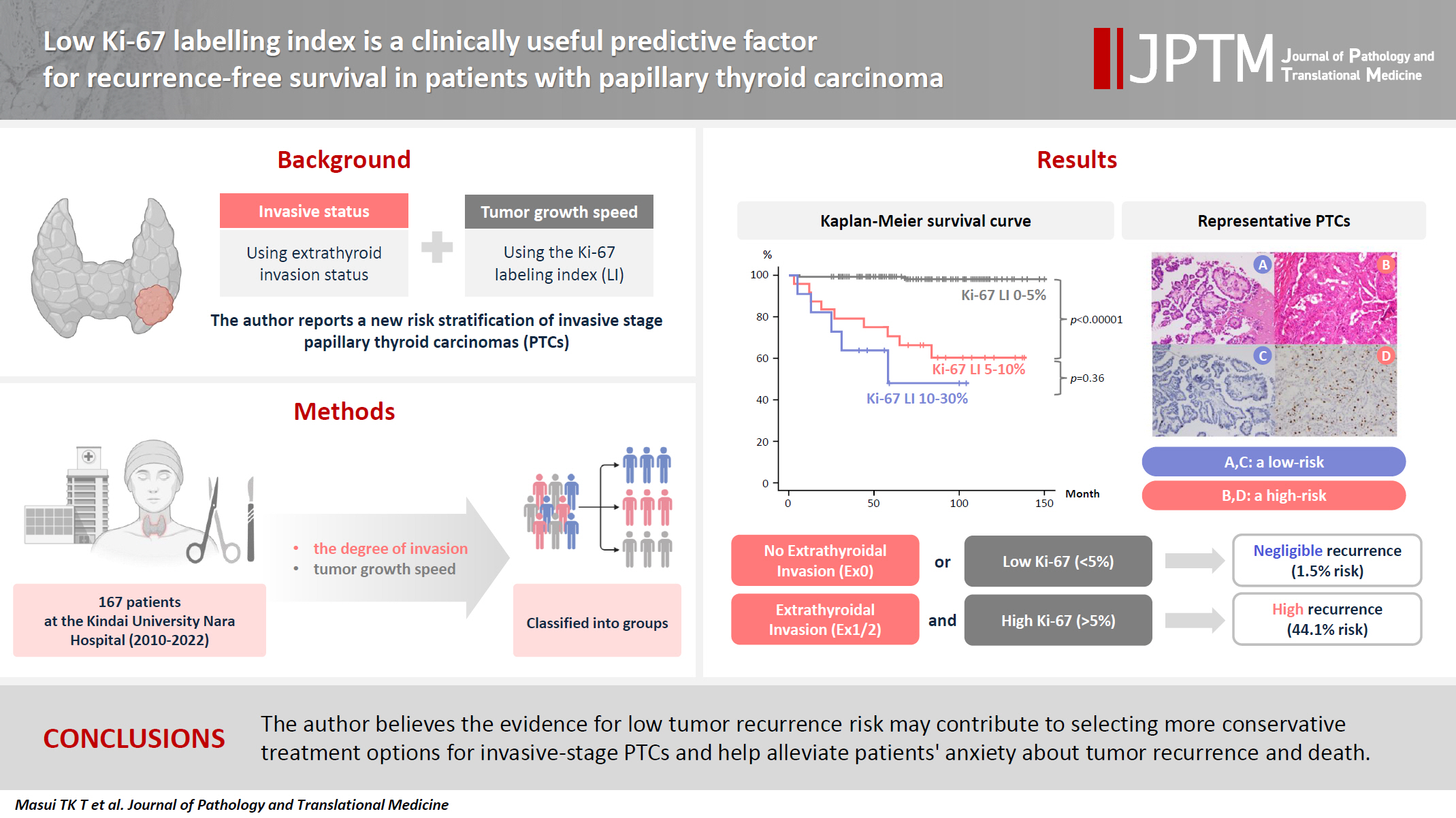
- Low Ki-67 labeling index is a clinically useful predictive factor for recurrence-free survival in patients with papillary thyroid carcinoma
- Takashi Masui, Katsunari Yane, Ichiro Ota, Kennichi Kakudo, Tomoko Wakasa, Satoru Koike, Hirotaka Kinugawa, Ryuji Yasumatsu, Tadashi Kitahara
- J Pathol Transl Med. 2025;59(2):115-124. Published online February 18, 2025
- DOI: https://doi.org/10.4132/jptm.2024.11.08
- 7,469 View
- 268 Download
- 2 Web of Science
- 3 Crossref
-
 Abstract
 PDF
PDF - Background
We report a new risk stratification of invasive stage papillary thyroid carcinomas (PTCs) by combining invasive status, using extrathyroid invasion (Ex) status, and tumor growth speed using the Ki-67 labeling index (LI). Methods: We examined tumor recurrence in 167 patients with PTC who were surgically treated at the Kindai University Nara Hospital between 2010 and 2022. The patients were classified according to the degree of invasion [negative (Ex0) or positive (Ex1, Ex2, and Ex3)] and tumor growth speed expressed with Ki-67 LI, as low (<5%) or high (>5%). This study confirmed previous findings that the disease-free survival (DFS) rate in PTCs significantly differed between patients with a high and low Ki-67 index. Results: When combining Ex status (negative or positive) and Ki-67 proliferation status (low or high), the DFS rate of invasion in the negative, low Ki-67 LI group was only 1.1%, while that of invasion in the positive, high Ki-67 LI was 44.1%. This study reports for the first time that recurrence risks can be stratified accurately when combining carcinoma’s essential two features of extrathyroid invasion status and tumor growth speed. Conclusions: We believe the evidence for low tumor recurrence risk may contribute to use of more conservative treatment options for invasive-stage PTCs and help alleviate patient anxiety about tumor recurrence and death. -
Citations
Citations to this article as recorded by
- Research Progress on the Correlation between Three Biomarkers, Ki-67, CAIX and VEGF and Clear Cell Renal Cell Carcinoma
锦容 马
Advances in Clinical Medicine.2025; 15(09): 326. CrossRef - Immunophenotypic Panel for Comprehensive Characterization of Aggressive Thyroid Carcinomas
Mihail Ceausu, Mihai Alin Publik, Dana Terzea, Carmen Adina Cristea, Dumitru Ioachim, Dana Manda, Sorina Schipor
Cells.2025; 14(19): 1554. CrossRef - High Ki-67 labeling index correlates with aggressive clinicopathological features in papillary thyroid carcinoma: a retrospective study
Defi Nurlia Erdian, Maria Francisca Ham, Dina Khoirunnisa, Agnes Stephanie Harahap
Thyroid Research.2025;[Epub] CrossRef
- Research Progress on the Correlation between Three Biomarkers, Ki-67, CAIX and VEGF and Clear Cell Renal Cell Carcinoma
- Fatty acid synthetase expression in triple-negative breast cancer
- Jin Hee Park, Hye Seung Han, So Dug Lim, Wook Youn Kim, Kyoung Sik Park, Young Bum Yoo, Seung Eun Lee, Wan-Seop Kim
- J Pathol Transl Med. 2022;56(2):73-80. Published online January 21, 2022
- DOI: https://doi.org/10.4132/jptm.2021.10.27

- 8,911 View
- 208 Download
- 11 Web of Science
- 11 Crossref
-
Abstract
PDF
- Background
Triple-negative breast cancer (TNBC) has a relatively poor prognosis. Research has identified potential metabolic targets, including fatty acid metabolism, in TNBC. The absence of effective target therapies for TNBC led to exploration of the role of fatty acid synthetase (FASN) as a potential target for TNBC therapy. Here, we analyzed the expression of FASN, a representative lipid metabolism–related protein, and investigated the association between FASN expression and Ki-67 and the programmed death ligand 1 (PD-L1) biomarkers in TNBC.
Methods
Immunohistochemical expression of FASN was analyzed in 166 patients with TNBC. For analytical purposes, patients with 0–1+ FASN staining were grouped as low-grade FASN and patients with 2–3+ FASN staining as high-grade FASN.
Results
FASN expression was observed in 47.1% of TNBC patients. Low and high expression of FASN was identified in 75.9% and 24.1%, respectively, and no statistically significant difference was found in T category, N category, American Joint Committee on Cancer stage, or recurrence rate between the low and high-FASN expression groups. Ki-67 proliferation level was significantly different between the low and high-FASN expression groups. FASN expression was significantly related to Ki-67 as the level increased. There was no significant difference in PD-L1 positivity between the low- and high-FASN expression groups.
Conclusions
We identified FASN expression in 166 TNBC patients. The Ki-67 proliferation index was positively correlated with FASN level, indicating higher proliferation activity as FASN increases. However, there was no statistical association with PD-L1 SP142, the currently FDA-approved assay, or FASN expression level. -
Citations
Citations to this article as recorded by- FASN at the crossroads of tumor metabolism, immune evasion, and therapy resistance: Mechanistic insights and therapeutic opportunities
Xuefeng Jiang, Guotao Fang, Wen Li, Yusheng Liu, Gang Chen, Silvio E. Perea, Yasser Perera, Rong Ma, Xiaofei Hu, Xinan Long
Critical Reviews in Oncology/Hematology.2026; 220: 105155. CrossRef - Serum fatty acid profiles across clinicopathologic heterogeneity in breast cancer: an exploratory study
Patryk T. Mucha, Agata Jedrzejewska, Alicja Pakiet, Patrycja Jablonska, Jacek Zielinski, Weronika Szczecinska, Miroslawa Puskulluoglu, Adriana Mika, Ewa M. Slominska, Marta Tomczyk
Therapeutic Advances in Medical Oncology.2026;[Epub] CrossRef - Lipid metabolism involved in progression and drug resistance of breast cancer
Wenxiang Fu, Aijun Sun, Huijuan Dai
Genes & Diseases.2025; 12(4): 101376. CrossRef - Unveiling the impact of lipid metabolism on triple-negative breast cancer growth and treatment options
Xin-xian Cai, Zhe-zhong Zhang, Xiao-xiao Yang, Wen-rui Shen, Liu-wei Yuan, Xi Ding, Ying Yu, Wen-yu Cai
Frontiers in Oncology.2025;[Epub] CrossRef - Protein biomarkers for diagnosis of breast cancer
Emeka Eze Joshua Iweala, Doris Nnenna Amuji, Faith Chinasaokwu Nnaji
Scientific African.2024; 25: e02308. CrossRef - Microarray analysis points to LMNB1 and JUN as potential target genes for predicting metastasis promotion by etoposide in colorectal cancer
Jiafei Liu, Hongjie Yang, Peng Li, Yuanda Zhou, Zhichun Zhang, Qingsheng Zeng, Xipeng Zhang, Yi Sun
Scientific Reports.2024;[Epub] CrossRef - The signature of extracellular vesicles in hypoxic breast cancer and their therapeutic engineering
Baiheng Zhu, Kehao Xiang, Tanghua Li, Xin Li, Fujun Shi
Cell Communication and Signaling.2024;[Epub] CrossRef - NFYA promotes malignant behavior of triple-negative breast cancer in mice through the regulation of lipid metabolism
Nobuhiro Okada, Chihiro Ueki, Masahiro Shimazaki, Goki Tsujimoto, Susumu Kohno, Hayato Muranaka, Kiyotsugu Yoshikawa, Chiaki Takahashi
Communications Biology.2023;[Epub] CrossRef - Role of EGFR and FASN in breast cancer progression
Suchi Chaturvedi, Mainak Biswas, Sushabhan Sadhukhan, Avinash Sonawane
Journal of Cell Communication and Signaling.2023; 17(4): 1249. CrossRef - Bioinformatics Method Was Used to Analyze the Highly Expressed Gene FAM83A of Breast Cancer in Young Women
Yongzhe Tang, Hao Wang, Qi He, Yuanyuan Chen, Jie Wang, Fahd Abd Algalil
Applied Bionics and Biomechanics.2022; 2022: 1. CrossRef - NCAPH promotes proliferation as well as motility of breast cancer cells by activating the PI3K/AKT pathway
Ting Zhang, Peng Li, Wanying Guo, Qipeng Liu, Weiqiang Qiao, Miao Deng
Physiology International.2022;[Epub] CrossRef
- FASN at the crossroads of tumor metabolism, immune evasion, and therapy resistance: Mechanistic insights and therapeutic opportunities
- Molecular and Clinicopathological Features of Gastrointestinal Stromal Tumors in Vietnamese Patients
- Quoc Dat Ngo, Quoc Thang Pham, Dang Anh Thu Phan, Anh Vu Hoang, Thi Ngoc Ha Hua, Sao Trung Nguyen
- J Pathol Transl Med. 2019;53(6):361-368. Published online September 16, 2019
- DOI: https://doi.org/10.4132/jptm.2019.08.27

- 9,321 View
- 160 Download
- 2 Web of Science
- 2 Crossref
-
Abstract
PDF
 Supplementary Material
 - Background
Gastrointestinal stromal tumors (GISTs) are the most frequent mesenchymal neoplasms of the gastrointestinal tract. Management of GIST patients is currently based on clinicopathological features and associated genetic changes. However, the detailed characteristics and molecular genetic features of GISTs have not yet been described in the Vietnamese population.
Methods
We first identified 155 patients with primary GIST who underwent surgery with primary curative intent between 2011 and 2014 at University Medical Center at Ho Chi Minh City, Vietnam. We evaluated the clinicopathological features and immunohistochemical reactivity to p53 and Ki-67 in these patients. Additionally, KIT genotyping was performed in 100 cases.
Results
The largest proportion of GISTs was classified as high-risk (43.2%). Of the 155 GISTs, 52 (33.5%) were positive for Ki-67, and 58 (37.4%) were positive for p53. The expression of Ki-67 and p53 were correlated with mitotic rate, tumor size, risk assessment, and tumor stage. Out of 100 GIST cases, KIT mutation was found in 68%, of which 62 (91.2%) were found in exon 11, two (2.9%) in exon 9, and four (5.8%) in exon 17. No mutation in exon 13 was identified. Additionally, KIT mutations did not correlate with any clinicopathological features.
Conclusions
The expression of Ki-67 and p53 were associated with high-risk tumors. Mutations in exon 11 were the most commonly found, followed by exon 17 and exon 9. Additionally, KIT mutation status was not correlated with any recognized clinicopathological features. -
Citations
Citations to this article as recorded by- Ki67 for evaluating the prognosis of gastrointestinal stromal tumors: A systematic review and meta‑analysis
Ji Li, An-Ran Wang, Xiao-Dong Chen, Hong Pan, Shi-Qiang Li
Oncology Letters.2022;[Epub] CrossRef - Endoscopic ultrasound‐guided fine‐needle aspiration cytology in the diagnosis of the gastrointestinal stromal tumor of the stomach
José‐Fernando Val‐Bernal, Elena Yllera, María Moris, Ihab Abdulkader Nallib, Angel Vázquez‐Boquete, María Martino
Diagnostic Cytopathology.2020; 48(9): 833. CrossRef
- Ki67 for evaluating the prognosis of gastrointestinal stromal tumors: A systematic review and meta‑analysis
- Serous Adenocarcinoma of Fallopian Tubes: Histological and Immunohistochemical Aspects
- Natalia Hyriavenko, Mykola Lyndin, Kateryna Sikora, Artem Piddubnyi, Ludmila Karpenko, Olha Kravtsova, Dmytrii Hyriavenko, Olena Diachenko, Vladyslav Sikora, Anatolii Romaniuk
- J Pathol Transl Med. 2019;53(4):236-243. Published online April 11, 2019
- DOI: https://doi.org/10.4132/jptm.2019.03.21
- 9,527 View
- 133 Download
- 4 Web of Science
- 6 Crossref
-
Abstract
PDF
- Background
Although primary cancer of the fallopian tubes is a relatively rare type of tumor in female reproductive organs, its mortality is quite high. It is important to identify molecular and biological markers of this malignancy that determine its specific phenotype.
Methods
The study was carried out on samples received from 71 female patients with primary cancer of the fallopian tubes. The main molecular and biological properties, including hormone status (estrogen receptor [ER], progesterone receptor [PR]), human epidermal growth factor receptor (HER2)/neu expression, proliferative potential (Ki-67), apoptosis (p53, Bcl-2), and pro-angiogenic (vascular endothelial growth factor) quality of serous tumors were studied in comparison with clinical and morphological characteristics.
Results
ER and PR expression is accompanied by low grade neoplasia, early clinical disease stage, and absence of lymphogenic metastasis (p < .001). HER2/neu expression is not typical for primary cancer of the fallopian tubes. Ki-67 expression is characterized by an inverse correlation with ER and PR (p < .05) and is associated with lymphogenic metastasis (p < .01). p53+ status correlates with high grade malignancy, tumor progression, metastasis, negative ER/PR (p < .001), and negative Bcl-2 status (p < .05). Positive Bcl-2 status is positively correlated with ER and PR expression and low grade malignancy.
Conclusions
Complex morphologic (histological and immunohistochemical) study of postoperative material allows estimation of the degree of malignancy and tumor spread to enable appropriate treatment for each case. -
Citations
Citations to this article as recorded by- Clinical and Morphological Analysis of Odontogenic Tumors and Tooth Developmental Anomalies
Yе.V. Kuzenko, S.M. Hermanchuk, O.O. Mykhno, D.H. Tsepochko, O.V. Kuzenko, A.Yu. Olishkevych
Kharkiv Dental Journal.2025; : 275. CrossRef - Rare non-serous fallopian tube cancers: institutional experience and literature review
Dmitrii Sumtsov, Georgyi Sumtsov, Nataliia Hyriavenko, Mykola Lyndin, Kateryna Sikora, Nataliia Kalashnik, Svitlana Smiian, Igor Gladchuk
Wiener Medizinische Wochenschrift.2024; 174(9-10): 199. CrossRef - UŞAQLIQ BORULARININ BİRİNCİLİ XƏRÇƏNGİ: DİAQNOSTİKASI VƏ MÜALİCƏSİNİN NƏTİCƏLƏRİ
D.G. Sumtsov, G.O. Sumtsov, N.I. Hyriavenko, S.A. Smiian, N.V. Kalashnyk, K.O. Sikora, N.M. Rozhkovska, I.Z. Gladchuk
Azerbaijan Medical Journal.2023; (4): 75. CrossRef - FEATURES OF ENDOMETRIUM STRUCTURE IN ALCOHOL-ABUSING HIV-INFECTED INDIVIDUALS
M. Lytvynenko
Inter Collegas.2021; 8(1): 52. CrossRef - Concurrent Clostridial Enteritis and Oviductal Adenocarcinoma with Carcinomatosis in an Adult Alpaca (Vicugna pacos)
Mandy Womble, Megan E. Schreeg, Allison Hoch, Enoch B. de Souza Meira, Derek Foster, Christopher Premanandan, Tatiane T. Negrão Watanabe
Journal of Comparative Pathology.2021; 189: 52. CrossRef - Problems of primary fallopian tube cancer diagnostics during and after surgery
D.G. Sumtsov, I.Z. Gladchuk, G.O. Sumtsov, N.I. Hyriavenko, M.S. Lyndin, V.V. Sikora, V.M. Zaporozhan
REPRODUCTIVE ENDOCRINOLOGY.2021; (59): 66. CrossRef
- Clinical and Morphological Analysis of Odontogenic Tumors and Tooth Developmental Anomalies
- Potential Role for a Panel of Immunohistochemical Markers in the Management of Endometrial Carcinoma
- Amany Salama, Mohammad Arafa, Eman ElZahaf, Abdelhadi Mohamed Shebl, Azmy Abd El-Hameed Awad, Sylvia A. Ashamallah, Reda Hemida, Anas Gamal, Abd AlRahman Foda, Khaled Zalata, El-Said M. Abdel-Hady
- J Pathol Transl Med. 2019;53(3):164-172. Published online February 28, 2019
- DOI: https://doi.org/10.4132/jptm.2019.02.12
- 13,040 View
- 391 Download
- 14 Web of Science
- 11 Crossref
-
Abstract
PDF
- Background
In order to improve the efficacy of endometrial carcinoma (EC) treatment, identifying prognostic factors for high risk patients is a high research priority. This study aimed to assess the relationships among the expression of estrogen receptors (ER), progesterone receptors (PR), human epidermal growth factor receptor 2 (HER2), Ki-67, and the different histopathological prognostic parameters in EC and to assess the value of these in the management of EC.
Methods
We examined 109 cases of EC. Immunohistochemistry for ER, PR, HER2, and Ki-67 were evaluated in relation to age, tumor size, International Federation of Gynecology and Obstetrics (FIGO) stage and grade, depth of infiltration, cervical and ovarian involvement, lymphovascular space invasion (LVSI), and lymph node (LN) metastasis.
Results
The mean age of patients in this study was 59.8 ± 8.2 years. Low ER and PR expression scores and high Ki-67 expression showed highly significant associations with non-endometrioid histology (p = .007, p < .001, and p < .001, respectively) and poor differentiation (p = .007, p < .001, and p <. 001, respectively). Low PR score showed a significant association with advanced stage (p = .009). Low ER score was highly associated with LVSI (p = .006), and low PR scores were associated significantly with LN metastasis (p = .026). HER2 expression was significantly related to advanced stages (p = .04), increased depth of infiltration (p = .02), LVSI (p = .017), ovarian involvement (p = .038), and LN metastasis (p = .038). There was a close relationship between HER2 expression and uterine cervical involvement (p = .009). Higher Ki-67 values were associated with LN involvement (p = .012).
Conclusions
The over-expression of HER2 and Ki-67 and low expression of ER and PR indicate a more malignant EC behavior. An immunohistochemical panel for the identification of high risk tumors can contribute significantly to prognostic assessments. -
Citations
Citations to this article as recorded by- Tissue Microarray for Gynecological Pathology Studies: A Mini-Review
Mohammad Arafa, Abd AlRahman Foda, Amany Salama, Ola Shalaby, Muna Al-Jabri, Fatma Al Hinai, Afrah Al-Rashdi, Samya Al-Husaini, Suaad Al-Badi
Journal of Microscopy and Ultrastructure.2026; 14(1): 1. CrossRef - Clinicopathological Correlation of Hormone Receptors, Angiogenesis, and Tumor Budding in Endometrial Carcinoma: A Tertiary Care Center Study
Senjuti Dasgupta, Arpita Das, Ujjwal Bandyopadhyay
The Journal of Obstetrics and Gynecology of India.2025;[Epub] CrossRef - Multiparameter MRI-based radiomics analysis for preoperative prediction of type II endometrial cancer
Yingying Cao, Wei Zhang, Xiaorong Wang, Xiaojing Lv, Yaping Zhang, Kai Guo, Shuai Ren, Yuan Li, Zhongqiu Wang, Jingya Chen
Heliyon.2024; 10(12): e32940. CrossRef - Correlation of PD-L1 expression with different clinico-pathological and immunohistochemical features of ovarian surface epithelial tumors
Asem Shalaby, Ola Shalaby, Hazem Abdullah, Mohamed Rachid Boulassel, Mohammad Arafa
Clinical and Translational Oncology.2024; 27(2): 699. CrossRef - Estrogen/Progesterone Receptor Loss, CTNNB1 and KRAS Mutations Are Associated With Local Recurrence or Distant Metastasis in Low-Grade Endometrial Endometrioid Carcinoma
Rajni Chibbar, Sabrina Foerstner, Janarathnee Suresh, Richa Chibbar, Alexandre Piche, Deeksha Kundapur, Rani Kanthan, Vijayanand Kundapur, Cheng Han Lee, Anita Agrawal, Raymond Lai
Applied Immunohistochemistry & Molecular Morphology.2023; 31(3): 181. CrossRef - Exploring the Prognostic and Predictive Roles of Ki-67 in Endometrial Cancer
Laura Paleari, Mariangela Rutigliani, Oriana D’Ecclesiis, Sara Gandini, Irene Maria Briata, Tania Buttiron Webber, Nicoletta Provinciali, Andrea DeCensi
International Journal of Translational Medicine.2023; 3(4): 479. CrossRef - Analysis of human epidermal growth factor receptor 2 immunohistochemical expression in high-grade endometrial carcinomas and its association with variable clinical outcomes
Malames M. Faisal, Marwa M. Shakweer, Ghada Refaat, Khaled S. Mohammed, Tarek I. ElMallawy, Magda H. Nasreldin, Laila M. Farid, Mariam B. Abouelkhair
Egyptian Journal of Pathology.2023; 43(2): 119. CrossRef - Correlation of PD-L1 immunohistochemical expression with microsatellite instability and p53 status in endometrial carcinoma
Mohammad Arafa, Abdelhadi Mohamed Shebl, Amany Salama, Eman ElZahaf, Sylvia A. Ashamallah, Abd AlRahman Foda, AzmyAbd El-Hameed Awad, Asem Shalaby
European Journal of Obstetrics & Gynecology and Reproductive Biology: X.2022; 16: 100172. CrossRef - Immunohistochemical Expression of Oestrogen and Epidermal Growth Factor Receptors in Endometrial Cancerous in Sudanese Patients
Salwa Abdalraheem Abubaker, Mohamed Elfatih Abdelwadoud, Mutaz Mohamed Ali, Hadia Alhaj Ahmad, Abuobieda Mohamed Khlafalla, Osman Mohammed Elmahi, Hisham Ali Waggiallah
Journal Of Biochemical Technology.2021; 12(1): 58. CrossRef - Expression of ER/PR Receptor, Her-2/neu, Ki67 and p53 in Endometrial Carcinoma: Clinicopathological Implication and Prognostic Value
V. B. Shivkumar, Manisha A. Atram, Nitin M. Gangane
Indian Journal of Gynecologic Oncology.2020;[Epub] CrossRef - Immunohistochemical study of ER, PR, p53 and Ki67 expression in patients with endometrial adenocarcinoma and atypical endometrial hyperplasia
Rachana Lakhe, Ravi M Swami, Preeti Doshi, Manjiri N Karandikar, Ravindra Nimbargi
IP Archives of Cytology and Histopathology Research.2020; 5(4): 274. CrossRef
- Tissue Microarray for Gynecological Pathology Studies: A Mini-Review
- Diagnostic Significance of Cellular Neuroglial Tissue in Ovarian Immature Teratoma
- Yun Chai, Chang Gok Woo, Joo-Young Kim, Chong Jai Kim, Shin Kwang Khang, Jiyoon Kim, In Ah Park, Eun Na Kim, Kyu-Rae Kim
- J Pathol Transl Med. 2017;51(1):49-55. Published online October 14, 2016
- DOI: https://doi.org/10.4132/jptm.2016.09.19
- 20,559 View
- 512 Download
- 6 Web of Science
- 8 Crossref
-
Abstract
PDF
- Background
Immature teratoma (IT) is a tumor containing immature neuroectodermal tissue, primarily in the form of neuroepithelial tubules. However, the diagnosis of tumors containing only cellular neuroglial tissue (CNT) without distinct neuroepithelial tubules is often difficult, since the histological characteristics of immature neuroectodermal tissues remain unclear. Here, we examined the significance of CNT and tried to define immature neuroectodermal tissues by comparing the histological features of neuroglial tissues between mature teratoma (MT) and IT.
Methods
The histological features of neuroglial tissue, including the cellularity, border between the neuroglial and adjacent tissues, cellular composition, mitotic index, Ki-67 proliferation rate, presence or absence of tissue necrosis, vascularity, and endothelial hyperplasia, were compared between 91 MT and 35 IT cases.
Results
CNTs with a cellularity grade of ≥ 2 were observed in 96% of IT cases and 4% of MT cases (p < .001); however, CNT with a cellularity grade of 3 in MT cases was confined to the histologically distinct granular layer of mature cerebellar tissue. Moreover, CNT in IT exhibited significantly higher rates of Ki-67 proliferation, mitoses, and necrosis than those in MT (p < .001). Furthermore, an infiltrative border of neuroglial tissue and glomeruloid endothelial hyperplasia were significantly more frequent in IT cases than in MT cases (p < .001).
Conclusions
Our results suggest that if CNT with a cellularity grade of ≥ 2 is not a component of cerebellar tissue, such cases should be diagnosed as IT containing immature neuroectodermal tissue, particularly if they exhibit an infiltrative border, mitoses, necrosis, and increased Ki-67 proliferation. -
Citations
Citations to this article as recorded by- An Atypical Presentation of a Pediatric Mature Teratoma: A Case Report and Review of the Literature
Ahmed M Othman, Abdulaziz A Abu Alnasr, Reem E Kordi, Shahad A Abu Alnasr
Cureus.2024;[Epub] CrossRef - Immature Teratoma: Diagnosis and Management—A Review of the Literature
Liviu Moraru, Melinda-Ildiko Mitranovici, Diana Maria Chiorean, Marius Coroș, Raluca Moraru, Ioan Emilian Oală, Sabin Gligore Turdean
Diagnostics.2023; 13(9): 1516. CrossRef - Congenital Immature Grade ΙΙΙ Teratoma of the Neck: A Case Report
Nazneen Liaqat, Israr Ud Din, Zeeshan Ali, Majid Rashid, Afsheen Liaqat
Cureus.2023;[Epub] CrossRef - Benign ovarian teratoma in the dog with predominantly nervous tissue: A case report
P Makovicky, AV Makarevich, P Makovicky, A Seidavi, L Vannucci, K Rimarova
Veterinární medicína.2022; 67(2): 99. CrossRef - Fascin as a Useful Marker for Identifying Neural Components in Immature Teratomas of Human Ovary and Those Derived From Murine Embryonic Stem Cells
Ryunosuke Umehara, Atsushi Kurata, Masakatsu Takanashi, Hirotsugu Hashimoto, Koji Fujita, Toshitaka Nagao, Masahiko Kuroda
International Journal of Gynecological Pathology.2019; 38(4): 377. CrossRef - Cerebellar Differentiation in Ovarian Teratoma: A Report of 6 Cases
Colin J.R. Stewart, Maxine L. Crook
International Journal of Gynecological Pathology.2018; 37(4): 316. CrossRef - Mitotic activity of epithelia of ectoand entodermal types in spontaneous and experimental teratomas of mice
Pavel A. Dyban
Medical academic journal.2018; 18(4): 42. CrossRef - Ovarian cystectomy in the treatment of apparent early-stage immature teratoma
Ting Zhao, Yan Liu, Xiao Wang, Hao Zhang, Yuan Lu
Journal of International Medical Research.2017; 45(2): 771. CrossRef
- An Atypical Presentation of a Pediatric Mature Teratoma: A Case Report and Review of the Literature
- The Predictive Value of Pathologic Features in Pituitary Adenoma and Correlation with Pituitary Adenoma Recurrence
- Jee Soon Kim, Youn Soo Lee, Min Jung Jung, Yong Kil Hong
- J Pathol Transl Med. 2016;50(6):419-425. Published online October 6, 2016
- DOI: https://doi.org/10.4132/jptm.2016.06.30
- 11,477 View
- 239 Download
- 18 Web of Science
- 19 Crossref
-
Abstract
PDF
- Background
The 2004 World Health Organization classification introduced atypical pituitary adenoma (aPA), which was equivocally defined as invasion with increased mitotic activity that had a Ki-67 labeling index (LI) greater than 3%, and extensive p53 immunoreactivity. However, aPAs that exhibit all of these features are rare and the predictive value for recurrence in pituitary adenomas (PAs) remains uncertain. Thus, we sought to characterize pathological features of PAs that correlated with recurrence.
Methods
One hundred and sixty-seven cases of surgically resected PA or aPA were retrieved from 2011 to 2013 in Seoul St. Mary’s Hospital. Among them, 28 cases were confirmed to be recurrent, based on pathologic or radiologic examination. The pathologic characteristics including mitosis, invasion, Ki-67 LI and p53 immunoreactivity were analyzed in relation to recurrence.
Results
Analysis of the pathologic features indicated that only Ki-67 LI over 3% was significantly associated with tumor recurrence (p = .02). The cases with at least one pathologic feature showed significantly higher recurrence rates (p < .01). Analysis indicated that cases with two pathologic features, Ki-67 LI over 3% and extensive p53 immunoreactivity 20% or more, were significantly associated with tumor recurrence (p < .01).
Conclusions
Based on these results, PA tumor recurrence can be predicted by using mitosis, invasion, Ki-67 LI (3%), or extensive p53 immunoreactivity (≥ 20%). Assessment of these features is recommended for PA diagnosis for more accurate prediction of recurrence. -
Citations
Citations to this article as recorded by- Long-term outcomes of adjuvant proton radiotherapy (PRT) for residual pituitary adenoma (PA) in adults – a retrospective, single institute experience
Fabian J. K. Allmendinger, Maximilian Deng, Sebastian Regnery, Lars Wessel, Katharina Kozyra, Felix Englert, Ricarda Wickert, Jannik Walter, Lucas Mose, Thomas Tessonnier, Sandro M. Krieg, Jürgen Debus, Laila König, Tanja Adena-Eichkorn
Journal of Neuro-Oncology.2026;[Epub] CrossRef - Experience using temozolomide in the treatment of aggressive pituitary adenomas
P. L. Kalinin, L. I. Astafyeva, I. V. Chernov, G. L. Kobyakov, D. V. Fomichev, Yu. Yu. Trunin
Russian journal of neurosurgery.2025; 26(4): 54. CrossRef - The Value of ER∝ in the Prognosis of GH- and PRL-Secreting PitNETs: Clinicopathological Correlations
Roxana-Ioana Dumitriu-Stan, Iulia-Florentina Burcea, Valeria Nicoleta Nastase, Raluca Amalia Ceaușu, Anda Dumitrascu, Laurentiu Catalin Cocosila, Alexandra Bastian, Sabina Zurac, Marius Raica, Catalina Poiana
International Journal of Molecular Sciences.2023; 24(22): 16162. CrossRef - Ki-67/MIB-1 and Recurrence in Pituitary Adenoma
Kent Tadokoro, Colten Wolf, Joseph Toth, Cara Joyce, Meharvan Singh, Anand Germanwala, Chirag Patel
Journal of Neurological Surgery Part B: Skull Base.2022; 83(S 02): e580. CrossRef - Association of PTTG1 expression with invasiveness of non-functioning pituitary adenomas
Su Jung Kum, Hye Won Lee, Soon Gu Kim, Hyungsik Park, Ilseon Hwang, Sang Pyo Kim
Journal of Pathology and Translational Medicine.2022; 56(1): 22. CrossRef - A Preoperative MRI-Based Radiomics-Clinicopathological Classifier to Predict the Recurrence of Pituitary Macroadenoma Within 5 Years
Yu Zhang, Yuqi Luo, Xin Kong, Tao Wan, Yunling Long, Jun Ma
Frontiers in Neurology.2022;[Epub] CrossRef - Endoscopic Endonasal Pituitary Surgery For Nonfunctioning Pituitary Adenomas: Long-Term Outcomes and Management of Recurrent Tumors
Anne-Laure Bernat, Pénélope Troude, Stefano Maria Priola, Ahmad Elsawy, Faisal Farrash, Ozgur Mete, Shereen Ezzat, Sylvia L. Asa, John De Almeida, Allan Vescan, Eric Monteiro, Joao Paulo Almeida, Gelareh Mohammed Zadeh, Fred Gentili
World Neurosurgery.2021; 146: e341. CrossRef - A Nomogram for Preoperatively Predicting the Ki-67 Index of a Pituitary Tumor: A Retrospective Cohort Study
Xiangming Cai, Junhao Zhu, Jin Yang, Chao Tang, Feng Yuan, Zixiang Cong, Chiyuan Ma
Frontiers in Oncology.2021;[Epub] CrossRef - Comparative Proteomic Study Shows the Expression of Hint-1 in Pituitary Adenomas
Carolina Carrillo-Najar, Daniel Rembao-Bojórquez, Martha L. Tena-Suck, Sergio Zavala-Vega, Noemí Gelista-Herrera, Miguel A. Ramos-Peek, Juan L. Gómez-Amador, Febe Cazares-Raga, Fidel de la Cruz Hernández-Hernández, Alma Ortiz-Plata
Diagnostics.2021; 11(2): 330. CrossRef - Prediction of recurrence in solid nonfunctioning pituitary macroadenomas: additional benefits of diffusion-weighted MR imaging
Ching-Chung Ko, Tai-Yuan Chen, Sher-Wei Lim, Yu-Ting Kuo, Te-Chang Wu, Jeon-Hor Chen
Journal of Neurosurgery.2020; 132(2): 351. CrossRef - Pituitary tumors: epidemiology and clinical presentation spectrum
Marta Araujo-Castro, Víctor Rodríguez Berrocal, Eider Pascual-Corrales
Hormones.2020; 19(2): 145. CrossRef - Ki67 in endocrine neoplasms: to count or not to count, this is the question! A systematic review from the English language literature
E. Guadagno, E. D’Avella, P. Cappabianca, A. Colao, M. Del Basso De Caro
Journal of Endocrinological Investigation.2020; 43(10): 1429. CrossRef - Study of Simple Immunohistochemical Cytocolorimetric Assay Application for More Accurate Assessment of Prognosis in Patients with Pituitary Adenomas
Pavel V. Nikitin, Marina V. Ryzhova, Lyudmila V. Shishkina, Svetlana V. Shugay, Irina V. Zubova
World Neurosurgery.2019; 122: e1047. CrossRef - The Prognostic Roles of the Ki-67 Proliferation Index, P53 Expression, Mitotic Index, and Radiological Tumor Invasion in Pituitary Adenomas
Rovshan Hasanov, Berna İmge Aydoğan, Saba Kiremitçi, Esra Erden, Sevim Güllü
Endocrine Pathology.2019; 30(1): 49. CrossRef - Residual Tumor Confers a 10-Fold Increased Risk of Regrowth in Clinically Nonfunctioning Pituitary Tumors
Jelena Maletkovic, Asmaa Dabbagh, Dongyun Zhang, Abdul Zahid, Marvin Bergsneider, Marilene B Wang, Michael Linetsky, Noriko Salamon, William H Yong, Harry V Vinters, Anthony P Heaney
Journal of the Endocrine Society.2019; 3(10): 1931. CrossRef - Atypical pituitary adenoma: a clinicopathologic case series
Martin J. Rutkowski, Ryan M. Alward, Rebecca Chen, Jeffrey Wagner, Arman Jahangiri, Derek G. Southwell, Sandeep Kunwar, Lewis Blevins, Han Lee, Manish K. Aghi
Journal of Neurosurgery.2018; 128(4): 1058. CrossRef - Both invasiveness and proliferation criteria predict recurrence of non-functioning pituitary macroadenomas after surgery: a retrospective analysis of a monocentric cohort of 120 patients
Julie Lelotte, Anne Mourin, Edward Fomekong, Alex Michotte, Christian Raftopoulos, Dominique Maiter
European Journal of Endocrinology.2018; 178(3): 237. CrossRef - Letter to the Editor. Atypical pituitary adenoma
Lauren E. Rotman, T. Brooks Vaughan, James R. Hackney, Kristen O. Riley
Journal of Neurosurgery.2018; 129(6): 1657. CrossRef - Molecular targeted therapies in adrenal, pituitary and parathyroid malignancies
Anna Angelousi, Georgios K Dimitriadis, Georgios Zografos, Svenja Nölting, Gregory Kaltsas, Ashley Grossman
Endocrine-Related Cancer.2017; 24(6): R239. CrossRef
- Long-term outcomes of adjuvant proton radiotherapy (PRT) for residual pituitary adenoma (PA) in adults – a retrospective, single institute experience
- Interobserver Variability of Ki-67 Measurement in Breast Cancer
- Yul Ri Chung, Min Hye Jang, So Yeon Park, Gyungyub Gong, Woo-Hee Jung, The Korean Breast Pathology Ki- Study Group
- J Pathol Transl Med. 2016;50(2):129-137. Published online February 15, 2016
- DOI: https://doi.org/10.4132/jptm.2015.12.24
- 15,190 View
- 134 Download
- 34 Web of Science
- 33 Crossref
-
Abstract
PDF
- Background
As measurement of Ki-67 proliferation index is an important part of breast cancer diagnostics, we conducted a multicenter study to examine the degree of concordance in Ki-67 counting and to find factors that lead to its variability. Methods: Thirty observers from thirty different institutions reviewed Ki-67–stained slides of 20 different breast cancers on whole sections and tissue microarray (TMA) by online system. Ten of the 20 breast cancers had hot spots of Ki-67 expression. Each observer scored Ki-67 in two different ways: direct counting (average vs. hot spot method) and categorical estimation. Intraclass correlation coefficient (ICC) of Ki-67 index was calculated for comparative analysis. Results: For direct counting, ICC of TMA was slightly higher than that of whole sections using average method (0.895 vs 0.858). The ICC of tumors with hot spots was lower than that of tumors without (0.736 vs 0.874). In tumors with hot spots, observers took an additional counting from the hot spot; the ICC of whole sections using hot spot method was still lower than that of TMA (0.737 vs 0.895). In categorical estimation, Ki-67 index showed a wide distribution in some cases. Nevertheless, in tumors with hot spots, the range of distribution in Ki-67 categories was decreased with hot spot method and in TMA platform. Conclusions: Interobserver variability of Ki-67 index for direct counting and categorical estimation was relatively high. Tumors with hot spots showed greater interobserver variability as opposed to those without, and restricting the measurement area yielded lower interobserver variability. -
Citations
Citations to this article as recorded by- Dynamic biomarkers in hormone receptor-positive/HER2-negative breast cancer trials: a new hope for precision oncology
Giuseppe Di Grazia, Rodrigo Sánchez-Bayona, Climent Casals-Pascual, Tomás Pascual, Daniele Generali, Alessandra Gennari, Paolo Vigneri, Nadia Harbeck, Javier Cortés, Aleix Prat, Francesco Schettini
npj Breast Cancer.2026;[Epub] CrossRef - Low-Proliferative Invasive Mucinous Carcinoma of the Breast in an Octogenarian: A Case Report Highlighting Pathology-Guided De-escalation
Rae-Anne Kastle, Christopher M Ahmad, Rachana Tadakamalla, Hima Patel, Amer Abboud
Cureus.2026;[Epub] CrossRef - Low E-cadherin expression is associated with poor prognosis in pulmonal adenocarcinoma
Fiete Gehrisch, Kiara A. Schmid, Martina Kluth, Georgia Makrypidi-Fraune, Katharina Möller, Maximilian Lennartz, Veit Bertram, Florian Lutz, Stefan Steurer, Philipp Busch, Birgit Hantzsch-Kuhn, Martin Reck, Till Olchers, David Benjamin Ellebrecht, Christo
Scientific Reports.2026;[Epub] CrossRef - Evaluating the Association of Ki-67 with Oncotype DX Recurrence Score in Early-Stage ER-Positive/HER2-Negative Breast Cancer
Dimitrios Dragoumis, George Kapetsis, Konstantinos Louis, Dimitrios Maniatis, Eleni Mpalampou, Konstantinos Bouloukos, Xenophon Xenakis, Nikolaos Papaioannou, Styliani Parpoudi, Grigorios Pesmatzoglou, Anna Sachoulidou, Eleftherios Sfakianakis, Sofia Tria
Cancers.2026; 18(11): 1731. CrossRef - Comparative analysis of Ki-67 labeling index morphometry using deep learning, conventional image analysis, and manual counting
Mohammad Rizwan Alam, Kyung Jin Seo, Kwangil Yim, Phoebe Liang, Joe Yeh, Chifu Chang, Yosep Chong
Translational Oncology.2025; 51: 102159. CrossRef - Machine Learning-Based Approaches for Breast Density Estimation from Mammograms: A Comprehensive Review
Khaldoon Alhusari, Salam Dhou
Journal of Imaging.2025; 11(2): 38. CrossRef - Letter re: A critical appraisal of the DATA trial analysis on the prognostic and predictive value of the luminal-like subtype
M. Rizk, K. Mokbel
ESMO Open.2025; 10(5): 105067. CrossRef - Clinico-Histomorphological and Mib-1 Analysis of Recurrent Meningiomas: A Retrospective Study
Sujata Sarangi, Asha Shenoy, Ashvini Kolhe, Kanchan Kothari
Asian Journal of Neurosurgery.2025; 20(04): 785. CrossRef - Emerging Diagnostics and Therapies in Neuroendocrine Neoplasms: A Critical Review
Jorge H. Hernandez-Felix, Monica Isabel Meneses-Medina, Rachel Riechelmann, Jonathan Strosberg, Rocio Garcia-Carbonero, Jaydira del Rivero
Cancers.2025; 17(22): 3632. CrossRef - Ki-67 Testing in Breast Cancer: Assessing Variability With Scoring Methods and Specimen Types and the Potential Subsequent Impact on Therapy Eligibility
Therese Bocklage, Virgilius Cornea, Caylin Hickey, Justin Miller, Jessica Moss, Mara Chambers, S. Emily Bachert
Applied Immunohistochemistry & Molecular Morphology.2024; 32(3): 119. CrossRef - Interobserver agreement and diagnostic challenges of Congo red staining for amyloid detection on fat pad aspiration biopsies
Levent Trabzonlu, T. Leif Helland, Melanie C. Kwan, Nathalie Kumiega, M. Lisa Zhang, Ivan Chebib, Vanda F. Torous
Journal of the American Society of Cytopathology.2024; 13(5): 359. CrossRef - Assessment of the Predictive Role of Ki-67 in Breast Cancer Patients’ Responses to Neoadjuvant Chemotherapy
Ghizlane Rais, Rania Mokfi, Farah Boutaggount, Meryem Maskrout, Soundouss Bennour, Chaymae Senoussi, Fadoua Rais
European Journal of Breast Health.2024; : 199. CrossRef - Improving the accuracy of reporting Ki-67 IHC by using an AI tool
Sahil Ajit Saraf, Aahan Singh, Wai Po Kevin Teng, Sencer Karakaya, M. Logaswari, Kaveh Taghipour, Rajasa Jialdasani, Li Yan Khor, Kiat Hon Lim, Sathiyamoorthy Selvarajan, Vani Ravikumar, Md Ali Osama, Priti Chatterjee, Santosh KV
Heliyon.2024; 10(22): e40193. CrossRef - Predictive Value of Ki-67 Index in Evaluating Sporadic Vestibular Schwannoma Recurrence: Systematic Review and Meta-analysis
Kunal Vakharia, Hirotaka Hasegawa, Christopher Graffeo, Mohammad H. A. Noureldine, Salomon Cohen-Cohen, Avital Perry, Matthew L. Carlson, Colin L. W. Driscoll, Maria Peris-Celda, Jamie J. Van Gompel, Michael J. Link
Journal of Neurological Surgery Part B: Skull Base.2023; 84(02): 119. CrossRef - Venous invasion and lymphatic invasion are correlated with the postoperative prognosis of pancreatic neuroendocrine neoplasm
Sho Kiritani, Junichi Arita, Yuichiro Mihara, Rihito Nagata, Akihiko Ichida, Yoshikuni Kawaguchi, Takeaki Ishizawa, Nobuhisa Akamatsu, Junichi Kaneko, Kiyoshi Hasegawa
Surgery.2023; 173(2): 365. CrossRef - Automated Molecular Subtyping of Breast Carcinoma Using Deep Learning Techniques
S. Niyas, Ramya Bygari, Rachita Naik, Bhavishya Viswanath, Dhananjay Ugwekar, Tojo Mathew, J Kavya, Jyoti R Kini, Jeny Rajan
IEEE Journal of Translational Engineering in Health and Medicine.2023; 11: 161. CrossRef - Grade Progression and Intrapatient Tumor Heterogeneity as Potential Contributors to Resistance in Gastroenteropancreatic Neuroendocrine Tumors
Diana Grace Varghese, Jaydira Del Rivero, Emily Bergsland
Cancers.2023; 15(14): 3712. CrossRef - Diagnostic Role and Prognostic Impact of PSAP Immunohistochemistry: A Tissue Microarray Study on 31,358 Cancer Tissues
Laura Sophie Tribian, Maximilian Lennartz, Doris Höflmayer, Noémi de Wispelaere, Sebastian Dwertmann Rico, Clara von Bargen, Simon Kind, Viktor Reiswich, Florian Viehweger, Florian Lutz, Veit Bertram, Christoph Fraune, Natalia Gorbokon, Sören Weidemann, C
Diagnostics.2023; 13(20): 3242. CrossRef - AI-Powered Segmentation of Invasive Carcinoma Regions in Breast Cancer Immunohistochemical Whole-Slide Images
Yiqing Liu, Tiantian Zhen, Yuqiu Fu, Yizhi Wang, Yonghong He, Anjia Han, Huijuan Shi
Cancers.2023; 16(1): 167. CrossRef - Expression of estrogen and progesterone receptors, HER2 protein and Ki-67 proliferation index in breast carcinoma in both tumor tissue and tissue microarray
UP Hacısalihoğlu, MA Dogan
Biotechnic & Histochemistry.2022; 97(4): 298. CrossRef - Diffusive Ki67 and vimentin are associated with worse recurrence-free survival of upper tract urothelial carcinoma: A retrospective cohort study from bench to bedside
Che Hsueh Yang, Wei Chun Weng, Yen Chuan Ou, Yi Sheng Lin, Li Hua Huang, Chin Heng Lu, Tang Yi Tsao, Chao Yu Hsu, Min Che Tung
Urologic Oncology: Seminars and Original Investigations.2022; 40(3): 109.e21. CrossRef - Should Ki-67 be adopted to select breast cancer patients for treatment with adjuvant abemaciclib?
P. Tarantino, H.J. Burstein, N.U. Lin, I.E. Krop, E.P. Winer, S.J. Schnitt, E.P. Hamilton, S.A. Hurvitz, H.S. Rugo, G. Curigliano, S.M. Tolaney
Annals of Oncology.2022; 33(3): 234. CrossRef - A novel deep classifier framework for automated molecular subtyping of breast carcinoma using immunohistochemistry image analysis
Tojo Mathew, S. Niyas, C.I. Johnpaul, Jyoti R. Kini, Jeny Rajan
Biomedical Signal Processing and Control.2022; 76: 103657. CrossRef - Deep learning for the standardized classification of Ki-67 in vulva carcinoma: A feasibility study
Matthias Choschzick, Mariam Alyahiaoui, Alexander Ciritsis, Cristina Rossi, André Gut, Patryk Hejduk, Andreas Boss
Heliyon.2021; 7(7): e07577. CrossRef - Oncotype DX Predictive Nomogram for Recurrence Score Output: The Novel System ADAPTED01 Based on Quantitative Immunochemistry Analysis
Fabio Marazzi, Roberto Barone, Valeria Masiello, Valentina Magri, Antonino Mulè, Angela Santoro, Federica Cacciatori, Luca Boldrini, Gianluca Franceschini, Francesca Moschella, Giuseppe Naso, Silverio Tomao, Maria Antonietta Gambacorta, Giovanna Mantini,
Clinical Breast Cancer.2020; 20(5): e600. CrossRef - Study of Ki-67 index in the molecular subtypes of breast cancer: Inter-observer variability and automated scoring
Divya Meermira, Meenakshi Swain, Swarnalata Gowrishankar
Indian Journal of Cancer.2020; 57(3): 289. CrossRef - Improving the accuracy of gastrointestinal neuroendocrine tumor grading with deep learning
Darshana Govind, Kuang-Yu Jen, Karen Matsukuma, Guofeng Gao, Kristin A. Olson, Dorina Gui, Gregory. E. Wilding, Samuel P. Border, Pinaki Sarder
Scientific Reports.2020;[Epub] CrossRef - Practical approaches to automated digital image analysis of Ki-67 labeling index in 997 breast carcinomas and causes of discordance with visual assessment
Ah-Young Kwon, Ha Young Park, Jiyeon Hyeon, Seok Jin Nam, Seok Won Kim, Jeong Eon Lee, Jong-Han Yu, Se Kyung Lee, Soo Youn Cho, Eun Yoon Cho, Irina V. Lebedeva
PLOS ONE.2019; 14(2): e0212309. CrossRef - Evaluation of Ki-67 Index in Core Needle Biopsies and Matched Breast Cancer Surgical Specimens
Soomin Ahn, Junghye Lee, Min-Sun Cho, Sanghui Park, Sun Hee Sung
Archives of Pathology & Laboratory Medicine.2018; 142(3): 364. CrossRef - Assessment of Ki-67 for Predicting Effective Prognosis in Breast Cancer Subtypes
Sangjung Park, Sunyoung Park, Jungho Kim, Sungwoo Ahn, Kwang Hwa Park, Hyeyoung Lee
Biomedical Science Letters.2018; 24(1): 9. CrossRef - Quantitative tumor heterogeneity assessment on a nuclear population basis
Anne‐Sofie Wessel Lindberg, Knut Conradsen, Rasmus Larsen, Michael Friis Lippert, Rasmus Røge, Mogens Vyberg
Cytometry Part A.2017; 91(6): 574. CrossRef - A comparison of Ki-67 counting methods in luminal Breast Cancer: The Average Method vs. the Hot Spot Method
Min Hye Jang, Hyun Jung Kim, Yul Ri Chung, Yangkyu Lee, So Yeon Park, William B. Coleman
PLOS ONE.2017; 12(2): e0172031. CrossRef - A Novel Breast Cancer Index for Prediction of Distant Recurrence in HR+ Early-Stage Breast Cancer with One to Three Positive Nodes
Yi Zhang, Brock E. Schroeder, Piiha-Lotta Jerevall, Amy Ly, Hannah Nolan, Catherine A. Schnabel, Dennis C. Sgroi
Clinical Cancer Research.2017; 23(23): 7217. CrossRef
- Dynamic biomarkers in hormone receptor-positive/HER2-negative breast cancer trials: a new hope for precision oncology
- PHH3 as an Ancillary Mitotic Marker in Gastrointestinal Stromal Tumors
- Yooju Shin, Jiyeon Hyeon, Boram Lee, Sang Yun Ha, Min Eui Hong, In Gu Do, Kyoung-Mee Kim
- J Pathol Transl Med. 2015;49(1):23-29. Published online January 15, 2015
- DOI: https://doi.org/10.4132/jptm.2014.10.08
- 12,099 View
- 82 Download
- 9 Web of Science
- 9 Crossref
-
Abstract
PDF
- Background
Counting mitoses is subjective and time-consuming. The adjunctive diagnostic utility of a recently reported mitotic marker, phosphohistone H3 (PHH3), was investigated in gastrointestinal stromal tumors (GISTs). Methods: We reviewed 77 GISTs for several proliferative indices. These included the mitotic count per 50 high power fields (HPFs), the immunohistochemical Ki- 67 labeling index and the immunohistochemical PHH3 mitotic index (MI). For comparison, Spearman’s rank correlation and interclass correlation coefficient were used. Results: Mitotic counts ranged from 0–138 (mean, 7.57±2.34) and the PHH3 MI ranged from 0–126 per 50 HPFs (mean, 9.61±2.27). We found a positive correlation between mitotic counts and PHH3 MI (r=0.810, p<.001). The inter-observer correlation coefficient for three participants was 0.975 for mitotic counts and 0.940 for the PHH3 MI. When using the PHH3 MI instead of mitotic counts in the Armed Forces Institute of Pathology (AFIP) stratification criteria, 10 cases were reclassified. In one patient with a mitotic count of 2 and a PHH3 MI of 6 per 50 HPFs, distant metastasis occurred. Conclusions: In GISTs, the PHH3 MI correlated adequately with mitotic counts and can be used as a useful adjunctive to count mitotic figures efficiently. -
Citations
Citations to this article as recorded by- Potential of Proliferative Markers in Pancreatic Cancer Management: A Systematic Review
Aryan Salahi‐Niri, Paniz Zarand, Negar Mansouri, Parvaneh Rastgou, Omid Yazdani, Romina Esbati, Fatemeh Shojaeian, Behnaz Jahanbin, Zhaleh Mohsenifar, Hamid Asadzadeh Aghdaei, Farid Azmoudeh Ardalan, Seyed Amir Ahmad Safavi‐Naini
Health Science Reports.2025;[Epub] CrossRef - A retrospective study on expression and clinical significance of PHH3, Ki67 and P53 in bladder exophytic papillary urothelial neoplasms
Gaoxiu Qi, Jinmeng Liu, Shuqi Tao, Wenyuan Fan, Haoning Zheng, Meihong Wang, Hanchao Yang, Yongting Liu, Huancai Liu, Fenghua Zhou
PeerJ.2023; 11: e15675. CrossRef - Loss of Slfn3 induces a sex-dependent repair vulnerability after 50% bowel resection
Emilie E. Vomhof-DeKrey, Jack T. Lansing, Diane C. Darland, Josey Umthun, Allie D. Stover, Christopher Brown, Marc D. Basson
American Journal of Physiology-Gastrointestinal and Liver Physiology.2021; 320(2): G136. CrossRef - Phosphohistone H3 (PHH3) as a surrogate of mitotic figure count for grading in meningiomas: a comparison of PHH3 (S10) versus PHH3 (S28) antibodies
Napaporn Puripat, Kongsak Loharamtaweethong
Virchows Archiv.2019; 474(1): 87. CrossRef - Gastrointestinal Stromal Tumors Risk Stratification Utilizing Phospho-Histone H3 Evaluated by Manual Counting and Computer-Assisted Image Analysis
Cao Jin, Yan Huang, Mansoor Nasim, Yihe Yang, Lili Lee
International Journal of Surgical Pathology.2019; 27(7): 706. CrossRef - The utility of phosphohistone H3 in early prediction of benign and borderline phyllodes tumor recurrence
AymenM El-Saka, MohamedA Mlees, YomnaA Zamzam
Egyptian Journal of Pathology.2019; 39(2): 402. CrossRef - Identification of Phosphohistone H3 Cutoff Values Corresponding to Original WHO Grades but Distinguishable in Well-Differentiated Gastrointestinal Neuroendocrine Tumors
Min Jeong Kim, Mi Jung Kwon, Ho Suk Kang, Kyung Chan Choi, Eun Sook Nam, Seong Jin Cho, Hye-Rim Park, Soo Kee Min, Jinwon Seo, Ji-Young Choe, Hyoung-Chul Park
BioMed Research International.2018; 2018: 1. CrossRef - Tumor Digital Masking Allows Precise Patient Triaging: A Study Based on Ki-67 Scoring in Gastrointestinal Stromal Tumors
Piotr Lewitowicz, Jaroslaw Matykiewicz, Magdalena Chrapek, Dorota Koziel, Agata Horecka-Lewitowicz, Martyna Gluszek-Osuch, Iwona Wawrzycka, Stanisław Gluszek
Scanning.2018; 2018: 1. CrossRef - The mitosis‐specific marker phosphohistone‐H3 (PHH3) is an independent prognosticator in uterine smooth muscle tumours: an outcome‐based study
Kin‐Long Chow, Ka‐Yu Tse, Ching‐Lung Cheung, Ka‐Wing Wong, Annie N Y Cheung, Richard W C Wong, Alice N H Chan, Nancy W F Yuen, Hextan Y S Ngan, Philip P C Ip
Histopathology.2017; 70(5): 746. CrossRef
- Potential of Proliferative Markers in Pancreatic Cancer Management: A Systematic Review
- IMP3, a Promising Prognostic Marker in Clear Cell Renal Cell Carcinoma
- Ji Young Park, Misun Choe, Yuna Kang, Sang Sook Lee
- Korean J Pathol. 2014;48(2):108-116. Published online April 28, 2014
- DOI: https://doi.org/10.4132/KoreanJPathol.2014.48.2.108
- 10,054 View
- 45 Download
- 4 Crossref
-
Abstract
PDF
Background Insulin-like growth factor II mRNA-binding protein 3 (IMP3) has been reported as a prognostic biomarker in various cancers. To validate IMP3 as a prognostic biomarker in renal cell carcinoma (RCC), we investigated the expression of IMP3, p53, and Ki-67, and their associations with clinicopathologic outcomes.
Methods We studied 148 clear cell RCCs (CCRCCs) from patients who underwent radical nephrectomy. The expression levels of IMP3, p53, and Ki-67 were assessed by immunohistochemical staining and the clinical and pathologic parameters were retrospectively reviewed.
Results Twenty-nine percent of CCRCCs expressed IMP3. Forty-one percent of IMP3-immunopositive tumors developed metastases, while only 11.4% of IMP3-negative tumors developed metastases (p<.001). A Kaplan-Meier curve showed that patients with IMP3-immunopositive tumors had lower metastasis-free survival and cancer-specific survival than did those with IMP3-immunonegative tumors (p<.001 and p<.001, respectively). Expression of high Ki-67 proliferation index was also associated with a higher metastatic rate. In the multivariate Cox regression analysis, pT stage and IMP3-positivity were independently associated with disease-specific survival.
Conclusions IMP3 is an independent prognostic biomarker for patients with CCRCC to predict metastasis and poor outcome.
-
Citations
Citations to this article as recorded by- IMP3 Immunohistochemical Expression Is Related with Progression and Metastases in Xenografted and Cutaneous Melanomas
Natividad Martin-Morales, Miguel Padial-Molina, Isabel Tovar, Virginea De Araujo Farias, Pedro Hernández-Cortés, Esperanza Ramirez-Moreno, Mercedes Caba-Molina, Justin Davis, Alejandro Carrero Castaño, Jose Mariano Ruiz de Almodovar, Pablo Galindo-Moreno,
Pathobiology.2024; 91(2): 132. CrossRef - circRARS synergises with IGF2BP3 to regulate RNA methylation recognition to promote tumour progression in renal cell carcinoma
Yuenan Liu, Kailei Chen, Yi Shou, Sen Li, Jun Wang, Qingyang Zhang, Ziwei Huang, Jiaju Xu, Mingfeng Li, Di Liu, Huageng Liang, Hongmei Yang, Xiaoping Zhang
Clinical and Translational Medicine.2023;[Epub] CrossRef - Prognostic value of insulin‑like growth factor 2 mRNA‑binding protein 3 and vascular endothelial growth factor‑A in patients with primary non‑small‑cell lung cancer
Jiannan Liu, Ying Liu, Wenjing Gong, Xiangshuo Kong, Congcong Wang, Shuhua Wang, Aina Liu
Oncology Letters.2019;[Epub] CrossRef - Epithelial‑mesenchymal transition in colorectal carcinoma cells is mediated by DEK/IMP3
Shuping You, Yun Guan, Weihong Li
Molecular Medicine Reports.2017;[Epub] CrossRef
- IMP3 Immunohistochemical Expression Is Related with Progression and Metastases in Xenografted and Cutaneous Melanomas
- Prognostic Significance of Heat Shock Protein 70 Expression in Early Gastric Carcinoma
- Youngran Kang, Woon Yong Jung, Hyunjoo Lee, Wonkyung Jung, Eunjung Lee, Bong Kyung Shin, Aeree Kim, Han Kyeom Kim, Baek-hui Kim
- Korean J Pathol. 2013;47(3):219-226. Published online June 25, 2013
- DOI: https://doi.org/10.4132/KoreanJPathol.2013.47.3.219
- 10,512 View
- 36 Download
- 8 Crossref
-
Abstract
PDF
Background Overexpression of heat shock protein 70 (HSP70) has been observed in many types of cancer including gastric adenocarcinomas, although the exact role of HSP70 in carcinogenesis remains unclear.
Methods The study analyzed a total of 458 radical gastrectomy specimens which were immunohistochemically stained with HSP70, p53, and Ki-67 antibodies.
Results The study determined that the expression of HSP70 was significantly increased in early gastric cancer (EGC) compared to advanced gastric cancer (p<0.001). The HSP70 expression was correlated with well-differentiated tumor type, intestinal type of Lauren classification and the lower pT and pN stage. Negative expression of Ki-67 and p53 expression was associated with poor prognosis. The study did not find any correlation between HSP70 and p53 expression. The study determined that HSP70 expression in the EGC subgroup was associated with a poor prognosis (p=0.009), as well as negative Ki-67 expression (p=0.006), but was not associated with p53. Based on multivariate analysis, HSP70 expression (p=0.024), negative expression of Ki-67, invasion depth and lymph node metastasis were determined to be independent prognostic markers.
Conclusions HSP70 is expressed in the early stages of gastric adenocarcinoma. In EGC, HSP70 is a poor independent prognostic marker and is correlated with a low proliferation index.
-
Citations
Citations to this article as recorded by- The Prognostic Importance of Ki-67 in Gastrointestinal Carcinomas: A Meta-analysis and Multi-omics Approach
Mahdieh Razmi, Fatemeh Tajik, Farideh Hashemi, Ayna Yazdanpanah, Fatemeh Hashemi-Niasari, Adeleh Divsalar
Journal of Gastrointestinal Cancer.2024; 55(2): 599. CrossRef - Clinicopathological significance of HSP70 expression in gastric cancer: a systematic review and meta-analysis
Xiaolu Wang, Li Xie, Lijing Zhu
BMC Gastroenterology.2021;[Epub] CrossRef - Beta-sheet-specific interactions with heat shock proteins define a mechanism of delayed tumor cell death in response to HAMLET
Aftab Nadeem, James C.S. Ho, Tuan Hiep Tran, Sanchari Paul, Victoria Granqvist, Nadege Despretz, Catharina Svanborg
Journal of Molecular Biology.2019; 431(14): 2612. CrossRef - Evolving paradigms on the interplay of mitochondrial Hsp70 chaperone system in cell survival and senescence
Shubhi Srivastava, Vinaya Vishwanathan, Abhijit Birje, Devanjan Sinha, Patrick D’Silva
Critical Reviews in Biochemistry and Molecular Biology.2019; 54(6): 517. CrossRef - Clinicopathologic significance and prognostic value of Ki-67 expression in patients with gastric cancer: a meta-analysis
Guanying Luo, Yunzhao Hu, Zhiqiao Zhang, Peng Wang, Zhaowen Luo, Jinxin Lin, Canchang Cheng, You Yang
Oncotarget.2017; 8(30): 50273. CrossRef - Extracellular HSP70-peptide complexes promote the proliferation of hepatocellular carcinoma cells via TLR2/4/JNK1/2MAPK pathway
Yi Zhe, Yan Li, Dan Liu, Dong-Ming Su, Jin-Gang Liu, Hang-Yu Li
Tumor Biology.2016; 37(10): 13951. CrossRef - The cytomegalovirus protein UL138 induces apoptosis of gastric cancer cells by binding to heat shock protein 70
Wenjing Chen, Kezhi Lin, Liang Zhang, Gangqiang Guo, Xiangwei Sun, Jing Chen, Lulu Ye, Sisi Ye, Chenchen Mao, Jianfeng Xu, Lifang Zhang, Lubin Jiang, Xian Shen, Xiangyang Xue
Oncotarget.2016; 7(5): 5630. CrossRef - Targeting the hsp70 gene delays mammary tumor initiation and inhibits tumor cell metastasis
J Gong, D Weng, T Eguchi, A Murshid, M Y Sherman, B Song, S K Calderwood
Oncogene.2015; 34(43): 5460. CrossRef
- The Prognostic Importance of Ki-67 in Gastrointestinal Carcinomas: A Meta-analysis and Multi-omics Approach
- The Utility of p16INK4a and Ki-67 as a Conjunctive Tool in Uterine Cervical Lesions
- Sangho Lee, Hyunchul Kim, Hyesun Kim, Chulhwan Kim, Insun Kim
- Korean J Pathol. 2012;46(3):253-260. Published online June 22, 2012
- DOI: https://doi.org/10.4132/KoreanJPathol.2012.46.3.253
- 12,657 View
- 117 Download
- 5 Crossref
-
Abstract
PDF
Background Immunohistochemical staining for p16INK4a and Ki-67 has been used to improve the accuracy in making a diagnosis of the uterine cervix cancer on biopsy. This study was conducted to examine the usefulness of these markers in the pathological diagnosis based on cervical biopsy.
Methods We selected a consecutive series of 111 colposcopically directed cervical punch biopsies. Using these biopsy samples, we performed an immunohistochemical staining for p16INK4a and Ki-67 to establish a diagnosis. The slides were circulated among four pathologists in a sequential order: the hematoxylin and eosin (H&E) slide, H&E slide and p16INK4a-stained slide, and H&E slide, p16INK4a- and Ki-67-stained slides.
Results The overall rates of the concordance in the first, the second, and the third diagnoses were 77.5%, 82.0%, and 82.0%, respectively. The rate of the concordance in the diagnosis of cervical intraepithelial neoplasm (CIN) 2/3 was increased from 62.2% to 73.0%. But there was a variability in the rate of the revision of the diagnosis between the pathologists. With the application of criteria for interpreting the expressions of p16INK4a and Ki-67, benign and CIN 1 lesions showed a p16INK4a expression score of 0 or 1. But CIN 2 and CIN 3 lesions showed a p16INK4a expression score of 2 and 3, respectively.
Conclusions The immunostain for p16INK4a and Ki-67 might be useful in reducing an inter-observer variability. But criteria for interpreting both markers should be strictly applied.
-
Citations
Citations to this article as recorded by- Possible role of negative human papillomavirus E6/E7 mRNA as a predictor of regression of cervical intraepithelial neoplasia 2 lesions in hr-HPV positive women
Maria Teresa Bruno, Nazario Cassaro, Salvatore Giovanni Vitale, Arianna Guaita, Sara Boemi
Virology Journal.2022;[Epub] CrossRef - Evaluation of p16, human papillomavirus capsid protein L1 and Ki-67 in cervical intraepithelial lesions: Potential utility in diagnosis and prognosis
Hanan AlSaeid Alshenawy
Pathology - Research and Practice.2014; 210(12): 916. CrossRef - Distribution of Human Papillomavirus 52 and 58 Genotypes, and Their Expression of p16 and p53 in Cervical Neoplasia
Tae Eun Kim, Hwal Woong Kim, Kyung Eun Lee
Korean Journal of Pathology.2014; 48(1): 24. CrossRef - Detection and pathological value of papillomavirus DNA and p16INK4A and p53 protein expression in cervical intraepithelial neoplasia
JINGBO WU, XIAO-JING LI, WEI ZHU, XIU-PING LIU
Oncology Letters.2014; 7(3): 738. CrossRef - p16INK4a Immunohistochemistry in Cervical Biopsy Specimens
Miriam Reuschenbach, Nicolas Wentzensen, Maaike G. Dijkstra, Magnus von Knebel Doeberitz, Marc Arbyn
American Journal of Clinical Pathology.2014; 142(6): 767. CrossRef
- Possible role of negative human papillomavirus E6/E7 mRNA as a predictor of regression of cervical intraepithelial neoplasia 2 lesions in hr-HPV positive women
- Expression of DNA Topoisomerase II-alpha as a Proliferating Marker in Urothelial Carcinoma of Urinary Bladder based on World Health Organization/International Society of Urological Pathology Consensus Classification: A Correlation with Expression of Ki-67 and Apoptosis
- Tae Jin Lee, Dong Ki Lee, Eon Sub Park, Jae Hyung Yoo
- Korean J Pathol. 2002;36(5):305-313.
- 2,170 View
- 23 Download
-
Abstract
PDF
- BACKGROUND
DNA topoisomerase II-alpha is linked with active cell proliferation in mammalian cells. The aim of this study was to examine the relationship between the expression of DNA topoisomerase II-alpha as a proliferating marker, and the expression of Ki-67 and apoptosis in urothelial carcinoma of urinary bladder based on World Health Organization/International Society of Urological Pathology (WHO/ISUP) consensus classification.
METHODS
73 urothelial carcinomas of the urinary bladder after transurethral resection and 25 carcinomas after radical cystectomy were investigated for histologic grading based on WHO and WHO/ISUP consensus classification. Formalin fixed, paraffin embedded tissue of 98 specimens from 73 patients were immunohistochemically stained for DNA topoisomerase II-alpha and Ki-67, and in situ TdT-mediated dUTP-biotin nick end labeling method for evaluation of apoptotic cells was performed. For each case, a DNA topoisomerase II-alpha, Ki-67, and apoptotic indices were determined.
RESULTS
The histologic grades of 73 cases based on the WHO grading system were 21.9% (16 cases) in grade 1, 65.8% (48 cases) in grade 2, and 12.3% (9 cases). 5.5% (4 cases) of papillary neoplasm of low malignant potential, 47.9% (35 cases) of urothelial carcinoma of low grade, and 46.6% (34 cases) in urothelial carcinoma of high grade were reclassified using the WHO/ISUP consensus classification. Histologic grades based on two grading systems were correlated to invasion and stage (p<0.05). DNA topoisomerase II-alpha, Ki-67, and apoptotic indices were correlated to histologic grades based on two grading system and invasion. Also, the correlation of DNA topoisomerase II-alpha and Ki-67 indices, and DNA topoisomerase II-alpha and apoptotic indices were significant, respectively.
CONCLUSIONS
DNA topoisomerase II-alpha appears to be an useful marker for assessing the proliferation potential of urothelial carcinoma of in the urinary bladder.
- p16INK4a, PTEN, E-cadherin, Bcl-2 and Ki-67 Expression in Prostate Cancer: Its Relationship with the Metastatic Potential and Known Prognostic Factors.
- Seok Ju Park, Woo Jung Sung, Mi Jin Kim
- Korean J Pathol. 2010;44(6):597-604.
- DOI: https://doi.org/10.4132/KoreanJPathol.2010.44.6.597
- 5,014 View
- 33 Download
- 3 Crossref
-
Abstract
PDF
- BACKGROUND
At present, adequate prognostic markers for prostate cancer progression are still lacking, in spite of intensive investigation. Accordingly, our study examined the relationship between expression of candidate biomarkers and metastasis in prostate cancer patients. Correlation of molecular markers with prostate-specific antigen (PSA) level, Gleason sum score and tumor stage were also evaluated.
METHODS
A total of 105 prostate tumor specimens and specimens from 19 cases of nodular hyperplasia were obtained through Yeungnam University Hospital from 2007 to 2008. Immunohistochemical analyses for p16INK4a, phosphatase and tensin homolog (PTEN), E-cadherin, Ki-67 and Bcl-2 were performed.
RESULTS
Overexpression of Bcl-2 was significantly related to bone (p = 0.006) and nodal metastases (p = 0.017). Other biomarkers were not related to metastatic potential. There were statistically significant relationships between increased PSA level and loss of expression of PTEN (p = 0.019) and E-cadherin (p = 0.001). High Ki-67 index was significantly correlated with nodal metastasis (p = 0.029) as well as with loss of p16INK4a expression (p = 0.002) and high Gleason score (p = 0.011).
CONCLUSIONS
High Gleason score, Bcl-2 overexpression and increased Ki-67 labeling have significant predictive value in assessing the potential for prostate cancer metastasis. In addition, a high Ki-67 index is related to high Gleason score and loss of p16INK4a expression. -
Citations
Citations to this article as recorded by- Over-expression of β-catenin is associated with high grade of prostatic cancer in Libyan patients
W. Said, F. Emaetig, K. El Gehani, T. Eldarat, A. Buhmeida, N. Enattah, A. Elzagheid, O. Al-Fituri
African Journal of Urology.2017; 23(2): 133. CrossRef - Bcl2 en cáncer avanzado de próstata y asociación con resistencia a la castración
R.F. Velázquez-Macías, F.E. De La Torre-Rendón, G. Ramos-Rodríguez, C.A. Calzada-Mendoza, R.M. Coral-Vázquez
Revista Mexicana de Urología.2016; 76(5): 288. CrossRef - Hedgehog signaling protein expression and its association with prognostic parameters in prostate cancer: A retrospective study from the view point of new 2010 anatomic stage/prognostic groups
Tae‐Jung Kim, Ji Youl Lee, Tae‐Kon Hwang, Chang Suk Kang, Yeong‐Jin Choi
Journal of Surgical Oncology.2011; 104(5): 472. CrossRef
- Over-expression of β-catenin is associated with high grade of prostatic cancer in Libyan patients
- The Expressions of E2F1 and p53 in Gastrointestinal Stromal Tumors and Their Prognostic Significance.
- Mi Jung Kwon, Eun Sook Nam, Seong Jin Cho, Hye Rim Park, Hyung Sik Shin, Jong Seok Lee, Chan Heun Park, Woon Geon Shin
- Korean J Pathol. 2009;43(3):212-220.
- DOI: https://doi.org/10.4132/KoreanJPathol.2009.43.3.212
- 5,165 View
- 34 Download
- 1 Crossref
-
Abstract
PDF
- BACKGROUND
E2F1 plays a critical role in the G1-to-S phase transition by inducing various genes that encode S phase-activating proteins and that modulate such diverse cellular functions as DNA synthesis, mitosis and apoptosis. The purpose of this study was to assess the E2F1 expression in relation to the clinicopathologic parameters and other tumor markers in gastrointestinal stromal tumors.
METHODS
Immunohistochemical stainings for obtaining the E2F1, p53, and Ki-67 labeling indices were performed on a tissue microarray of 72 gastrointestinal stromal tumor specimens. The clinicopathologic parameters that were analyzed including the risk grade system by Miettinen et al. and the disease-free survival (DFS) rate.
RESULTS
1) An E2F1 expression was correlated with a larger tumor size, a p53 expression and a shorter period of DFS (p=0.014, p=0.007, and p=0.039). 2) A p53 expression was significantly associated with a high risk grade, a larger tumor size, high mitotic counts and a shorter period of DFS (p=0.003, p=0.044, p<0.001, and p<0.0001). 3) A high-risk grade and the epithelioid type were significantly associated with a shorter period of DFS (p=0.0006 and p=0.0008).
CONCLUSIONS
E2F1, as well as p53, may be a potentially novel independent prognostic factor for predicting a worse outcome for those patients suffering with Gastrointestinal stromal tumors. -
Citations
Citations to this article as recorded by- Comparison of tissue microarray and full section in immunohistochemistry of gastrointestinal stromal tumors
Mi Jung Kwon, Eun Sook Nam, Seong Jin Cho, Hye Rim Park, Hyung Sik Shin, Jun Ho Park, Chan Heun Park, Won Jae Lee
Pathology International.2009; 59(12): 851. CrossRef
- Comparison of tissue microarray and full section in immunohistochemistry of gastrointestinal stromal tumors
- A Study of the Relationship between p53 Mutation and Proliferating Activities in Astrocytic Tumors.
- Geun Ho Lee, Jong Sang Choi
- Korean J Pathol. 1999;33(3):158-168.
- 1,917 View
- 11 Download
-
Abstract
PDF
- To evaluate the relationship between p53 protein expression and proliferating activity in astrocytic tumors, we performed a study using 37 cases of astrocytic tumors; 13 cases of low-grade astrocytoma (LGA), 9 cases of anaplastic astrocytoma (ANA), and 15 cases of glioblastoma multiforme (GM). The p53 protein expression was studied by immunohistochemical staining (IHC) with DO-7 monoclonal antibody in 37 cases and p53 mutation was detected by single strand conformational polymorphism (SSCP) using PCR products of 31 cases. Proliferating activities were detected by Ki-67 (MIB-1) and proliferating cell nuclear antigen (PCNA). Immunohistochemically, 24.3% (9/37) of astrocytic neoplasms showed p53 expression, which consisted of 7.7% (1/13) of LGA, 44.4% (4/9) of ANA, and 26.7% (4/15) of GM. The p53 expression was statistically significant between the tumor grades. p53 mutations on exon 5 were noted in 6 (19.4%) out of 31 cases of astrocytic tumors. Average indices of MIB-1 and PCNA were 1.5 2.6% and 7.0 10.1% in LGA, 10.0 12.7% and 23.7 23.2% in ANA, and 30.9 22.4% and 69.9 26.7% in GM, respectively. p53 positive group by IHC showed significantly higher average MIB-1 (26.2 23.5%) and PCNA index (56.7 30.3%) than those (12.1 18.3%, 27.6 29.6%) of p53 negative group (p<0.05). p53 mutation group also showed significantly higher MIB-1 (30.7 26.0%) and PCNA index (55.5 32.6%) than those without p53 mutation (10.8 16.5%, 24.2 28.7% respectively). These results showed that about one-fifth of astrocytic tumors have p53 abnormalities, which were related with higher proliferating activities than those without p53 abnormalities.
- The Expression of c-erbB-2, EGFR, p53 and Ki-67 in Ovarian Borderline Tumors and Carcinomas of the Ovary.
- Kyueng Whan Min, Moon Hyang Park
- Korean J Pathol. 2007;41(5):296-306.
- 2,827 View
- 63 Download
-
Abstract
PDF
- BACKGROUND
An ovarian surface epithelial tumor is a heterogenous disease, and various biological and molecular factors are important for its development and progression. Several findings support EGFR or c-erbB-2 as adverse prognostic indicators for an ovarian carcinoma.
METHODS
We reviewed the histological and clinical findings of 52 carcinomas (17 endometrioid, 16 serous, 13 mucinous and 6 clear cell tumors), and 26 borderline (10 serous and 16 mucinous) tumors. Expression of c-erbB-2, EGFR, p53, and Ki-67 was evaluated on paraffinembedded tissue from a primary ovarian tumor by immunohistochemical methods.
RESULTS
Expression of c-erbB-2 was found in 7.6% of tumors and expression of EGFR was found in 9.6% of tumors by immunohistochemical analysis. No significance was found between cerbB- 2 and EGFR expression as indicators of a poor prognosis. The expression of p53 and Ki-67 (>50%) correlated with the grade and type of tumor in the ovarian cancers. p53 and Ki- 67 overexpression (>50%) was absent in the borderline ovarian tumors, whereas ovarian carcinomas showed expression of both p53 and Ki-67.
CONCLUSION
Expression of c-erbB- 2, EGFR, p53, and Ki-67 as determined by immunohistochemical analysis did not correlate with prognostic significance. However, p53 and Ki-67 expression may be used as markers to predict aggressive behavior, and to differentiate between malignant and borderline epithelial ovarian tumors. Further large-scale studies are required to clarify the significance of c-erbB-2 and EGFR expression in ovarian tumors.
- The Role of MIB-1 Expression and Apoptosis in Experimental Crescentic Glomerulonephritis.
- Nam Hoon Kim, Wan Seop Kim, Jung Woo Noh, Moon Hyang Park
- Korean J Pathol. 1999;33(4):231-242.
- 2,241 View
- 14 Download
-
Abstract
PDF
- It has been postulated that programmed cell death via apoptosis may be critical for remodelling of glomeruli after inflammatory injury. To understand the regulatory mechanism of apoptosis in experimental crescentic glomerulonephritis (CGN), we examined the MIB-1 score (proliferation index, PI) and apoptotic index during the progression of experimental CGN to end-stage renal failure. CGN was induced in New Zealand White rabbits by administration of guinea pig anti-GBM IgG after sensitization with guinea pig IgG and their kidneys were analyzed for the development of crescents through sequential renal biopsies. Serum creatinine levels progressively increased in a time course until day 45. The PI in glomeruli, tubular epithelial cells, and interstitium progressively increased during the progression of experimental CGN. The mean numbers of MIB-1 positive intraglomerular nuclei (PI) were significantly correlated with degrees of crescent formation and the numbers of apoptotic cells in the glomeruli, tubules, and interstitium. Significant apoptosis was present from day 1 (15.8 10.16 cells/glomerular cross section) and increased in number with the proliferative lesions as glomerular inflammation continued. Moreover, apoptosis increased during the resolution of the glomerular inflammation, and many apoptotic cells were present in the sclerotic lesions in day 17 (18.6 12.99 cells/glomerular cross section). As glomerular inflammation subsided, cellular crescents progressed to fibrous crescents with a reduction of cellularity by day 45. On day 45, the glomerular PI and the numbers of apoptotic cells were markedly decreased. The correlations found in CGN between the creatinine level and the percentage of crescents, between the percentage of crescent and PI, and between the PI and number of apoptotic cells support the hypothesis that there is a change in the glomerular and tubulo-interstitial apoptosis under pathologic conditions. These findings indicate that apoptosis plays an essential role in the resolution of intra- and extraglomerular inflammation and in the elimination of glomerular cells within the sclerotic regions for progressive CGN. The regulation of the apoptotic phenomenon and increased PI during CGN may be important in the progression of glomerular inflammation and the development of pathologic glomerular sclerosis.
- Studies on the Expression of the p16 (INK4A), p53, and Ki-67 Labeling Index in Inflammatory and Neoplastic Diseases of the Uterine Cervix.
- Jong Sil Lee, Jeong Gyu Shin, Gyung Hyuck Ko, Jeong Hee Lee, Hwal Woong Kim
- Korean J Pathol. 2004;38(4):238-243.
- 2,549 View
- 55 Download
-
Abstract
PDF
- BACKGROUND
Prior studies of p16, p53, and Ki-67 expression have suggested that these markers may be preferentially expressed in cervical neoplasms. The purpose of this study was to assess the expression and clinical significance of p16, p53 proteins, and the Ki-67 labeling index in the cervical lesions.
METHODS
We analyzed 54 uterine cervical specimens obtained by surgical biopsy. The expression of p16, p53 proteins, and Ki-67 was evaluated by immunohistochemical methods. The immunohistochemical findings were then correlated with the histologic diagnosis.
RESULTS
Positive scores for p16, p53, and Ki-67 were seen in 75% (6/8), 0% (0/8), and 13% (1/8) of low grade intraepithelial lesions (LSIL), respectively, and 100% (23/23), 17% (4/23), and 74% (17/23) of high grade intraepithelial lesions (HSIL), respectively, and 100% (10/10), 20% (2/10), and 70% (7/10) of invasive squamous cell carcinomas, respectively. Both normal epithelium and inflammatory lesions scored negative for these three markers in all of the 13 cases. p16 and Ki-67 expression correlated with the severity of uterine cervix lesions.
CONCLUSIONS
p16 and Ki-67 are complementary surrogate biomarkers for cervical squamous intraepithelial neoplasia. However, immunohistochemical expression for p53 has no correlation with the grade of cervical squamous intraepithelial neoplasia.
- Caspase 3 and Ki-67 Immunoreactivity and Its Correlation with Frequency of Apoptosis in Gastric Adenomas and Carcinomas.
- Jin Hee Sohn, Seoung Wan Chae, Kyung Chan Choi, Hyung Sik Shin
- Korean J Pathol. 2001;35(4):286-290.
- 2,296 View
- 28 Download
-
Abstract
PDF
- BACKGROUND
Apoptosis, also known as programmed cell death, is under genetic control and is mediated by apoptosis-specific genes, certain oncogenes and tumor suppressor genes. Caspase 3, a group of cystein proteases, is involved in the induction of apoptosis and has been considered to be correlated with apoptosis. Therefore, we tried to define whether caspase 3 is expressed in gastric adenoma and carcinoma, and correlated with apoptosis.
METHODS
The apoptotic index and caspase 3 and Ki-67 immunoreactivity were observed in 25 gastric adenomas, 31 early gastric carcinomas (EGC) and 64 advanced gastric carcinomas (AGC) by in situ labelling and immunohistochemistry. RESULTS: The mean number of apoptotic bodies and caspase 3 immunoreactivity were significantly increased from adenoma through EGC to AGC. Ki-67 immunoreactivity was more increased in AGC than in adenoma and EGC. And the number of apoptotic bodies were positively correlated with caspase 3 and Ki-67 immunoreactivity, and caspase 3 immunoreactivity was negatively correlated with Ki-67 immunoreactivity even though they were statistically insignificant.
CONCLUSIONS
Our results suggest that caspase 3 activation is important for inducing apoptosis, and both caspase 3 and apoptosis are increased along the tumor progression.
- Immunohistochemical Markers for Metastasis in Clear Cell Renal Cell Carcinoma.
- Kyungeun Kim, Cheryn Song, Jae Y Ro, Hanjong Ahn, Yong Mee Cho
- Korean J Pathol. 2008;42(2):81-86.
- 2,695 View
- 25 Download
-
Abstract
PDF
- BACKGROUND
Renal cell carcinoma (RCC) is notorious for its high metastatic potential, and 30% of RCC patients present with metastatic disease at the initial presentation and 50% of them will develop metastasis or recurrence after radical surgery.
METHODS
In an attempt to identify the best predictive marker(s) for metastasis in patients with clear cell RCCs (CRCCs), we examined the expression patterns of 7 metastasis/prognosis-related markers by constructing a tissue microarray including primary CRCC specimens from 30 metastatic and 60 nonmetastatic CRCCs. The markers we studied were Ki-67, MUC1, CD44s, PTEN, gelsolin, CA9 and p53.
RESULTS
The expressions of Ki-67, PTEN, CD44s, gelsolin and p53 were increased, whereas those of MUC1 and CA9 were decreased in the metastatic CRCCs compared with the non-metastatic CRCCs. The receiver operating characteristic curve-area under the curve (AUC) value of Ki-67 was 0.671, which was the highest among the 7 markers. The optimal cut-off value, sensitivity and specificity of the Ki-67 expression were 1.67%, 86.7% and 41.7%, respectively.
CONCLUSIONS
These results demonstrate that the Ki-67 expression was increased in metastatic CRCCs, and it had the highest predictive value among the 7 markers. This suggests that Ki-67 could be an excellent predictive marker for metastasis in CRCC patients.
- Correlation between Clinical Outcome and Proliferation Index in Diffuse Large B-Cell Lymphoma.
- Sung Shin Park, Joo ryung Huh, Seung Sook Lee, Yun Koo Kang, Dae Seog Heo, Chul Woo Kim
- Korean J Pathol. 1999;33(7):475-482.
- 2,576 View
- 17 Download
-
Abstract
PDF
- The diffuse large B-cell lymphoma category of the Revised European American Classification of Lymphoid Neoplasms (REAL) encompasses different morphologic lymphoma subtypes in a single entity, especially the diffuse large cell (DLC) and the immunoblastic (IBL) subtypes by Working Formulation (WF). The aim of this study is to determine the influence of the morphologic subdivision within this category with respect to clinical outcome and proliferative index using Ki-67 immunostainig combined with image analysis. We retrospectively reviewed 74 patients from 1990 to 1996, who were diagnosed with diffuse large B-cell lymphoma. All cases were reclassified according to REAL and Working Formulation (WF), and Ki-67 immunostaining was performed in all the cases. Fifty-eight cases (78.4%) were classified as DLC and 16 cases (21.6%) as IBL, according to WF. Twenty one cases (28.4%) showed nodal involvement and 53 cases (71.6%), extranodal involvement. All cases were found to display a variable degree of nuclear Ki-67 staining. A proliferative index of 50% or higher identified a group of patients (77%) who had poor clinical results. Overall survival was significantly reduced in these patients displaying high Ki-67 associated proliferative index compared to those with a low proliferative index (p=0.007). 5-year survival estimates were 93% in the low proliferative index group and 55% in the high proliferative index group. A multivariate regression analysis incorporating commonly used clinical prognostic factors confirmed the independent effect of proliferation index on survival. Moreover, all of the 16 IBL cases showed Ki-67 positivity of 50% or higher, which correlates with the poor clinical outcome compared to 70.7% of DLC (p=0.014). We conclude that subdivision of the diffuse large B-cell lymphoma category of the REAL classification is necessary in terms of prognostic significance in correlation with Ki-67 proliferative index.
- Diffuse Leiomyomatosis of the Uterus: A Brief Case Report.
- Su Jin Kim, Mee Sook Roh
- Korean J Pathol. 2005;39(1):63-65.
- 3,660 View
- 97 Download
-
Abstract
PDF
- Diffuse leiomyomatosis of the uterus is a rare condition that is distinguished from the uterine leiomyoma due to the diffuse involvement of the myometrium by numerous, ill-defined, smooth muscle nodules. We present here a case of diffuse uterine leiomyomatosis in a 34-year-old woman. The hysterectomy revealed a symmetrically enlarged uterus containing numerous, small, ill-defined leiomyomatous nodules. Microscopically, the nodules were composed of compact fascicles and interweaving bundles of uniform benign smooth muscle cells. On the immunohistochemical staining, the progesterone receptor level was higher in the leiomyomatosis than in the adjacent normal myometrial tissue, but the estrogen receptor level and Ki-67 labeling index were equal in both areas. At the twelve months follow-up, this patient has been doing very well with no evidence of pelvic or intraabdominal recurrence of disease.
- Expressions and Diagnostic Usefulness of MIB-1 and p53 in Uterine Smooth Muscle Tumors.
- Mi Jin Kim, Yong Jin Kim, Seung Ho LeeSeungHo
- Korean J Pathol. 2001;35(6):524-530.
- 2,495 View
- 10 Download
-
Abstract
- BACKGROUND
Controversy still remains concerning the criteria for the categorization of uterine smooth muscle tumors by conventional histologic examination. Various ancillary techniques have been used to improve diagnostic accuracy.
METHODS
Immunohistochemical study of MIB-1 and p53 was performed on 10 usual leiomyomas (UL), 13 cellular leiomyomas (CL), 5 bizarre leiomyomas (BL), 2 cases of intravenous leiomyomatosis (IL), 5 smooth muscle tumors of uncertain malignant potential (STUMP) and 8 leiomyosarcomas (LMS), to investigate the diagnostic value of MIB-1 and p53 in uterine smooth muscle tumors.
RESULTS
The MIB-1 labelling index was low in ULs and their variants (mean 5.67+/-5.53), but it was increased in STUMPs (17.67+/-6.51) and markedly increased in LMSs (35.71+/-11.35). In ULs and their variants, no immunostaining for p53 was noted except in one case of BL, while 2 (40%) of 5 STUMPs and 3 (38%) of 8 LMSs showed positive reactions for p53. There were significant differences among leiomyoma, STUMP and LMS in the MIB-1 labelling index and p53 expression.
CONCLUSIONS
These results suggest that both abnormal expressions of p53 and a high MIB-1 labelling index are frequently associated with leiomyosarcoma. Our data also indicate that the classification system of Kempson and Hendrickson is well correlated with the MIB-1 labelling index.
- Ki-67 Labelling Index and Bax Expression According to the Capsular Invasion in the Follicular Neoplasms of the Thyroid.
- Hee Kyung Kim, Dong Wha Lee, So Young Jin, Dong Won Kim
- Korean J Pathol. 2001;35(6):531-535.
- 2,419 View
- 10 Download
-
Abstract
- BACKGROUND
There have been a few studies concerning the differential diagnosis between follicular adenomas and minimally invasive follicular carcinomas, but it is difficult to exclude the possibility of minute capsular and/or vascular invasion throughout the capsular areas as a whole.
METHODS
We examined the diagnostic usefulness of Ki-67 labelling index and bax expression for the differential diagnosis of follicular adenomas and minimally invasive follicular carcinomas.
RESULTS
The result of immunohistochemical staining with Ki-67 and bax antibodies were analyzed in 58 cases of follicular neoplasms from 1996 to 1999. Of 58 cases, 35 were follicular adenomas and 23 were minimally invasive follicular carcinomas. The Ki-67 labelling index was significantly higher in minimally invasive follicular carcinoma of the thyroid (Ki-67 labelling index, 1.62+/-0.35%) than follicular adenoma (0.46+/-0.21%) (P<0.05). Of the follicular adenomas, Ki-67 labelling index of the tumor with 5 cm or more in diameter was 0.38+/-0.13%, while that of the tumor with less than 5 cm was 0.51+/-0.24%. Of the minimally invasive follicular carcinoma, Ki-67 labelling index of the tumor with 5 cm more was 1.30+/-0.07%, while that of the tumor with less than 5 cm was 1.65+/-0.37%. Diffuse bax expression was seen in 27 of 35 cases of follicular adenomas and 2 of 23 cases of minimally invasive follicular carcinoma (P<0.05).
CONCLUSIONS
Our findings suggest Ki-67 labelling index and the degree of bax expression are useful markers for the differential diagnosis between the follicular adenoma and the minimally invasive follicular carcinoma of the thyroid.
- Differentiation, Proliferative Index, and Caspase 3 Expression Rate in the Immunohistochemical Stains of Medulloblastoma as Prognostic Factors.
- Sung Eun Kim, Woo Ick Yang, Tai Seung Kim
- Korean J Pathol. 2001;35(6):536-543.
- 2,178 View
- 10 Download
-
Abstract
- BACKGROUND
Medulloblastoma is a highly malignant neuroepithelial tumor of the childhood, less frequently, of adults, located in the posterior cranial fossa. It shows multiple lines of differentiation, expressing neuronal, glial, mesenchymal and ectodermal markers. The prognostic significance of cell differentiation has been studied, but received little agreement. In highly malignant tumors, very high proliferative index has been demonstrated. A major contributor to cell loss in medulloblastoma is reported to be apoptosis. In medulloblstoma, a linear relation between apoptotic index and proliferative index has not been convincingly demonstrated.
METHODS
We analyzed the immunohistochemical features, proliferative indices and apoptotic indices in medulloblastoma patients with regard to their clinical courses. Clinical features of 58 patients with medulloblastoma were reviewed. The presence of glial fibrillary acidic protein, synaptophysin, vimentin, and epithelial membrane antigen were examined with immunohistochemical method. The proliferative index (Ki-67) and caspase 3 expressing rate were calculated.
RESULTS
There was no significant correlation between the prognosis and the degree of cell differentiation. The positive correlation was noted between proliferative index and apoptotic index in a tumor mass.
CONCLUSIONS
Only proliferative index could be used as a prognostic factor.
- The Pattern of Cell Proliferation and Apoptosis in Human Embryonic and Fetal Brain.
- Suk Jin Choi, Jung Ran Kim
- Korean J Pathol. 2002;36(1):38-44.
- 2,340 View
- 16 Download
-
Abstract
PDF
- BACKGROUND
Cell proliferation and apoptosis account for the major morphogenetic mechanisms during development of the central nervous system. We investigated these processes in developing human brains.
METHODS
We examined human embryonic and fetal brains. Cell proliferation was analysed by classical histology and MIB-1 immunohistochemistry; cell death was investigated by the TdT-mediated dUTP-biotin nick end labelling method.
RESULTS
Most proliferating cells were observed in the ventricular zone (VZ) in the 3rd-10th week of gestational age (GA), and in both the VZ and the subventricular zone (SV) in the 19-24th week of GA. The proliferation index of the VZ was highest in the 8th week of GA and then decreased as the GA advanced. Apoptotic cells were observed in the VZ as early as the 5th week of GA. They were also observed in the intermediate zone in the 19-24th week of GA, although they were significantly lower in amount compared to that in the VZ and SV.
CONCLUSIONS
These results suggest that apoptosis occurring early in the embryonic period is related to a cellular mechanism which selects and determines the cells that are committed to migration and differentiation during the development of the human brain.
- Correlation of Ki-67, p53 and bcl-2 Expression with Grade and Behavior of Ependymoma.
- Jae Ho Han, Tai Seung Kim
- Korean J Pathol. 1999;33(9):723-728.
- 2,661 View
- 29 Download
-
Abstract
PDF
- Ependymomas constitute no more than 5 to 7% of all primary CNS neoplasm and their biologic behavior is difficult to predict by microscopic appearances. Recently, many studies have attempted to correlate biologic behavior with tumor proliferation index, tumor suppressor gene and oncogene using immunohistochemical stains. We evaluated 25 cases of surgically resected intracranial ependymomas for the proliferation activity using Ki-67, and expression of p53 and bcl-2 protein with regard to the prognosis. The cases were divided into 17 ependymomas (WHO Grade II), 3 papillary ependymomas (WHO Grade II), and 5 anaplastic ependymomas. Clinically, the patients were divided into two groups, recurrent (18 cases) or non-recurrent (7 cases). The Ki-67 proliferation index was significantly higher in the recurrent group (p<0.05) and in the younger ages (correlation index=0.534). Although Ki-67 proliferation index was higher in anaplastic ependymoma, it was not significant statistically (p>0.05). p53 protein expression tended to increase in the patients who had anaplastic ependymoma and in the recurrent group. bcl-2 expression was not correlated with histologic grade or recurrence of the tumor. We conclude that Ki-67 proliferation index and p53 expression are important markers for predicting biologic behavior of ependymoma.
- An Analysis of p53, bcl-2 and Ki-67 Expressions, and Apoptosis in Rectal Cancer: Their Correlation with the Tumor Response after Preoperative Radiochemotherapy.
- Jinyoung Yoo, Su Zy Kim, Hyeon Min Cho, Sung Whan Kim, Hyung Min Chin, Jung Yong Lee, Jun Ki Kim, Seok Jin Kang, Chung Soo Chun, Chang Suk Kang
- Korean J Pathol. 2005;39(4):222-228.
- 2,261 View
- 22 Download
-
Abstract
PDF
- Background
: Preoperative radiochemotherapy (RCT) has been administered for locally advanced rectal cancer to increase the therapeutic benefits, and to preserve the sphincter in low-lying tumors, however, tumor responses after RCT are variable. Methods : Apoptotic index (AI), and expressions of Ki-67, p53 and bcl-2 were analyzed in pretreatment biopsies from 69 patients with rectal cancer by immunohistochemistry. Tumor response was graded in surgically resected specimens by using a three-scale grading system: no response (NR), partial remission (PR) and complete remission (CR). Results : CR was identified in 19 cases (28%), PR in 24 cases (35%), and NR in 26 cases (38%) of 69 cases. p53 protein was expressed in 49 cases (71%), whereas bcl-2 was in 42 cases (61%). The pretreatment Ki-67 labeling index was 65.4+/-3.4%. The tumor response was not associated with any of these markers. Tumors with CR/PR showed a higher AI (0.84+/-.84%/0.66+/-.52%) than that of tumors with NR (0.58+/-0.54%). There was a significant correlation between tumor response and the histologic differentiation (p=0.008) or recurrence (p=0.039). Conclusions : The AI revealed a tendency to increase in tumors with CR/PR, while expressions of p53 and bcl-2, and Ki-67 labeling index had little direct association with tumor response.
- Correlation between p53 Immunohistochemical Expression, DNA Ploidy and Ki-67 Expression in Gastric Carcinoma.
- Young Lyun Oh, Joung Ho Han, Young Hyeh Ko, Cheol Keun Park, Hwoe J Ree
- Korean J Pathol. 1997;31(12):1264-1271.
- 2,490 View
- 18 Download
-
Abstract
PDF
- We examined the p53 protein overexpression and evaluated its correlation with pathobiological variables, including: (1) patient age, sex, tumor size, histological type and grade, invasion depth, vascular invasion, perineural invasion and lymph node status; (2) the Ki-67 labeling index in 100 gastric carcinomas; and (3) the DNA ploidy pattern, S phase fraction (SPF), and the proliferation index (PI) in 84 cases using flow cytometry. The positive rate of p53 staining was 48% and the p53 immunoreactivity was independent of variable clinicopathologic factors. No correlation was made between the Ki-67 labeling index with p53 immunostaining and DNA ploidy parameters. Aneuploidy rate was slightly higher in the p53 positive group (55.6%) than the p53 negative group (44.4%)(p=0.097). The mean values of SPF and PI were significantly higher in the p53 protein positive group. Aneuploidy was more often observed in the intestinal type (p=0.038), advanced gastric carcinoma (p=0.015) and lymph node positive group(p=0.039). The above results suggest that although the p53 protein overexpression has no significant correlation with pathological factors and the Ki-67 labeling index, it may play an important role in tumor cell proliferation. Since p53 protein overexpression was slightly higher in the aneuploidy group showing significant correlation with poor prognostic parameters, it is thought that re-evaluation of the p53 mutation by molecular biological study is needed.
- Correlation of Clinical Stage and Presumptive Prognostic Factors in Renal Cell Carcinoma.
- Jin Ye Yoo, Hye Jae Cho
- Korean J Pathol. 1999;33(11):1061-1066.
- 2,155 View
- 16 Download
-
Abstract
PDF
- Renal cell carcinoma is the most common primary cancer of the kidney. The tumor stage is a reliable prognostic marker in renal cell carcinoma which is significantly associated with patient survival. But assessment of other prognostic factors has produced varying and often conflicting results. We reevaluated the significance of varied prognostic parameters in 33 cases of renal cell carcinoma; clinical stage, cell type, histologic pattern, DNA ploidy, Ki-67 labeling index, and bcl-2 oncoprotein expression. We could not statistically prove that DNA ploidy and bcl-2 expression were related to any examined parameters. Cell type was not related to clinical stage nor nuclear grade but there was a significant correlation (p=0.002) between cell type and histologic pattern. Nuclear grade (p=0.007) and Ki-67 labeling index (p=0.036) were significantly related to clinical stage, suggesting their value as complementary prognostic markers for renal cell carcinoma.
- Ganglion Cell Tumors.
- Sung Hye Park, Harry V Vinters
- Korean J Pathol. 2002;36(3):167-174.
- 2,425 View
- 16 Download
-
Abstract
PDF
- BACKGROUND
In ganglion cell tumos, immunohistochemical characteristics and malignant changes of neuronal cells and the usefulness of the MIB-1 (Ki67) indices for granding ganglion cell tumors and abnormalities of the adjacent nonneoplastic cortex have been issued.
METHODS
The clinicopathologic features of 34 surgically resected ganglion cell tumors (32 gangliogliomas and 2 gangliocytomas) were retrospectively analysed, and immunohistochemical characteristics and malignant changes of neuronal cells and the usefulness of the MIB-1 (Ki67) indices for grading ganglion cell tumors and abnormalities of the adjacent normal cortex were investigated using various immunohistochemical studies.
RESULTS
According to the Daumas-Duport grading system, there were 24 (70.6%) grade II, 8 (23.5%) grade III, and two (5.9%) grade IV cases. Malignant transformation was present only in the glial (7 cases) or both glial and neuronal (3 cases) components. The MIB-1 indices were statistically significant (p<0.001): grade II was 0.0-1.05% (0.27+/-0.3%), grade III was 0.8-8.02% (2.8+/-3.2%), and grade IV was 3.0-4.99% (3.99+/-1.0). Anaplasia and MIB-1 positivity was observed among the neurons in the three cases. Perikaryal cytoplasmic expression or surface punctate accentuation of synaptophysin were noted only in the neoplastic neurons in some cases. Fifteen out of 20 cases, which included the nonneoplastic cerebral cortex, displayed mild cortical dysplasia (microdysgenesis).
CONCLUSIONS
The neuronal component also showed malignant transformations with proliferating activity. In our study, synaptophysin-immunoreactive patterns of neoplastic neurons were unique. The MIB-1 indices were helpful for grading ganglion cell tumos. Only mild cortical dysplasia was present in the normal cortex adjacent to the tumor.
- Altered Expression of DNA Topoisomerase IIalpha, Ki-67, p53 and p27 in Non-Hodgkin's Lymphoma.
- Kyeong Min Lee, Mee Young Sol, Hyun Jeong Kang, Dong Hoon Shin, Kyung Un Choi, Hwal Woong Kim, Jee Yeon Kim, Do Youn Park, Chang Hun Lee
- Korean J Pathol. 2005;39(5):332-337.
- 2,578 View
- 19 Download
-
Abstract
PDF
- BACKGROUND
Topoisomerase II (TOPO II) is an enzyme that separates intertwined chromosomes during DNA synthesis by transiently breaking and joining DNA strands. The level of TOP II is one of the determinants of cellular sensitivity to anti-tumor drugs in non-Hodgkin's lymphoma patients. The alpha form of TOPO II has been recently used as a marker of cellular proliferation. High levels of TOPO IIalpha are expressed in aggressive and proliferative tumors.
METHODS
This study was designed to evaluate the relationship between TOPO IIalpha expression and clinicopathological parameters including age, gender, the serum LDH level, the serum beta2-microglobulin level and stage, or expressions, of Ki-67, p53 and p27, in non-Hodgkin's lymphoma. We analyzed forty-one biopsied tissue specimens from patients with non-Hodgkin's lymphoma.
RESULTS
The expression of TOPO IIalpha increased with the clinical stage and it was correlated with Ki-67 and p53 expressions. However, TOPO IIalpha expression did not have any significant correlation with age, gender, the serum LDH level, the serum 2-microglobulin level and the p27 expression.
CONCLUSIONS
TOPO IIalpha expression is a useful marker of cellular proliferation and it may serve as a prognostic factor of a tumor's progression and aggressiveness in non-Hodgkin's lymphomas.
- p53 Expression and Ki-67 Labeling Index in Brain Tumor with Special Reference to Tumor and Histologic Grade.
- Duck Hwan Kim, Yeon Lim Suh, Dong Ik Shin, Hyung Jin Shin, Jong Hyun Kim
- Korean J Pathol. 1998;32(2):81-87.
- 3,864 View
- 87 Download
-
Abstract
PDF
- Mutation in the p53 suppressor gene is the most common genetic alteration found in human cancers including primary brain tumors. Ki-67 labeling index(LI) is known to be a marker of proliferating activity. The purpose of this study was to verify whether an immunohistochemical expression of p53 antibody and Ki-67 LI could be related to different clinicopathologic parameters including histologic grade, size, invasiveness and recurrence of the brain tumors. Materials were based on the 147 surgically resected brain tumors during the last two years. Of the 147 brain tumors, there were 35 astrocytic tumors, 35 meningiomas, 10 oligodendrogliomas, 7 craniopharyngiomas, 5 dysembryoplastic neuroepithelial tumors, 4 medulloblastomas, 5 ependymomas, 23 pituitary adenomas, 9 schwannomas, and 14 other brain tumors. The p53 expression and Ki-67 LI were higher in malignant brain tumors including astrocytic tumors, medulloblastoma, PNET and gliosarcoma. The p53 positivity was correlated with histologic grades and tumor recurrence. The brain tumors with a high Ki-67 LI(>6%) also showed a close relationship to a higher histologic grading, radiological invasiveness and recurrence. There was no evident correlation with the age and tumor size with p53 expression and Ki-67 LI. These results suggest that p53 overexpression and high proliferation potential of the tumor cells are associated with the higher histologic grade and aggressive clinical course in the central nervous system tumors.
- Expression of Thymidylate Synthase in Non-Small Cell Lung Cancer.
- Jinyoung Yoo, Suzi Kim, Byoung Yong Shim, Sung Hwan Kim, So Hyang Song, Deog Gon Cho, Meyung Im Ahn, Chi Hong Kim, Kyu Do Cho, Seok Jin Kang, Hoon Kyo Kim
- Korean J Pathol. 2005;39(6):412-417.
- 2,430 View
- 14 Download
-
Abstract
PDF
- BACKGROUND
Thymidylate synthase (TS) catalyzes the methylation of deoxyuridine monophosphate (dUMP) to deoxythymidine monophosphate (dTMP), and this is an essential step in DNA biosynthesis. The present investigation was designed to determine the expression of TS in the patients with non-small cell lung cancer (NSCLC) and to assess the possible associations between the TS status and the p53 or proliferative index (PI).
METHODS
The archival tumor tissues from 56 previously untreated NSCLC patients were examined by immunohistochemistry for TS, p53 and Ki-67.
RESULTS
Forty-one men and 15 women (age range: 35 to 79 years, mean age: 62 years) were included in this study. The TS expression was high in 40 patients (71.4%) and low in 16 patients (28.6%). The aberrant expression of p53 was detected in 35 patients (62.5%). The mean PI for all the patients was 31.4+/-12.1. The TS-high tumors tended to be more poorly differentiated (p=0.069). The TS expression by a semiquantitative fourscale grading system was significantly correlated with the PIs (p=0.003). No correlation was established between the TS expression and the p53 status (p=0.806) or survival (p=0.951). CONCLUSIONS: TS was not confirmed to be a useful marker for determining the prognosis of NSCLC patients. However, our data suggest that the tumor cells with higher TS expression have a higher proliferative activity.
- Histological and Immunohistochemical Characteristics of Keratoacanthoma and Well Differentiated Squamous Cell Carcinoma.
- Lucia Kim, Seung Kyung Hann, Kwang Gil Lee
- Korean J Pathol. 2000;34(2):145-153.
- 2,824 View
- 50 Download
-
Abstract
PDF
- Keratoacanthoma (KA) is a rapidly growing cutaneous tumor and is difficult to distinguish from squamous cell carcinoma (SCC) on histomorphology alone. In the present study, we compared the clinicopathological and immunohistochemical findings using antibodies for p21 (WAF-1/CIP1), p53, laminin, and Ki-67 proteins in 27 cases of KA and 31 cases of well differentiated SCC. KA was characterized by occurrence in sun-exposed skin, shorter duration, and smaller size. Expression of p21 protein was increased according to the maturation and involution of KA, but there was no statistical significance. Expression of Ki-67 was decreased in the involutional stage of KA than in SCC (p<0.05). But p53 protein and Ki-67 were expressed in the cells at the periphery of both KA and SCC. The immunoexpression pattern of p53 and Ki-67 suggests that proliferative activities of the two lesions are similar. Laminin was present in the infiltrative cell nests of fully developed and involutional stages of KA, and it may be related to the healing process of KA. In conclusion, it is suggested that the degree of proliferative activity in the proliferative and fully developed stages of KA is similar to that of SCC and that p21 gene expression represents the differentiation property of KA and may be associated with its regression.
- Expressions of Vascular Endothelial Growth Factor (VEGF), Phospholipase C-gammal and Ki-67 in Squamous Cell Carcinoma and High Grade Squamous Intraepithelial Lesion of Uterine Cervix.
- Ki Kwon Kim, Jung Ran Kim
- Korean J Pathol. 1998;32(4):290-297.
- 2,442 View
- 17 Download
-
Abstract
PDF
- Angiogenesis is an early event in tumorigenesis and facilitates tumor progression and metastasis. This study was performed to evaluate the expression of vascular endothelial growth factor (VEGF), phospholipase C-gamma1 (PLC-gamma1) and Ki-67, and to assess the relationship between them in cervical squamous cell neoplasia. The materials were fifty cervical squamous cell lesions, consisted of thirty HSIL (6 moderate dysplasia, 11 severe dysplasia, 13 carcinoma in situ), and twenty invasive squamous cell carcinoma (ISCC) cases. Immunohistochemical stain for VEGF, PLC-gamma1 and Ki-67 were done. Expression rate of VEGF was significantly higher in ISCC than in HSIL (p=0.012). PLC-gamma1 expression was significantly higher in ISCC than in HSIL (p=0.004). Ki-67 labelling index was significantly higher in ISCC than in HSIL (p=0.001) and higher in VEGF-positive tumors than in VEGF-negative tumors (p=0.018), but there were no significant differences between PLC-gamma1 expression and Ki-67 labelling index (p>0.05), and between PLC-gamma1 and VEGF (p>0.05). This study suggests that PLC-gamma1 and VEGF may play an important role in tumor cell proliferation and invasion, and these may be a useful marker to predict the possibility of invasion in cervical cancer.
- Argyrophilic Nucleolar Organizer Region and Expression of Ki-67 in Malignant Lymphoma.
- Hee Jung Lee, An Hi Lee, Kyo Young Lee, Chang Suk Kang, Sang In Shim, Byung Kee Kim
- Korean J Pathol. 2000;34(4):257-263.
- 2,214 View
- 14 Download
-
Abstract
PDF
- Nucleolar organizer regions (NORs) are loops of DNA which occur in the nucleoli of cells which possess ribosomal RNA genes. The numbers and areas of NORs have been thought to be related to cellular activities. We aimed to investigate the direct relationship between the parameters of AgNORs and cellular proliferative activity using immunohistochemical method with the monoclonal antibody Ki-67, which demonstrates proliferating nuclei. The sequential technique for the simultaneous Ki-67 immunostaining and NOR staining was applied to the same slides of a series of non-Hodgkin's lymphomas (NHL) of the low, intermediate, and high grade type. The number of AgNOR per cell was counted and mean NOR percentage nuclear area (NPNA) was measured by morphometry in both the Ki-67 positive and Ki-67 negative nuclei. The increased immunoreactivity for Ki-67 was found in the high grade than in the low grade non-Hodgkin's lymphoma. This was reflected in the two areas of the palatine tonsils and lymph nodes, the positive cell counts being higher in the follicle center nuclei than in those in the interfollicular compartment. In general the numbers and NPNA of AgNORs were higher in the Ki-67 positive nuclei than in those lacking the antigen in malignant lymphomas as well as in control. The AgNORs numbers and NPNA in controls were the highest in the Ki-67 positive cells in the follicular area and the lowest in the Ki-67 negative cells in the perifollicular area. In malignant lymphomas the numbers and NPNA of AgNORs tended to increase in proportion to their grade in both the Ki-67 positive and negative cells. The numbers of AgNORs of the high grade and the NPNA of the intermediate and the high grade were significantly higher in Ki-67 positive cells than in Ki-67 negative ones. With this double staining method it was now possible to confirm that numbers and NPNA of AgNORs were directly related to the cellular proliferative activity. In maligant lymphoma, among the several parameters of the AgNORs, NPNA in Ki-67 positive cells is proposed to be the most useful marker in determining the prognosis of the patient.
- p21 Protein Expression and Cell Proliferation Activity in Human Multistep Hepatocarcinogenesis.
- Kye Weon Kwon, Young Nyun Park, Chan Il Park
- Korean J Pathol. 2000;34(5):325-330.
- 2,205 View
- 19 Download
-
Abstract
PDF
- p21 is a universal inhibitor of cyclin-dependent kinase (cdk) and of cell-cycle progression. p21 expression is variable according to the type of tissue and the pathologic condition. To study the role of p21 in the multistep hepatocarcinogenesis, the expression of p21, p53 and Ki-67 was investigated in 53 cases of inactive liver cirrhosis, 4 cases of low grade dysplastic nodules, 3 cases of high grade dysplastic nodules, 7 cases of early hepatocellular carcinomas (HCCs), 27 cases of small HCCs (< or =3 cm), and 52 cases of advanced HCCs (>3 cm). p21 expression was not detected in liver cirrhosis, low grade dysplastic nodules, high grade dysplastic nodules and early HCCs which were mitotically inactive. p21 expression was significantly increased in small HCCs and advanced HCCs which were mitotically active. p21 expression was significantly correlated with Ki-67 labelling indices. p53 protein was not expressed in liver cirrhosis, dysplastic nodules, and early HCCs. The expression of p53 protein was, however, significantly increased in small and advanced HCCs. The p21 expression was not correlated with p53 expression. Therefore, p21 is suggested to play a role in the mitotically active small and advanced HCCs, but not in the mitotically inactive lesion of dysplastic nodules and early HCC in multistep hepatocarcinogenesis. These findings suggest that homeostatic mechanism of growth control is not totally destroyed in HCC.
- Sinonasal Low-Grade Adenocarcinoma: Report of Three Cases with the Clinicopathologic and Immunohistochemical Findings.
- Joon Seon Song, Shin Kwang Khang, Jooryung Huh, Bong Jae Lee, Kyung Ja Cho
- Korean J Pathol. 2006;40(3):235-240.
- 2,435 View
- 23 Download
-
Abstract
PDF
- Low-grade adenocarcinomas that primarily arise within the sinonasal tract are uncommon tumors. We report here on three cases of primary sinonasal low-grade adenocarcinomas. The patients were 2 females and 1 male with ages of 48, 57 and 64, respectively. Microscopically, the tumors had a well developed tubulopapillary growth pattern that consisted of columnar or pseudostratified cells with eosinophilic cytoplasm, round to oval nuclei and rare mitotic activity. On immunohistochemistry, the tumor cells were strongly positive for cytokeratin 7, but they were negative for cytokeratin 20, CDX-2 and p53. The Ki-67 labeling index was very low (mean: 1.9%). Two patients developed recurrent tumors at the primary site after the initial surgery, but all the patients are presently alive without metastasis 6 years 8 months, 8 years 8 months, and 11 months after the initial diagnosis. When considering the progress of these tumors, we think that it's important to understand the pathology of this entity to avoid underdiagnosis because a complete excision is required for effective treatment.
- Prognostic Implications of Ki-67 Labelling Index and p53, bcl-2 Protein Expression in the Primary Central Nervous System Lymphoma.
- Jae Ho Han, Woo Ick Yang, Tai Seung Kim
- Korean J Pathol. 2000;34(6):456-464.
- 2,414 View
- 12 Download
-
Abstract
PDF
- It has been claimed that CNS lymphoma, a rare neoplasm accounting for only a small fraction of malignant brain tumors, occurs with increasing frequency in immunologically normal as well as immunocompromised individuals. We investigated the prognostic value of Ki-67 index, p53, and bcl-2 oncoprotein expression in relation to the clinicopathological parameters in the primary CNS lymphoma patients. The tumors were graded by Kiel classification and the Working formulation and included 33 high-grade, 4 intermediate-grade, and 5 low-grade lymphomas. The phenotype was determined in 38 cases: 30 were B cell type and 8 were T cell type. All cases displayed variable degrees of nuclear Ki-67 staining from 1.0% to 92.0% (mean 51.1%). A highly significant correlation was established between the proportion of Ki-67 positive cells and the classification into grades (p=0.0002) and phenotypes (p=0.0002). Overexpression of p53 and bcl-2 protein was found in 37.1% and 51.4% of 35 patients, respectively. And p53 expression was significantly increased in B cell type (p=0.02). On Kaplan-Meier survival curve, the phenotype, grade of tumors, and p53 and bcl-2 protein expression were not correlated with overall survival. On multivariate analyses, overall survival was independently influenced by Ki-67 index. In conclusion, it is suggested that Ki-67 proliferating index is the most important marker for predicting biologic behavior of the primary CNS lymphoma.
- Flow Cytometric DNA Analysis in Papillary Carcinoma of Thyroid Gland: comparison with Ki-67 immunohistochemical staining.
- Mee Joo, Hye Je Cho
- Korean J Pathol. 1996;30(11):959-965.
- 2,137 View
- 18 Download
-
Abstract
PDF
- Nuclear DNA content was measured using a flow cytometric method to analyze 36 paraffin- embedded and 7 fresh tissues of 43 papillary carcinomas of thyroid gland. DNA aneuploidy was found in 3 cases(6.9%) and diploidy in 40 cases(93.1%). But there were no suggestive findings in clinical history, and cytological and morphological features for aneuploidy. In 40 diploid cases, S-phase fraction(SPF) were analyzed with regard to sex, age, tumor size, presence or absence of capsular invasion, lymph node involvement and ground glass nuclei. Among the multiple factors, only the tumor size, especially the larger sized-group(above 2cm in tumor diameter) was found to have a statistically significant higher SPF than the smaller sized-group (p<0.05). And high SPF groups relatively well corresponded to the high risk group. Thirty nine cases of papillary carcinoma have also been evaluated for proliferative activity with Ki-67 monoclonal antibody. The average Ki-67 labeling index was 0.36% in total cases, and that of the aneuploid cases was 0.73%, which was higher than that of the diploid cases(0.33%). So. We think that the low aneuploid rate and low Ki-67 labeling index relatively well represent the usual good clinical course of this tumor and the high SPF is a suggestive finding for a high risk group.
- Analysis of Expression of p63 in Cervical Neoplasia Comparing with Other Immunohistochemical Markers .
- Min Yeong Kim, Sam Hyun Cho, Moon Hyang Park
- Korean J Pathol. 2003;37(5):333-341.
- 3,185 View
- 96 Download
-
Abstract
PDF
- BACKGROUND
The reproducibility in grading a cervical intraepithelial neoplasia (CIN) are not perfect. The aim of this study was to assess the value of the immunohistochemical expression of p63 and the other biomarkers for grading a CIN (dysplasia and in situ carcinoma), and diagnosing invasive carcinomas.
METHODS
Sixty six cervical specimens were immunostained with the monoclonal antibodies against p63, Ki-67, p27Kip1, and p53 to determine the localization.
RESULTS
The p63 positive cells are well linked with squamous cell maturation and the degree of dysplasia. In mild dysplasia, the p63 positive cells were localized to the basal and parabasal cells, which gradually extended into the middle and upper layers in moderate and severe dysplasia. p63 expression was strong in immature squamous epithelium and invasive squamous cells, but was constantly absent in an adenocarcinoma. The Ki-67 positive cells were scattered from the parabasal cells to the superficial cells in accordance with the degree of dysplasia. p27Kip1 expression was noted in the intermediate cells in the normal cervix. In CIN, the p27Kip1 positive nuclei tended to extend to the basal cells, but it showed no diagnostic consistency in an invasive carcinoma. p53 expression was also variable.
CONCLUSION
p63 is a useful diagnostic adjunct for grading CIN as well as for detecting microinvasion and squamous differentiation in invasive carcinoma. However, immunohistochemical expressions for the p27Kip1 and p53 have no correlation with the grade of CIN and squamous cell carcinoma.
- Immunohistochemical Characteristics of Biliary Tract Carcinoma and Its Precancerous Lesions.
- Jiyoung Kim, Youngnyun Park, Hogeun Kim
- Korean J Pathol. 1998;32(11):985-992.
- 2,535 View
- 10 Download
-
Abstract
- Carcinomas of the biliary tract are known to be more common in East Asia than in Western countries, but their exact histopathological characteristics and tumorigenesis are not well elucidated. To examine the histological and immunohistochemical characteristics of the biliary tract carcinomas according to their anatomical sites and to elucidate their tumorigenesis, we performed histological review and immunohistochemical study in a total of 135 cases of biliary tract carcinomas; 24 intrahepatic bile duct, 34 gallbladder, 51 common bile duct, and 26 periampullary carcinomas. Precancerous lesions were associated with 5 (20.8%) cases of intrahepatic duct carcinomas (dysplasia 5), 7 (20.6%) cases of gallbladder carcinomas (adenoma 5, dysplasia 2), 10 (19.6%) cases of common bile duct carcinomas (adenoma 7, dysplasia 3), and 2(7.7%)cases of periampullary carcinomas (adenoma 2). Immunohistochemically, c-erbB-2 expression in gallbladder carcinoma (21/34, 62%) was significantly higher than that of intrahepatic (8/24, 33%). Ki-67 indices were higher in common bile duct carcinomas (19%) than those of intrahepatic bile duct (14%) or periampullary carcinomas (12%). Overexpression of p53 gene product in the periampullary carcinomas (20/22, 77%) was higher than that of intrahepatic (12/24, 50%) or common bile duct carcinoma (26/51, 51%). In the precancerous lesions the c-erbB-2 expression was present in 29% of the gallbladder, 20% of the intrahepatic, 10% of the common bile duct precancerous lesions and none of the 2 cases of adenomas in the periampullary region. The p53 overexpression in the precancerous lesions was frequent, ranging from 43% to 60%. These results suggest that a mechanism involving p53 gene mutation and c-erbB-2 gene activation is present in the tumorigenesis in a significant number of the biliary tract carcinomas and they may be the early events in the tumorigenesis of the biliary tract carcinomas.
- Expression of p53 Protein and Ki-67 in Atypical Ductal Hyperplasia, Ductal Carcinoma in Situ, and Microinvasive Ductal Carcinoma of the Breast.
- Yi Kyeong Chun, Hye Sun Kim, Yee Jeong Kim, Sung Ran Hong, Hy Sook Kim, Byung Jun Park, Sung Su Kang, Ji Hyun Lee, Sung Kong Lee, Sun Hee Sung, Woon Sup Han
- Korean J Pathol. 2000;34(9):665-672.
- 2,641 View
- 23 Download
-
Abstract
PDF
- Mutation of the p53 gene is one of the most common genetic alterations in invasive breast carcinoma. However, it is unclear that the mutation usually occurs in noninvasive breast lesions. It might be expected that there is a correlation between histologic progression of breast lesions and proliferative rate. We investigated the expression of p53 protein and Ki-67 labelling index (LI) using immunohistochemistry in 16 ductal carcinoma in situ with microinvasion (DCIS-Mi), 56 DCIS, 15 atypical ductal hyperplasia (ADH), and 7 intraductal hyperplasia (IDH). Expression of p53 protein was detected in 33.9% of DCIS and 56.3% of DCIS-Mi and was confined exclusively in Van Nuys DCIS group 2 and 3. In ADH and IDH, no expression of p53 protein was found. There was no significant correlation between Van Nuys DCIS groups and Ki-67 LI. In conclusion, p53 mutation may be involved in the neoplastic progression from ADH to DCIS and is directly related to high nuclear grade and associated necrosis of DCIS.
- The prognostic significance of tumor angiogenesis, proliferating cell nuclear antigen(PCNA), and the Ki-67 index in carcinoma of the uterine cervix.
- Chan Pil Park, Seung Yon Lee, Moon Hyang Park
- Korean J Pathol. 1997;31(1):1-14.
- 2,207 View
- 14 Download
-
Abstract
PDF
- Angiogenesis, the induction of new capillaries and venules, is associated with tumor growth. This study was designed to determine whether cervical carcinomas are angiogenic, and to investigate whether tumor angiogenesis can serve as a prognostic factor in cervical carcinoma. Surgical specimens of 47 cervical carcinomas were immunohistochemically stained specifically for endothelial cells with factor VIII-related antigen to identify all vessels. Microvessels were counted from photographs of 200x microscopic fields. In addition, thirty-seven cases were studied by immunohistochemical means using the monoclonal antibodies for PCNA and for Ki-67 to determine tumor cell proliferation rates in cervical carcinomas. The microvessel count(MVC), the PCNA labelling index, and the Ki-67 index were calculated and compared with known prognostic factors and disease free survival rates in cervical carcinomas. A wide range in the MVC count(range 12-100 mean=38.2+/-19.2), the PCNA labeling index(8-69% mean=33.6+/-15.2%), and in the extent of Ki-67 staining(0-43% mean=10.3+/-10.5%) was observed, indicating considerable variation of tumor angiogenic activity and tumor growth rates. This study showed statistically significant correlations in disease free survival rates with both lymph node status and the microvessel count. However, there was no significant difference in disease free survival rates between tumor stage, age, the PCNA labelling index, and the Ki-67 index.
- Significance of Circumferential Resection Margin Involvement Following Esophagectomy for Esophageal Cancer.
- Mee Sook Roh, Jae Ik Lee, Phil Jo Choi
- Korean J Pathol. 2004;38(1):23-28.
- 2,238 View
- 15 Download
-
Abstract
PDF
- BACKGROUND
This study was performed to examine the significance of the circumferential resection margin (CRM) involvement by a tumor on the postoperative survival after esophageal cancer surgery.
METHODS
Fifty nine resected cases of esophageal cancers were retrospectively reviewed. The presence of a tumor either at, or within 1 mm of, the CRM was recorded. By an immunohistochemical study for Ki-67, the Ki-67 differential grades (Ki-67 DG) were defined according to the differences between the Ki-67 labeling indices of the central and of peripheral areas of the tumor nearest to the CRM: Ki-67 DG 0 (< or =10%) and Ki-67 DG 1 (>10%). The CRM involvement was correlated with the clinicopathological factors, Ki-67 DG and survival data.
RESULTS
CRM involvement was found in 26 (44.1%) of the 59 cases. There were significant differences in the cases, both with and without CRM involvement of tumor cells, in relation to lymph node metastasis, lymphovascular, perineural invasions and tumor stage (p<0.05). Ten (38.3%) of the 26 with, and 3 (9.1%) of 33 cases without, CRM involvement, showed Ki-67 DG 1 (p=0.007). The 3-year survivals of patients with and without CRM involvement were 26.8 and 61.8%, respectively (p=0.003).
CONCLUSIONS
These results show that the CRM involvement status may be used as a predictor of survival after esophageal cancer surgery, and CRM involvement is more an indicator of an advanced disease than of an incomplete resection.
- The Relationship of Ki-67, Proliferating Cell Nuclear Antigen, AgNORs and p53 Protein Expression in Astrocytoma.
- Dong Ja Kim, Jae Weon Lim, Yoon Kyung Sohn
- Korean J Pathol. 1999;33(1):25-31.
- 2,095 View
- 17 Download
-
Abstract
PDF
- The grading of astrocytoma has traditionally relied on histological assessment, but there are some differences in their parameters, application, and reproducibility. Recently, numerous studies have attempted to correlate biological aggresiveness with tumor proliferation index using new immunohistochemical methods. The purpose of this study is to correlate the histopathological grades of astrocytoma with the expression of Ki-67, PCNA, p53 protein and AgNORs. The paraffin sections of 41 consecutive cases of astrocytomas were examined. Histologically the tumors were graded as three groups under the St. Anne-Mayo system and showed 14 cases in grade II, 15 cases in grade III and 12 cases in grade IV (glioblastoma multiforme). As a result, the Ki-67 labelling index and p53 protein expression tended to increase with increasing grade of malignancy. But the univariate analysis showed that there was no significant difference between the tumor grades (p>0.05). The PCNA labelling index and number of AgNORs revealed striking differences between the grade II and grade III astrocytomas (p<0.05). We concluded that the PCNA labelling index and AgNORs counting are useful markers for differentiation between grade II and III astrocytomas.
- The Clinicopathological Characteristics of Gastrointestinal Neuroendocrine Tumors; An Analysis of 65 Cases.
- Hyunjoo Lee, Jungwoo Choi, Jung Suk An, Hyunchul Kim, Bong Kyung Shin, Aeree Kim, Hankyeom Kim, Insun Kim
- Korean J Pathol. 2007;41(3):149-157.
- 2,473 View
- 27 Download
-
Abstract
PDF
- Background
: This study was designed to investigate gastrointestinal neuroendocrine tumors with an emphasis on their clinicopathological characteristics.
Methods
: Sixty-five cases were reviewed and classified as typical carcinoid (TC), atypical carcinoid (AC), large cell neuroendocrine carcinoma (LCNEC) and small cell carcinoma (SmCC). We performed immunohistochemistry to characterize the expression of the immunoreactivity for synaptophysin, chromogranin, gastrin, somatostatin, thyroid transcription factor-1, p53 and Ki-67.
Results
: Most commonly, the tumors were located in the rectum (54%), followed by the stomach (23%) and colon (9%). Histologically, the tumors were classified as 49 TCs, 4 ACs, 6 LCNECs and 6 SmCCs. Most tumors were stained positive for synaptophysin and/or chromogranin. Four LCNECs and one SmCC were p53-positive. The carcinoids revealed a low level (<5%) of reactivity for Ki-67, while > or =30% of the cells showed reactivity for Ki-67 in the majority of LCNECs and SmCCs. Six patients with metastatic carcinoids were older than those patients without metastasis (64 vs 48 years, respectively, p=0.004). Furthermore, the size of tumors was larger for the patients with metastatic carcinoids than for the patients with nonmetastatic carcinoids (2.3 vs 0.5 cm, respectively, p=0.005).
Conclusion
: Old age, large tumor size and muscle invasion are associated with high grade neuroendocrine tumor and lymph node metastasis for those patients with carcinoids.
 First
First Prev
Prev


