 E-submission
Articles
- Page Path
- HOME > J Pathol Transl Med > Volume 58(6); 2024 > Article
-
Original Article
Histopathologic classification and immunohistochemical features of papillary renal neoplasm with potential therapeutic targets -
Jeong Hwan Park1,2
 , Su-Jin Shin3, Hyun-Jung Kim4, Sohee Oh5, Yong Mee Cho6
 -
Journal of Pathology and Translational Medicine 2024;58(6):321-330.
DOI: https://doi.org/10.4132/jptm.2024.07.31
Published online: September 12, 2024
1Department of Pathology, Seoul National University College of Medicine, Seoul, Korea
2Department of Pathology, Seoul Metropolitan Government-Seoul National University Boramae Medical Center, Seoul, Korea
3Department of Pathology, Gangnam Severance Hospital, Yonsei University College of Medicine, Seoul, Korea
4Department of Pathology, Inje University Sanggye Paik Hospital, Seoul, Korea
5Medical Research Collaborating Center, Seoul Metropolitan Government-Seoul National University Boramae Medical Center, Seoul, Korea
6Department of Pathology, Asan Medical Center, University of Ulsan College of Medicine, Seoul, Korea
- Corresponding Author: Yong Mee Cho, MD, PhD, Department of Pathology, Asan Medical Center, University of Ulsan College of Medicine, 88 Olympic-ro 43-gil, Songpa-gu, Seoul 05505, Korea Tel: +82-2-3010-5965, Fax: +82-2-3010-7898, E-mail: yongcho@amc.seoul.kr
© The Korean Society of Pathologists/The Korean Society for Cytopathology
This is an Open Access article distributed under the terms of the Creative Commons Attribution Non-Commercial License (http://creativecommons.org/licenses/by-nc/4.0) which permits unrestricted non-commercial use, distribution, and reproduction in any medium, provided the original work is properly cited.


Figure & Data
References
Citations
- Tissue-Based Biomarkers Important for Prognostication and Management of Genitourinary Tumors, Including Surrogate Markers of Genomic Alterations
Leonie Beauchamp, Shreeya Indulkar, Eric Erak, Mohammad Salimian, Andres Matoso
Surgical Pathology Clinics.2025; 18(1): 175. CrossRef - Papillary renal neoplasm with reverse polarity: a case report and literature review
Diego Gonzalez, Kris Kokoneshi, Sam Kwon, Ryan Thomas Mathews, Ryan Michael Antar, Maher Ali, Abiye Kassa, Michael Whalen
Frontiers in Oncology.2025;[Epub] CrossRef
 PubReader
 ePub Link
-
 Cite this Article
- Cite this Article
-
- Close
- Download Citation
- Close
- Figure
-
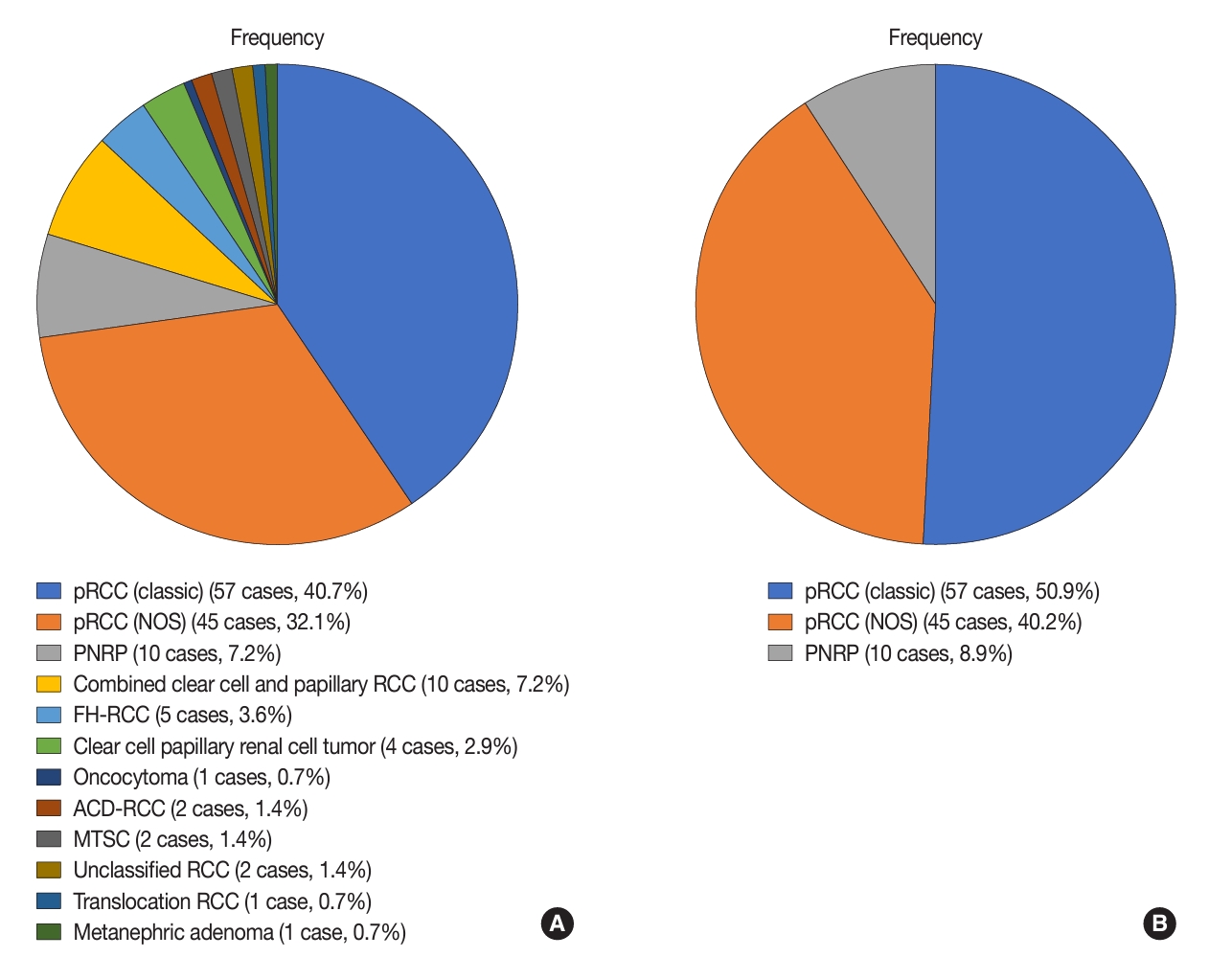
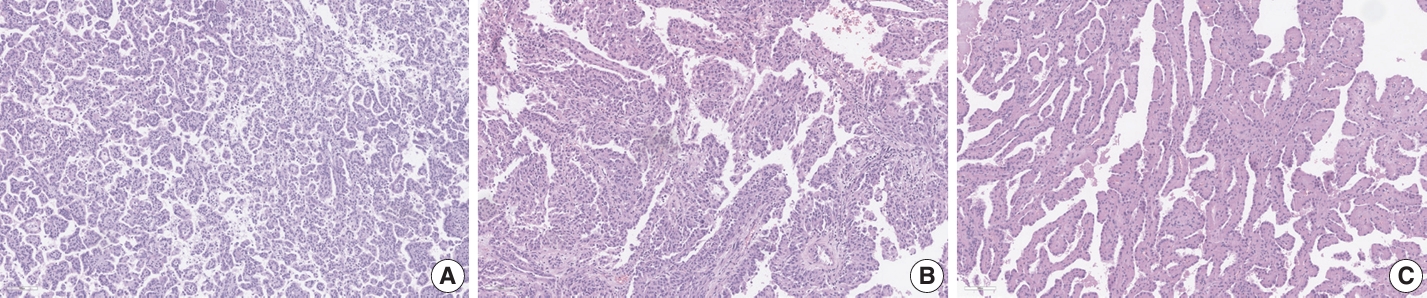
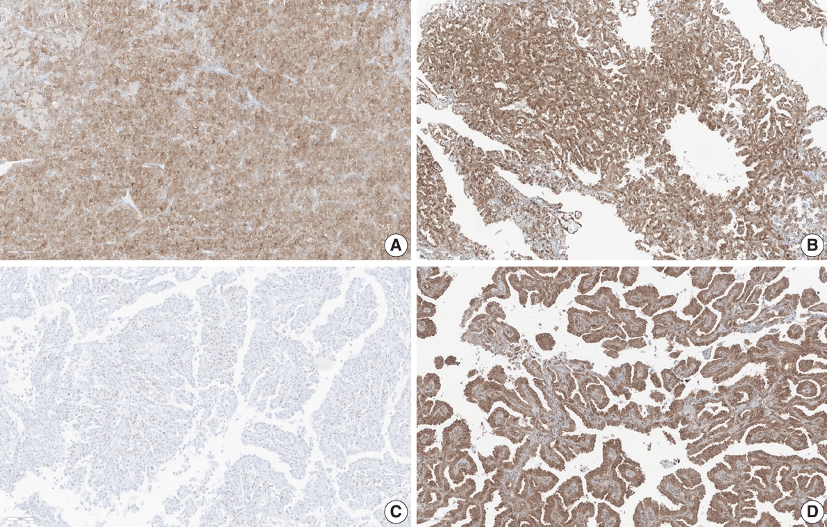


Fig. 1.
Fig. 2.
Fig. 3.
Graphical abstract
| pRCC (classic) (n = 57) | pRCC (NOS) (n = 45) | PNRP (n = 10) | p-value | |
|---|---|---|---|---|
| Age (yr) | 59.3 ± 12.2 | 61.3 ± 11.5 | 55.9 ± 9.6 | .374 |
| Sex | > .99 | |||
| Male | 43 (75.4) | 34 (75.6) | 8 (80.0) | |
| Female | 14 (24.6) | 11 (24.4) | 2 (20.0) | |
| WHO/ISUP grade | < .001 |
|||
| 1/2 | 50 (87.7) | 22 (48.9) | 10 (100) | |
| 3/4 | 7 (12.3) | 23 (51.1) | 0 | |
| Hemorrhage | .820 | |||
| Absent | 41 (71.9) | 31 (68.9) | 8 (80.0) | |
| Present | 16 (28.1) | 14 (31.1) | 2 (20.0) | |
| Necrosis | .285 | |||
| Absent | 44 (77.2) | 35 (77.8) | 10 (100) | |
| Present | 13 (22.8) | 10 (22.2) | 0 | |
| Sarcomatoid change | .338 | |||
| Absent | 56 (98.2) | 42 (93.3) | 10 (100) | |
| Present | 1 (1.8) | 3 (6.7) | 0 |
| pRCC (classic) (n = 57) | pRCC (NOS) (n = 45) | PNRP (n = 10) | p-value | |
|---|---|---|---|---|
| c-MET | 0.021 | |||
| Negative | 18 (32.1) | 24 (54.5) | 8 (80.0) | |
| Faint to weak membranous staining | 16 (28.6) | 13 (29.5) | 2 (20.0) | |
| Focal or diffuse moderate membranous staining | 19 (33.9) | 5 (11.4) | 0 | |
| Diffuse strong membranous staining | 3 (5.4) | 2 (4.6) | 0 | |
| p16 | 0.328 | |||
| Negative/patchy positivity | 57 (100) | 42 (95.5) | 10 (100) | |
| Block positivity | 0 | 2 (4.5) | 0 | |
| c-Myc | 0.199 | |||
| Negative | 52 (92.9) | 44 (100) | 10 (100) | |
| Postive | 4 (7.1) | 0 | 0 | |
| Ki-67 proliferation index (continuous) (mean ± SD) | 0.3 ± 0.5 | 0.8 ± 2.0 | 0.2 ± 0.3 | 0.081 |
| Ki-67 proliferation index (categorized) (%) | 0.080 | |||
| < 1 | 52 (91.2) | 35 (79.5) | 10 (100) | |
| 1–3 | 5 (8.8) | 4 (9.1) | 0 | |
| > 3 | 0 | 5 (11.4) | 0 | |
| p53 | NA | |||
| Normal pattern | 56 (100) | 45 (100) | 10 (100) | |
| Abnormal pattern | 0 | 0 | 0 | |
| STING | 0.020 |
|||
| Negative | 43 (75.4) | 33 (73.3) | 3 (30.0) | |
| Positive | 14 (24.6) | 12 (26.7) | 7 (70.0) |
| STING | Ki-67 proliferation index |
p-value | ||
|---|---|---|---|---|
| <1% (n = 97) | 1%–3% (n = 9) | > 3% (n = 5) | ||
| Negative (n = 78) | 70 (72.2) | 7 (77.8) | 1 (20.0) | .063 |
| Positive (n = 33) | 27 (27.8) | 2 (22.2) | 4 (80.0) | |
Values are presented as mean ± SD or number (%). pRCC, papillary renal cell carcinoma; NOS, not otherwise specified; PNRP, papillary neoplasm with reverse polarity; WHO/ISUP, World Health Organization/International Society of Urological Pathology; SD, standard deviation. Post-hoc Bonferroni analysis revealed statistical significance between two groups.
Values are presented as number (%) unless otherwise indicated. pRCC, papillary renal cell carcinoma; NOS, not otherwise specified; PNRP, papillary neoplasm with reverse polarity; SD, standard deviation; NA, not available; STING, stimulator of interferon genes. Post-hoc Bonferroni analysis revealed statistical significance between two groups.
Values are presented as number (%). STING, stimulator of interferon genes.

