 E-submission
Articles
- Page Path
- HOME > J Pathol Transl Med > Volume 47(3); 2013 > Article
-
Case Study
Multicystic Biliary Hamartoma of the Liver - Ji Soo Song, Sang Jae Noh1, Baik Hwan Cho2, Woo Sung Moon1
-
Korean Journal of Pathology 2013;47(3):275-278.
DOI: https://doi.org/10.4132/KoreanJPathol.2013.47.3.275
Published online: June 25, 2013
Department of Radiology, Research Institute of Clinical Medicine and Research Institute for Endocrine Sciences, Chonbuk National University Medical School, Jeonju, Korea.
1Department of Pathology, Research Institute of Clinical Medicine and Research Institute for Endocrine Sciences, Chonbuk National University Medical School, Jeonju, Korea.
2Department of Surgery, Research Institute of Clinical Medicine and Research Institute for Endocrine Sciences, Chonbuk National University Medical School, Jeonju, Korea.
- Corresponding Author: Woo Sung Moon, M.D. Department of Pathology, Chonbuk National University Medical School, 567 Baekje-daero, Deokjin-gu, Jeonju 561-756, Korea. Tel: +82-63-270-3086, Fax: +82-63-270-3135, mws@chonbuk.ac.kr
© 2013 The Korean Society of Pathologists/The Korean Society for Cytopathology
This is an Open Access article distributed under the terms of the Creative Commons Attribution Non-Commercial License (http://creativecommons.org/licenses/by-nc/3.0/) which permits unrestricted non-commercial use, distribution, and reproduction in any medium, provided the original work is properly cited.
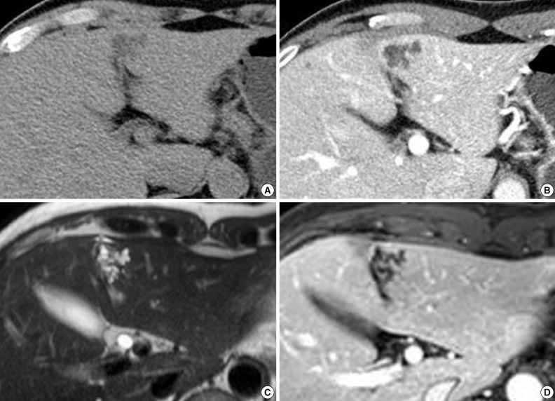
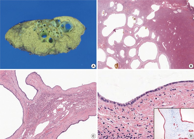
Figure & Data
References
Citations
- Multicystic biliary hamartoma with long-term gradual enlargement treated by laparoscopic partial hepatectomy
Satoshi Nishiwada, Tetsuya Tanaka, Yuki Kirihataya, Takeshi Takei, Tomomi Sadamitsu, Masato Takano, Masayoshi Sawai, Atsushi Yoshimura
Clinical Journal of Gastroenterology.2025; 18(3): 527. CrossRef - Technical Considerations in EEG Source Imaging
Benjamin H. Brinkmann
Journal of Clinical Neurophysiology.2024; 41(1): 2. CrossRef - A Case of Multicystic Biliary Hamartoma with a Marked Peribiliary Gland Component Successfully Treated by Purely Laparoscopic Anatomical Liver Resection
Keita Kai, Takao Ide, Tomokazu Tanaka, Kumpei Yukimoto, Hiroyuki Irie, Hirokazu Noshiro, Shinichi Aishima
Journal of Gastrointestinal Cancer.2023; 54(3): 996. CrossRef - Characteristics of multicystic biliary hamartoma: A case report
Jia Lian, Lixia Sun, Yankai Yang, Jun Li, Ye Zhang, Guiqiu Liu, Weijuan Hu
Frontiers in Surgery.2023;[Epub] CrossRef - Recurrent sepsis in a patient with biliary hamartomas
Maria Beatriz Santos, Magda Ponta Garça, Bárbara Vieira, Paulo Ávila, Alexandra Freitas
European Journal of Case Reports in Internal Medicine.2023;[Epub] CrossRef - Hamartoma multiquístico de vías biliares
Victoria Carmona, Iago Justo, Yolanda Rodríguez-Gil, Alberto Marcacuzco, Carmelo Loinaz, Carlos Jiménez
Cirugía Española.2022; 100(12): 800. CrossRef - Multicystic Biliary Hamartoma With Xanthogranulomatous Inflammation on 18F-FDG PET/CT
Nahomi Shono, Yoichi Otomi, Hideki Otsuka, Takayoshi Shinya, Masafumi Harada
Clinical Nuclear Medicine.2022; 47(10): 882. CrossRef - Intrahepatic multicystic biliary hamartoma: A case report
Chen-Yu Wang, Fu-Yang Shi, Wei-Feng Huang, Yan Tang, Ting Li, Guo-Lin He
World Journal of Clinical Cases.2022; 10(26): 9361. CrossRef - A Case of Multicystic Biliary Hamartoma Treated with Left Medial Sectionectomy
Naomi KUROKI, Tomoaki TANAKA, Takanobu SUGASE, Syoji TANIGUCHI, Takashi GOTO, Rintaro KOGA, Takumi KIWAKI, Hiroyuki TANAKA
Nihon Rinsho Geka Gakkai Zasshi (Journal of Japan Surgical Association).2022; 83(2): 395. CrossRef - Multicystic biliary hamartoma
Victoria Carmona, Iago Justo, Yolanda Rodríguez-Gil, Alberto Marcacuzco, Carmelo Loinaz, Carlos Jiménez
Cirugía Española (English Edition).2022; 100(12): 800. CrossRef - Case Report: Incidentally Discovered a Rare Cystic Lesion of Liver: Multicystic Biliary Hamartoma
Wentao Mu, Peng Su, Shanglei Ning
Pathology and Oncology Research.2021;[Epub] CrossRef - Bile Duct Hamartoma Mimicking Liver Metastasis in Suspected Porcelain Gallbladder: a Case Report
Gautham Krishnamurthy, Harjeet Singh, Sravya Deepika Ganti, Ganga Ram Verma
Journal of Gastrointestinal Cancer.2019; 50(4): 1022. CrossRef - A variant of multicystic biliary hamartoma presenting as an intrahepatic cystic neoplasm
Tetsuro Tominaga, Takafumi Abo, Naoe Kinoshita, Tomonori Murakami, Yasunori Sato, Yasuni Nakanuma, Kenich Harada, Junichi Masuda, Takeshi Nagayasu, Atsushi Nanashima
Clinical Journal of Gastroenterology.2015; 8(3): 162. CrossRef - Hamartoma biliar multiquístico intrahepático: presentación de un caso clínico
María Jezabel Fernández-Carrión, Ricardo Robles Campos, Asunción López Conesa, Roberto Brusadín, Pascual Parrilla Paricio
Cirugía Española.2015; 93(9): e103. CrossRef - Intrahepatic Multicystic Biliary Hamartoma: Presentation of a Case Report
María Jezabel Fernández-Carrión, Ricardo Robles Campos, Asunción López Conesa, Roberto Brusadín, Pascual Parrilla Paricio
Cirugía Española (English Edition).2015; 93(9): e103. CrossRef - Multicystic biliary hamartoma: A report of a rare entity and a review of the literature
Rachel E. Beard, Eric U. Yee, Koenraad J. Mortele, Khalid Khwaja
International Journal of Surgery Case Reports.2014; 5(12): 919. CrossRef - Multicystic biliary hamartoma mimicking intrahepatic cholangiocarcinoma: report of a case
Tomoaki Yoh, Ryuji Okamura, Hiroyuki Nakayama, Xue Lin, Yuya Nakamura, Tatsushi Kato
Clinical Journal of Gastroenterology.2014; 7(5): 418. CrossRef
 PubReader
 ePub Link
-
 Cite this Article
- Cite this Article
-
- Close
- Download Citation
- Close
- Figure
-


Fig. 1
Fig. 2
| Case No. | Author | Age/Sex | Location | Size (cm) | Clinical symptoms | Treatments |
|---|---|---|---|---|---|---|
| 1 | Zen et al. [1] | 59/M | Segment 4 | 4.2 | Abdominal pain | Left hepatectomy |
| 2 | Zen et al. [1] | 70/F | Segment 3 | 1.8 | Liver dysfunction | Segmentectomy |
| 3 | Zen et al. [1] | 69/F | Segment 3 | 2.8 | Elevation of liver enzyme | Segmentectomy |
| 4 | Kai et al. [3] | 55/M | Segment 6 | 5.0 | Incidental | Partial resection |
| 5 | Ryu et al. [2] | 45/M | Segment 7 | 2.0-3.5 (case nos. 5-7) | Incidental | Partial resection |
| 6 | Ryu et al. [2] | 58/M | Segment 3 | - | Incidental | Partial resection |
| 7 | Ryu et al. [2] | 55/F | Segment 6/7 | - | Incidental | Partial resection |
| 8 | Kobayashi et al. [4] | 30/M | Segment 6 | 3.6 | Incidental | Partial resection |
| Present case | 52/M | Segment 3 | 2.7 | Abdominal discomfort | Partial resection |
M, male; F, female.

