 E-submission
Search
- Page Path
- HOME > Search
- Recent topics on thyroid cytopathology: reporting systems and ancillary studies
- Mitsuyoshi Hirokawa, Ayana Suzuki
- J Pathol Transl Med. 2025;59(4):214-224. Published online June 30, 2025
- DOI: https://doi.org/10.4132/jptm.2025.04.18
- 6,459 View
- 351 Download
- 1 Web of Science
- 1 Crossref
-
 Abstract
 PDF
PDF - As fine-needle aspiration techniques and diagnostic methodologies for thyroid nodules have continued to evolve and reporting systems have been updated accordingly, we need to be up to date with the latest information to achieve accurate diagnoses. However, the diagnostic approaches and therapeutic strategies for thyroid nodules vary across laboratories and institutions. Several differences exist between Western and Eastern practices regarding thyroid fine-needle aspiration. This review describes the reporting systems for thyroid cytopathology and ancillary studies. Updated reporting systems enhance the accuracy, consistency, and clarity of cytology reporting, leading to improved patient outcomes and management strategies. Although a single global reporting system is optimal, reporting systems tailored to each country is acceptable. In such cases, compatibility must be ensured to facilitate data sharing. Ancillary methods include liquid-based cytology, immunocytochemistry, biochemical measurements, flow cytometry, molecular testing, and artificial intelligence, all of which improve diagnostic accuracy. These methods continue to evolve, and cytopathologists should actively adopt the latest methods and information to achieve more accurate diagnoses. We believe this review will be useful to practitioners of routine thyroid cytology.
-
Citations
Citations to this article as recorded by
- Importance of clinical, ultrasound and cytological characteristics in predicting malignancy in thyroid nodules with indeterminate cytology
Gabriel Gonçalves dos Santos, Wendy Muller Tirapani, Cínthia Minatel Riguetto, Icleia Siqueira Barreto, Denise Engelbrecht Zantut-Wittmann
Annals of Medicine.2026;[Epub] CrossRef
- Importance of clinical, ultrasound and cytological characteristics in predicting malignancy in thyroid nodules with indeterminate cytology
- Breast fine-needle aspiration cytology in the era of core-needle biopsy: what is its role?
- Ahrong Kim, Hyun Jung Lee, Jee Yeon Kim
- J Pathol Transl Med. 2025;59(1):26-38. Published online January 15, 2025
- DOI: https://doi.org/10.4132/jptm.2024.11.01
- Correction in: J Pathol Transl Med 2025;59(2):147
- 17,836 View
- 553 Download
- 6 Web of Science
- 8 Crossref
-
Abstract
PDF
- Fine-needle aspiration cytology (FNAC) has long been recognized as a minimally invasive, cost-effective, and reliable diagnostic tool for breast lesions. However, with the advent of core-needle biopsy (CNB), the role of FNAC has diminished in some clinical settings. This review aims to re-evaluate the diagnostic value of FNAC in the current era, focusing on its complementary use alongside CNB, the adoption of new approaches such as the International Academy of Cytology Yokohama System, and the implementation of rapid on-site evaluation to reduce inadequate sample rates. Advances in liquid-based cytology, receptor expression testing, molecular diagnostics, and artificial intelligence are discussed, highlighting their potential to enhance the diagnostic accuracy of FNAC. Despite challenges, FNAC remains a valuable diagnostic method, particularly in low-resource settings and specific clinical scenarios, and its role continues to evolve with technology.
-
Citations
Citations to this article as recorded by- Evaluation of Breast Lesions on Cytology Using International Academy of Cytology Yokohama Standardized Reporting System
Manish Jaiswal, Anurag Gupta, Tripti Verma, Pradyumn Singh, Rita Yadav, Akash Agarwal, Ashish Singhal, Nuzhat Husain, Shamrendra Narayan, Neha Singh
Diagnostic Cytopathology.2026; 54(3): 184. CrossRef - Personalizing therapies over the course of hormone receptor‐positive/HER2‐negative metastatic breast cancer
Akshara Singareeka Raghavendra, Senthil Damodaran, Carlos H. Barcenas, Suzanne A. Fuqua, Rachel M. Layman, Debu Tripathy
CA: A Cancer Journal for Clinicians.2026;[Epub] CrossRef - Transforming Breast Cancer Control in East Africa by Integrating Cytomorphology and Genetics Into National Policy
Josephine N Rioki, Mwangi Joseph, Rency Lel, Marshal Mweu, Lucy Muchiri
Cureus.2026;[Epub] CrossRef - Prélèvements mammaires percutanés
A. Ribrag, R. Foucher
EMC - Gynécologie.2026; 41(3): 1. CrossRef - Age and tumor size as independent predictors of malignancy in BI-RADS 4 and 5 breast lesions: A cross-sectional study in Vietnam
De Van Nguyen, Trung Van Pham, Tam Huu Dinh, Dung Ngoc Tran, Chung Thanh Dang, Dongling Wu
PLOS One.2026; 21(7): e0352690. CrossRef - Selective medical intelligence: Optimising AI-based breast cancer diagnosis classification through adaptive data filtering
Nicholas Christakis, Panagiotis Tirchas, Dimitris Drikakis
Neurocomputing.2026; 700: 134471. CrossRef - Diagnostic Performance of the International Academy of Cytology Yokohama System for Reporting Breast Fine-Needle Aspirates: A Cytology-Histopathology Correlation Study From Central India
Aditi Rathore, Archana Shrivastava, Maneesh Sulya
Cureus.2026;[Epub] CrossRef - Bulk-lysis protocols as a sensitive method for investigation of circulating CK19 cells in the peripheral blood of patients with breast cancer by flow cytometry
Daniella Serafin Couto Vieira, Laura Otto Walter, Maria Eduarda Cunha da Silva, Lisandra de Oliveira Silva, Heloísa Zorzi Costa, Chandra Chiappin Cardoso, Fernando Carlos de Lander Schmitt, Maria Cláudia Santos-Silva
Analytical Methods.2025; 17(23): 4771. CrossRef
- Evaluation of Breast Lesions on Cytology Using International Academy of Cytology Yokohama Standardized Reporting System
- Intravascular schwannoma as an extremely unusual cause of vein obstruction: a case report
- Luis Miguel Chinchilla-Tábora, Beatriz Segovia Blázquez, José María Sayagués, Marta Rodríguez González, Joaquín González-Rivero, José Antonio Muñoz León, Andrea Beatriz Jiménez Pérez, Idalia González Morais, Diego Bueno-Sacristán, María Dolores Ludeña
- J Pathol Transl Med. 2024;58(5):249-254. Published online July 3, 2024
- DOI: https://doi.org/10.4132/jptm.2024.05.15

- 5,574 View
- 294 Download
-
Abstract
PDF
- The blood vessel lumen is an extremely rare location for a benign peripheral nerve sheath tumor like schwannoma. Less than 10 cases have been previously reported. In this report, we present a case of a 68-year-old woman who had a soft tissue nodule at the posterior calf of her left leg during a physical examination. Pathological examination was performed after complete surgical excision. The patient underwent follow-up for 12 months after surgery without evidence of recurrence or any other complication. This is the first case of intravascular schwannoma reported as a cause of vein obstruction. Microscopically, the tumor was composed of Schwann spindle cells that were immunoreactive for S100 protein and SOX10. This tumor was surrounded by a well-defined vascular smooth muscle wall. Prospective series are required to improve the knowledge on the underlying mechanisms of intravascular schwannoma development.
- Postmortem lung and heart examination of COVID-19 patients in a case series from Jordan
- Maram Abdaljaleel, Isra Tawalbeh, Malik Sallam, Amjad Bani Hani, Imad M. Al-Abdallat, Baheth Al Omari, Sahar Al-Mustafa, Hasan Abder-Rahman, Adnan Said Abbas, Mahmoud Zureigat, Mousa A. Al-Abbadi
- J Pathol Transl Med. 2023;57(2):102-112. Published online March 14, 2023
- DOI: https://doi.org/10.4132/jptm.2023.01.30

- 8,652 View
- 176 Download
- 1 Crossref
-
Abstract
PDF
- Background
Coronavirus disease 2019 (COVID-19) has emerged as a pandemic for more than 2 years. Autopsy examination is an invaluable tool to understand the pathogenesis of emerging infections and their consequent mortalities. The aim of the current study was to present the lung and heart pathological findings of COVID-19–positive autopsies performed in Jordan.
Methods
The study involved medicolegal cases, where the cause of death was unclear and autopsy examination was mandated by law. We included the clinical and pathologic findings of routine gross and microscopic examination of cases that were positive for COVID-19 at time of death. Testing for severe acute respiratory syndrome coronavirus 2 (SARS-CoV-2) was confirmed through molecular detection by real-time polymerase chain reaction, serologic testing for IgM and electron microscope examination of lung samples.
Results
Seventeen autopsies were included, with male predominance (76.5%), Jordanians (70.6%), and 50 years as the mean age at time of death. Nine out of 16 cases (56.3%) had co-morbidities, with one case lacking such data. Histologic examination of lung tissue revealed diffuse alveolar damage in 13/17 cases (76.5%), and pulmonary microthrombi in 8/17 cases (47.1%). Microscopic cardiac findings were scarcely detected. Two patients died as a direct result of acute cardiac disease with limited pulmonary findings.
Conclusions
The detection of SARS-CoV-2 in postmortem examination can be an incidental or contributory finding which highlights the value of autopsy examination to determine the exact cause of death in controversial cases. -
Citations
Citations to this article as recorded by- Massive Splenic Infarction Following COVID-19 Infection, with Histopathological Observations, and Review of the Literature
Yousef Samir Amr, Aseel Qassem Al-Omari, Mousa Husein Saadeh, Samir Sami Amr
Open Journal of Pathology.2025; 15(02): 69. CrossRef
- Massive Splenic Infarction Following COVID-19 Infection, with Histopathological Observations, and Review of the Literature
- A machine-learning expert-supporting system for diagnosis prediction of lymphoid neoplasms using a probabilistic decision-tree algorithm and immunohistochemistry profile database
- Yosep Chong, Ji Young Lee, Yejin Kim, Jingyun Choi, Hwanjo Yu, Gyeongsin Park, Mee Yon Cho, Nishant Thakur
- J Pathol Transl Med. 2020;54(6):462-470. Published online August 31, 2020
- DOI: https://doi.org/10.4132/jptm.2020.07.11

- 8,491 View
- 132 Download
- 9 Web of Science
- 11 Crossref
-
Abstract
PDF
 Supplementary Material
 - Background
Immunohistochemistry (IHC) has played an essential role in the diagnosis of hematolymphoid neoplasms. However, IHC interpretations can be challenging in daily practice, and exponentially expanding volumes of IHC data are making the task increasingly difficult. We therefore developed a machine-learning expert-supporting system for diagnosing lymphoid neoplasms.
Methods
A probabilistic decision-tree algorithm based on the Bayesian theorem was used to develop mobile application software for iOS and Android platforms. We tested the software with real data from 602 training and 392 validation cases of lymphoid neoplasms and compared the precision hit rates between the training and validation datasets.
Results
IHC expression data for 150 lymphoid neoplasms and 584 antibodies was gathered. The precision hit rates of 94.7% in the training data and 95.7% in the validation data for lymphomas were not statistically significant. Results in most B-cell lymphomas were excellent, and generally equivalent performance was seen in T-cell lymphomas. The primary reasons for lack of precision were atypical IHC profiles for certain cases (e.g., CD15-negative Hodgkin lymphoma), a lack of disease-specific markers, and overlapping IHC profiles of similar diseases.
Conclusions
Application of the machine-learning algorithm to diagnosis precision produced acceptable hit rates in training and validation datasets. Because of the lack of origin- or disease- specific markers in differential diagnosis, contextual information such as clinical and histological features should be taken into account to make proper use of this system in the pathologic decision-making process. -
Citations
Citations to this article as recorded by- Discovery of Marker Genes in Adult T Cell Leukemia (ATL) Pathogenesis with Machine Learning Models and Performance Comparison
Sabire Kiliçarslan, Sait Can Yücebaş
Karadeniz Fen Bilimleri Dergisi.2025; 15(3): 1046. CrossRef - Revolutionizing Pathology with Artificial Intelligence: Innovations in Immunohistochemistry
Diana Gina Poalelungi, Anca Iulia Neagu, Ana Fulga, Marius Neagu, Dana Tutunaru, Aurel Nechita, Iuliu Fulga
Journal of Personalized Medicine.2024; 14(7): 693. CrossRef - Enhanced Immunohistochemistry Interpretation with a Machine Learning-Based Expert System
Anca Iulia Neagu, Diana Gina Poalelungi, Ana Fulga, Marius Neagu, Iuliu Fulga, Aurel Nechita
Diagnostics.2024; 14(17): 1853. CrossRef - Optimization of diagnosis and treatment of hematological diseases via artificial intelligence
Shi-Xuan Wang, Zou-Fang Huang, Jing Li, Yin Wu, Jun Du, Ting Li
Frontiers in Medicine.2024;[Epub] CrossRef - Artificial intelligence in lymphoma histopathology: a systematic review (Preprint)
Yao Fu, Zongyao Huang, Xudong Deng, Linna Xu, Yang Liu, Mingxing Zhang, Jinyi Liu, Bin Huang
Journal of Medical Internet Research.2024;[Epub] CrossRef - Real-Life Barriers to Diagnosis of Early Mycosis Fungoides: An International Expert Panel Discussion
Emmilia Hodak, Larisa Geskin, Emmanuella Guenova, Pablo L. Ortiz-Romero, Rein Willemze, Jie Zheng, Richard Cowan, Francine Foss, Cristina Mangas, Christiane Querfeld
American Journal of Clinical Dermatology.2023; 24(1): 5. CrossRef - Validation of a Machine Learning Expert Supporting System, ImmunoGenius, Using Immunohistochemistry Results of 3000 Patients with Lymphoid Neoplasms
Jamshid Abdul-Ghafar, Kyung Jin Seo, Hye-Ra Jung, Gyeongsin Park, Seung-Sook Lee, Yosep Chong
Diagnostics.2023; 13(7): 1308. CrossRef - Clinical approaches for integrating machine learning for patients with lymphoma: Current strategies and future perspectives
Dai Chihara, Loretta J. Nastoupil, Christopher R. Flowers
British Journal of Haematology.2023; 202(2): 219. CrossRef - Current Trend of Artificial Intelligence Patents in Digital Pathology: A Systematic Evaluation of the Patent Landscape
Muhammad Joan Ailia, Nishant Thakur, Jamshid Abdul-Ghafar, Chan Kwon Jung, Kwangil Yim, Yosep Chong
Cancers.2022; 14(10): 2400. CrossRef - Recent Application of Artificial Intelligence in Non-Gynecological Cancer Cytopathology: A Systematic Review
Nishant Thakur, Mohammad Rizwan Alam, Jamshid Abdul-Ghafar, Yosep Chong
Cancers.2022; 14(14): 3529. CrossRef - Diagnosis prediction of tumours of unknown origin using ImmunoGenius, a machine learning-based expert system for immunohistochemistry profile interpretation
Yosep Chong, Nishant Thakur, Ji Young Lee, Gyoyeon Hwang, Myungjin Choi, Yejin Kim, Hwanjo Yu, Mee Yon Cho
Diagnostic Pathology.2021;[Epub] CrossRef
- Discovery of Marker Genes in Adult T Cell Leukemia (ATL) Pathogenesis with Machine Learning Models and Performance Comparison
- A retrospective cytohistological correlation of fine-needle aspiration cytology with classification by the Milan System for Reporting Salivary Gland Cytopathology
- Ji Hyun Park, Yoon Jin Cha, Ja Yeong Seo, Jae Yol Lim, Soon Won Hong
- J Pathol Transl Med. 2020;54(5):419-425. Published online July 8, 2020
- DOI: https://doi.org/10.4132/jptm.2020.06.09

- 8,233 View
- 208 Download
- 12 Web of Science
- 15 Crossref
-
Abstract
PDF
- Background
Before publication of the new classification system named the Milan System for Reporting Salivary Gland Cytopathology (MSRSGC) in 2018, there was no standard classification for salivary gland lesions obtained by fine-needle aspiration (FNA). We therefore aimed to evaluate the diagnostic utility of this system by retrospectively reviewing FNA samples using the MSRSGC and to determine their risk of developing into neoplasms and becoming malignant.
Methods
Retrospective slide review and classification of salivary gland FNAs obtained over a 6-year period (2013–2018) at a single center were performed by two pathologists. The risks of neoplasm and malignancy for each category also were calculated.
Results
This study surveyed 374 FNAs (371 patients) performed over a six-year period and selected 148 cases that included documented surgical follow-up (39.6%). Among the surgically treated cases, the distributions of FNA categories were as follows: non-diagnostic (ND; 16.9%), non-neoplastic (NN; 2.7%), atypia of undetermined significance (AUS; 3.4%), benign (BN; 54.7%), salivary gland neoplasm of uncertain malignant potential (SUMP; 10.1%), suspicious for malignancy (SM; 6.8%), and malignant (M; 5.4%). The risk of malignancy (ROM) was 24.0% for ND, 0% for NN, 40.0% for AUS, 2.5% for BN, 46.7% for SUMP, 100% for SM, and 87.5% for M. The overall diagnostic accuracy was 95.9% (142/148 cases).
Conclusions
The newly proposed MSRSGC appears to be a reliable system for classification of salivary gland lesions according to the associated ROM. -
Citations
Citations to this article as recorded by- Optimizing preoperative assessment of salivary gland lesions: updates and practical implications in the Milan System 2nd edition
Patrizia Straccia, Vincenzo Fiorentino, Alessia Piermattei, Qianqian Zhang, Belen Padial-Urtueta, Federica Cianfrini, Antonino Mule’, Esther Diana Rossi
Virchows Archiv.2026; 488(6): 1203. CrossRef - The Impact of Lesion-Specific and Sampling-Related Factors on Success of Salivary Gland Fine-Needle Aspiration Cytology
Marcel Mayer, Mohammad Marwan Alfarra, Kathrin Möllenhoff, Marianne Engels, Christoph Arolt, Alexander Quaas, Philipp Wolber, Louis Jansen, Lisa Nachtsheim, Maria Grosheva, Jens Peter Klussmann, Sami Shabli
Head and Neck Pathology.2025;[Epub] CrossRef - The Myriad Spectrum of Salivary Gland Lesions: Cytohistological Correlation on Fine Needle Aspiration Cytology, Core Needle Biopsy, and Resections in a 5‐Year Single Institutional Experience of North India
Zachariah Chowdhury, Pallavi Majumdar, Sumeet Narain, Komal Lamba
Diagnostic Cytopathology.2025; 53(8): 391. CrossRef - Diagnostic Performance of the Milan System for Reporting Salivary Gland Cytopathology and a Proposed Algorithm for Fine-Needle Aspiration Cytology of Salivary Gland Lesions
Norihide Mochizuki, Hirotaka Fujita, Takuma Tajiri, Masataka Ueda, Makiko Kurata, Chie Inomoto, Tomoko Sugiyama, Daisuke Maki, Shuichi Shiraishi, Tomohisa Machida, Hitoshi Ito, Yohei Masugi, Naoya Nakamura
Acta Cytologica.2025; 69(4): 324. CrossRef - A study of fine needle aspiration cytology and histopathology correlation of salivary gland neoplasms in a tertiary care hospital: an observational study
Asima Malik, Ahlam Mushtaq, Salma Bhat, Suhail Naik
International Journal of Contemporary Pediatrics.2025; 13(1): 23. CrossRef - The Milan system for reporting salivary gland cytopathology – Assessment of utility and the risk of malignancy
Annu E. Prakash, Renu Sukumaran, Nileena Nayak, K. Lakshmi, Anitha Mathews, Jayasree Kattoor
Indian Journal of Cancer.2024; 61(3): 575. CrossRef - Salivary gland fine-needle aspiration biopsy: quality assurance results from a tertiary cancer center
Fanni Ratzon, Dominique L. Feliciano, Nora Katabi, Bin Xu, Oscar Lin, Xiao-Jun Wei
Journal of the American Society of Cytopathology.2023; 12(3): 206. CrossRef - Cytohistological correlation and risk stratification of salivary gland lesions using the Milan System for Reporting Salivary Gland Cytopathology: A tertiary care centre experience
Tarun Kumar, Prerna Tewari, Jitendra Singh Nigam, Shreekant Bharti, Surabhi, Ruchi Sinha, Punam Prasad Bhadani
Cytopathology.2023; 34(3): 225. CrossRef - Assessment of Risk of Malignancy of Fine-needle Aspiration Cytology in Salivary Gland Lesions Using the Milan System for Reporting Salivary Gland Cytopathology Categorization: A Systematic Review and Meta-analysis
Amit Kumar, Subhash Chandra, Bishnupati Singh, Swati Sharma, Ankita Tandon, Ajoy Kumar Shahi
The Journal of Contemporary Dental Practice.2023; 23(10): 1039. CrossRef - Milan Sınıflandırma Sistemi’ne Göre Değerlendirilen Tükürük Bezi İnce İğne Aspirasyon Sitolojilerinin Histopatolojik Tanı Uyumu
Özlem SARAYDAROĞLU, Selin YİRMİBEŞ
Uludağ Üniversitesi Tıp Fakültesi Dergisi.2023; 49(3): 285. CrossRef - Milan system for reporting salivary gland cytopathology: Adoption and outcomes in a community setting
Samih J. Nassif, Ali R. Sasani, Garrey T. Faller, Jennifer L. Harb, Jagdish K. Dhingra
Head & Neck.2022; 44(6): 1462. CrossRef - Nondiagnostic salivary gland FNAs are associated with decreased risk of malignancy compared with “all‐comer” patients: Analysis of the Milan System for Reporting Salivary Gland Cytopathology with a focus on Milan I: Nondiagnostic
Shu K. Lui, Troy Tenney, Patrick C. Mullane, Kartik Viswanathan, Daniel J. Lubin
Cancer Cytopathology.2022; 130(10): 800. CrossRef - Application of the Milan System for Reporting Salivary Gland Cytopathology: A systematic review and meta‐analysis
Zhaoyang Wang, Huan Zhao, Huiqin Guo, Changming An
Cancer Cytopathology.2022; 130(11): 849. CrossRef - Multiplexed single‐cell analysis of FNA allows accurate diagnosis of salivary gland tumors
Juhyun Oh, Tae Yeon Yoo, Talia M. Saal, Lisa Tsay, William C. Faquin, Jonathan C.T. Carlson, Daniel G. Deschler, Sara I. Pai, Ralph Weissleder
Cancer Cytopathology.2022; 130(8): 581. CrossRef - Cytologic analysis of vitreous fluids: A retrospective review of our 24 years of experience
Gabriel L. Collins, Elizabeth W. Hubbard, Christopher T. Clark, Lisa D. Duncan, Laurentia Nodit
Diagnostic Cytopathology.2021; 49(10): 1122. CrossRef
- Optimizing preoperative assessment of salivary gland lesions: updates and practical implications in the Milan System 2nd edition
- Adjunctive markers for classification and diagnosis of central nervous system tumors: results of a multi-center neuropathological survey in Korea
- Yoon Jin Cha, Se Hoon Kim, Na Rae Kim
- J Pathol Transl Med. 2020;54(2):165-170. Published online February 20, 2020
- DOI: https://doi.org/10.4132/jptm.2020.02.04
- 9,311 View
- 226 Download
- 1 Web of Science
- 1 Crossref
-
Abstract
PDFSupplementary Material
- Background
The revised 4th 2016 World Health Organization (WHO) classification of tumors of the central nervous system (CNS) classification has adopted integrated diagnosis encompassing the histology and molecular features of CNS tumors. We aimed to investigate the immunohistochemistry, molecular testing, and testing methods for diagnosis of CNS tumors in pathological labs of tertiary centers in Korea, and evaluate the adequacy of tests for proper diagnosis in daily practice.
Methods
A survey, composed of eight questions concerning molecular testing for diagnosis of CNS tumors, was sent to 10 neuropathologists working in tertiary centers in Korea.
Results
For diagnosis of astrocytic and oligodendroglial tumors, all 10 centers performed isocitrate dehydrogenase mutations testing and 1p/19q loss of heterozygosity. For glioneuronal tumors, immunohistochemistry (IHC) assays for synaptophysin (n = 9), CD34 (n = 7), BRAF(VE1) (n = 5) were used. For embryonal tumors, particularly in medulloblastoma, four respondents used IHC panel (growth factor receptor bound protein 2-associated protein 1, filamin A, and yes-associated protein 1) for molecular subclassification. Regarding meningioma, all respondents performed Ki-67 IHC and five performed telomerase reverse transcriptase promoter mutation.
Conclusions
Most tertiary centers made proper diagnosis in line with 2016 WHO classification. As classification of CNS tumors has evolved to be more complex and more ancillary tests are required, these should be performed considering the effect of necessity and justification. -
Citations
Citations to this article as recorded by- Exploring the role of epidermal growth factor receptor variant III in meningeal tumors
Rashmi Rana, Vaishnavi Rathi, Kirti Chauhan, Kriti Jain, Satnam Singh Chhabra, Rajesh Acharya, Samir Kumar Kalra, Anshul Gupta, Sunila Jain, Nirmal Kumar Ganguly, Dharmendra Kumar Yadav, Timir Tripathi
PLOS ONE.2021; 16(9): e0255133. CrossRef
- Exploring the role of epidermal growth factor receptor variant III in meningeal tumors
- Frozen Cytology of Meningeal Malignant Solitary Fibrous Tumor/Hemangiopericytoma
- Myunghee Kang, Na Rae Kim, Dong Hae Chung, Gie-Taek Yie
- J Pathol Transl Med. 2019;53(3):192-197. Published online April 11, 2019
- DOI: https://doi.org/10.4132/jptm.2019.03.20
- 9,521 View
- 160 Download
- 7 Web of Science
- 8 Crossref
-
Abstract
PDF
- A 51-year-old woman presented with severe dizziness. The brain magnetic resonance image revealed a 5.5 cm multiloculated mass with a thick rim in the left temporal lobe. Cytological examination of frozen diagnosis of the mass showed hypercellular sheets of round and rhabdoid cells in a hemorrhagic background, and two mitotic figures were observed. Histologically, the excised dura-based mass consisted of predominantly round cells with small foci of rhabdoid tumor cells in a pseudoalveolar pattern in a hemorrhagic background, and the cells showed nuclear positivity for signal transducer and activator of transcription 6 as well as frequent mitosis. The mass was diagnosed as a grade 3 solitary fibrous tumor (SFT)/hemangiopericytoma (HPC). The cytological diagnosis of SFT/HPC is challenging because of the heterogeneous cytological findings, such as histological heterogeneity, and because there are no standardized cytological criteria for malignant SFT/HPC. Cytological findings, such as singly scattered small cells, hypercellularity, rare ropy collagen, and round and rhabdoid cells with pseudoalveolar pattern, may assist in the diagnosis of malignant SFT/HPC.
-
Citations
Citations to this article as recorded by- A Rare Case of Cervical Solitary Fibrous Tumor in a Pediatric Patient: Case Report and Literature Review
Eleonora Becattini, Lorenzo Sgarbanti, Giuseppina Bevacqua, Valentina Grespi, Carlo Conti
NeuroSci.2025; 6(2): 49. CrossRef - Meningeal Solitary Fibrous Tumor: A Cytological Report With Emphasis on the Usefulness of Immunocytochemical Analysis for STAT6
Hiroyuki Okanishi, Mitsuaki Ishida, Naoto Kohno, Isako Kataoka, Mari Tomiuka, Mayumi Uragami, Shizuka Ono, Chihiro Deguchi, Reika Takeda, Yoshitaka Kurisu, Yoshinobu Hirose
Diagnostic Cytopathology.2025;[Epub] CrossRef - Cytologic features of mesenchymal, melanocytic and haematolymphoid tumours of the central nervous system and metastases
Carmen Bárcena, José A. Jiménez‐Heffernan
Cytopathology.2024; 35(5): 590. CrossRef - A Hemangiopericytoma in the External Auditory Canal: A Rare Clinical Presentation and Management
Vaibhavi Patil, Prasad Deshmukh, Sagar S Gaurkar , Ayushi Ghosh Moulic, Jasleen Kaur
Cureus.2024;[Epub] CrossRef - Scoring system for intraoperative diagnosis of intracranial schwannoma by squash cytology
Hirotaka Fujita, Takuma Tajiri, Tomohisa Machida, Nozomi Nomura, Suguru Toguchi, Hitoshi Itoh, Shinichiro Hiraiwa, Tomoko Sugiyama, Chie Inomoto, Masaaki Imai, Shinri Oda, Masami Shimoda, Naoya Nakamura
Cytopathology.2022; 33(2): 196. CrossRef - Occurrence of a solitary fibrous tumor adjacent to the resection bed of a high-grade meningioma: A case report
Coby Cunningham, Rocco Dabecco, Justin Davanzo
Interdisciplinary Neurosurgery.2021; 25: 101277. CrossRef - A case of solitary fibrous tumor arising in the meninge
Saori NAKANISHI, Naoto KURODA, Toshiko TAKAI, Mari KOJIMA, Misato OONOGI
The Journal of the Japanese Society of Clinical Cytology.2021; 60(4): 224. CrossRef - Intraoperative frozen cytology of intraosseous cystic meningioma in the sphenoid bone
Na Rae Kim, Gie-Taek Yie
Journal of Pathology and Translational Medicine.2020; 54(6): 508. CrossRef
- A Rare Case of Cervical Solitary Fibrous Tumor in a Pediatric Patient: Case Report and Literature Review
- Intraoperative Frozen Cytology of Central Nervous System Neoplasms: An Ancillary Tool for Frozen Diagnosis
- Myunghee Kang, Dong Hae Chung, Na Rae Kim, Hyun Yee Cho, Seung Yeon Ha, Sangho Lee, Jungsuk An, Jae Yeon Seok, Gie-Taek Yie, Chan Jong Yoo, Sang Gu Lee, Eun Young Kim, Woo Kyung Kim, Seong Son, Sun Jin Sym, Dong Bok Shin, Hee Young Hwang, Eung Yeop Kim, Kyu Chan Lee
- J Pathol Transl Med. 2019;53(2):104-111. Published online January 14, 2019
- DOI: https://doi.org/10.4132/jptm.2018.11.10
- 17,057 View
- 697 Download
- 11 Web of Science
- 9 Crossref
-
Abstract
PDF
- Background
Pathologic diagnosis of central nervous system (CNS) neoplasms is made by comparing light microscopic, immunohistochemical, and molecular cytogenetic findings with clinicoradiologic observations. Intraoperative frozen cytology smears can improve the diagnostic accuracy for CNS neoplasms. Here, we evaluate the diagnostic value of cytology in frozen diagnoses of CNS neoplasms.
Methods
Cases were selected from patients undergoing both frozen cytology and frozen sections. Diagnostic accuracy was evaluated.
Results
Four hundred and fifty-four cases were included in this retrospective single-center review study covering a span of 10 years. Five discrepant cases (1.1%) were found after excluding 53 deferred cases (31 cases of tentative diagnosis, 22 cases of inadequate frozen sampling). A total of 346 cases of complete concordance and 50 cases of partial concordance were classified as not discordant cases in the present study. Diagnostic accuracy of intraoperative frozen diagnosis was 87.2%, and the accuracy was 98.8% after excluding deferred cases. Discrepancies between frozen and permanent diagnoses (n = 5, 1.1%) were found in cases of nonrepresentative sampling (n = 2) and misinterpretation (n = 3). High concordance was observed more frequently in meningeal tumors (97/98, 99%), metastatic brain tumors (51/52, 98.1%), pituitary adenomas (86/89, 96.6%), schwannomas (45/47, 95.8%), high-grade astrocytic tumors (47/58, 81%), low grade astrocytic tumors (10/13, 76.9%), non-neoplastic lesions (23/36, 63.9%), in decreasing frequency.
Conclusions
Using intraoperative cytology and frozen sections of CNS tumors is a highly accurate diagnostic ancillary method, providing subtyping of CNS neoplasms, especially in frequently encountered entities. -
Citations
Citations to this article as recorded by- Qualitative and quantitative assessment of ex vivo human brain tumors using quantitative oblique back-illumination microscopy (qOBM)
Srinidhi Bharadwaj, Paloma Casteleiro Costa, Caroline Serafini, Brienna Heinsz, Alice Hsu, Nischita Kaza, Zhe Guang, Zhenmin Li, Jeffrey J. Olson, Kimberly Hoang, Stewart Neill, Francisco E. Robles
Biomedical Optics Express.2026; 17(4): 1936. CrossRef - Intraoperative Integrated Diagnostic System for Malignant Central Nervous System Tumors
Takahiro Hayashi, Kensuke Tateishi, Shinichiro Matsuyama, Hiromichi Iwashita, Yohei Miyake, Akito Oshima, Hirokuni Honma, Jo Sasame, Katsuhiro Takabayashi, Kyoka Sugino, Emi Hirata, Naoko Udaka, Yuko Matsushita, Ikuma Kato, Hiroaki Hayashi, Taishi Nakamur
Clinical Cancer Research.2024; 30(1): 116. CrossRef - A multicenter proof-of-concept study on deep learning-based intraoperative discrimination of primary central nervous system lymphoma
Xinke Zhang, Zihan Zhao, Ruixuan Wang, Haohua Chen, Xueyi Zheng, Lili Liu, Lilong Lan, Peng Li, Shuyang Wu, Qinghua Cao, Rongzhen Luo, Wanming Hu, Shanshan lyu, Zhengyu Zhang, Dan Xie, Yaping Ye, Yu Wang, Muyan Cai
Nature Communications.2024;[Epub] CrossRef - Advancements in Neurosurgical Intraoperative Histology
Ali A. Mohamed, Emma Sargent, Cooper Williams, Zev Karve, Karthik Nair, Brandon Lucke-Wold
Tomography.2024; 10(5): 693. CrossRef - Unveiling the potential application of intraoperative brain smear for brain tumor diagnosis in low-middle-income countries: A comprehensive systematic review
Muhammad Shakir, Ahmed Altaf, Hawra Hussain, Syed Muhammad Aqeel Abidi, Zoey Petitt, Mahnoor Tariq, Ahmed Gilani, S. Ather Enam
Surgical Neurology International.2023; 14: 325. CrossRef - A Comparative Study of Squash Smear Cytology Diagnosis and Radiological Diagnosis with Histopathology in Central Nervous System Lesions
B N Kumarguru, G Santhipriya, S Kranthi Kumar, R Ramesh Kumar, A S Ramaswamy, P Janakiraman
Journal of Cytology.2022; 39(1): 1. CrossRef - Intraoperative squash cytology provides a qualitative intraoperative diagnosis for cases in which frozen section yields a diagnosis of equivocal brain tumour
Hirotaka Fujita, Takuma Tajiri, Tomohisa Machida, Nozomi Nomura, Suguru Toguchi, Hitoshi Itoh, Shinichiro Hiraiwa, Tomoko Sugiyama, Masaaki Imai, Shinri Oda, Masami Shimoda, Naoya Nakamura
Cytopathology.2020; 31(2): 106. CrossRef - Intraoperative frozen cytology of intraosseous cystic meningioma in the sphenoid bone
Na Rae Kim, Gie-Taek Yie
Journal of Pathology and Translational Medicine.2020; 54(6): 508. CrossRef - Use of 5-Aminolevulinic Acid for Confirmation of Lesional Biopsy Sample in Presumed High-Grade Glioma
Victoria L. Watson, Jeffrey W. Cozzens
World Neurosurgery.2019; 132: 21. CrossRef
- Qualitative and quantitative assessment of ex vivo human brain tumors using quantitative oblique back-illumination microscopy (qOBM)
- Primary Peripheral Gamma Delta T-Cell Lymphoma of the Central Nervous System: Report of a Case Involving the Intramedullary Spinal Cord and Presenting with Myelopathy
- Jeemin Yim, Seung Geun Song, Sehui Kim, Jae Won Choi, Kyu-Chong Lee, Jeong Mo Bae, Yoon Kyung Jeon
- J Pathol Transl Med. 2019;53(1):57-61. Published online October 1, 2018
- DOI: https://doi.org/10.4132/jptm.2018.08.21
- 8,748 View
- 160 Download
- 5 Web of Science
- 4 Crossref
-
Abstract
PDF
- Primary central nervous system lymphoma of T-cell origin (T-PCNSL) is rare, and its clinicopathological features remain unclear. Peripheral T-cell lymphoma of γδ T-cell origin is an aggressive lymphoma mainly involving extranodal sites. Here, we report a case of γδ T-PCNSL involving the intramedullary spinal cord and presenting with paraplegia. A 75-year-old Korean woman visited the hospital complaining of back pain and lower extremity weakness. Magnetic resonance imaging revealed multifocal enhancing intramedullary nodular lesions in the thoracic and lumbar spinal cord. An enhancing nodular lesion was observed in the periventricular white matter of the lateral ventricle in the brain. There were no other abnormalities in systemic organs or skin. Laminectomy and tumor removal were performed. The tumor consisted of monomorphic, medium-to-large atypical lymphocytes with pale-to-eosinophilic cytoplasm. Immunohistochemically, the tumor cells were CD3(+), TCRβF1(-), TCRγ(+), CD30(-), CD4(-), CD8(-), CD56(+), TIA1(+), granzyme B(+), and CD103(+). Epstein-Barr virus in situ was negative. This case represents a unique T-PCNSL of γδ T-cell origin involving the spinal cord.
-
Citations
Citations to this article as recorded by- B-Cell Lymphoma Intramedullary Tumor: Case Report and Systematic Review
Daniel Gregório Gonsalves, Paulo Eduardo Albuquerque Zito Raffa, Gabriela Gerenutti de Sousa, Melissa Esposito Gomes Rigueiral, Iracema Araújo Estevão, Cesar Cozar Pacheco, Roger Thomaz Rotta Medeiros, Paulo Roberto Franceschini, Paulo Henrique Pires de A
Asian Journal of Neurosurgery.2023; 18(02): 231. CrossRef - Primary intramedullary spinal cord lymphoma misdiagnosed as longitudinally extensive transverse myelitis: a case report and literature review
Huizhen Ge, Li Xu, Huajie Gao, Suqiong Ji
BMC Neurology.2023;[Epub] CrossRef - Clinicopathologic and Genetic Features of Primary T-cell Lymphomas of the Central Nervous System
Jeemin Yim, Jiwon Koh, Sehui Kim, Seung Geun Song, Jeong Mo Bae, Hongseok Yun, Ji-Youn Sung, Tae Min Kim, Sung-Hye Park, Yoon Kyung Jeon
American Journal of Surgical Pathology.2022; 46(4): 486. CrossRef - Peripheral T-Cell Lymphomas Involving the Central Nervous System: A Report From the Czech Lymphoma Study Group Registry
Heidi Mocikova, Robert Pytlík, Katerina Benesova, Andrea Janikova, Juraj Duras, Alice Sykorova, Katerina Steinerova, Vit Prochazka, Vit Campr, David Belada, Marek Trneny
Frontiers in Oncology.2022;[Epub] CrossRef
- B-Cell Lymphoma Intramedullary Tumor: Case Report and Systematic Review
- Cytologic Diagnosis of Noninvasive Follicular Thyroid Neoplasm with Papillary-like Nuclear Features and Its Impact on the Risk of Malignancy in the Bethesda System for Reporting Thyroid Cytopathology: An Institutional Experience
- Milim Kim, Joung Eun Kim, Hyun Jeong Kim, Yul Ri Chung, Yoonjin Kwak, So Yeon Park
- J Pathol Transl Med. 2018;52(3):171-178. Published online April 3, 2018
- DOI: https://doi.org/10.4132/jptm.2018.04.03
- 12,911 View
- 210 Download
- 24 Web of Science
- 19 Crossref
-
Abstract
PDF
- Background
This study was performed to analyze cytologic diagnosis of noninvasive follicular thyroid neoplasm with papillary-like nuclear features (NIFTP) and its impact on the risk of malignancy (ROM) in the Bethesda System for Reporting Thyroid Cytopathology (TBSRTC).
Methods
Five thousand five hundred and forty-nine cases of thyroid fine-needle aspiration cytology (FNAC) diagnosed between 2012 and 2014 were included in this study. Diagnostic categories based on TBSRTC were compared with final surgical diagnoses, and the ROM in each category was calculated both when NIFTP was included in malignant lesions and when excluded from malignant lesions.
Results
Of the 5,549 thyroid FNAC cases, 1,891 cases underwent surgical resection. In final diagnosis, 1,700 cases were revealed as papillary thyroid carcinoma (PTC), and 25 cases were reclassified as NIFTP. The cytologic diagnoses of NIFTP were non-diagnostic in one, benign in five, atypia of undetermined significance (AUS) in 14, follicular neoplasm in two, and suspicious for malignancy in three cases. Collectively, NIFTP/encapsulated follicular variant of PTC (EFVPTC) were more frequently classified as benign, AUS, or follicular neoplasm and less frequently categorized as malignant compared to conventional PTCs. Exclusion of NIFTP from malignant diagnoses resulted in a slight decrease in malignancy rates in non-diagnostic, benign, AUS, follicular neoplasm, and suspicious for malignancy categories without any statistical significance.
Conclusions
The decrease in the ROM was not significant when NIFTP was excluded from malignant lesions. In thyroid FNACs, NIFTP/EFVPTCs were mostly classified into indeterminate categories. Therefore, it might be feasible to separate NIFTP/EFVPTC from conventional PTC on FNAC to guide clinicians to conservative management for patients with NIFTP/EFVPTC. -
Citations
Citations to this article as recorded by- High Rates of Unnecessary Surgery for Indeterminate Thyroid Nodules in the Absence of Molecular Test and the Cost-Effectiveness of Utilizing Molecular Test in an Asian Population: A Decision Analysis
Man Him Matrix Fung, Ching Tang, Gin Wai Kwok, Tin Ho Chan, Yan Luk, David Tak Wai Lui, Carlos King Ho Wong, Brian Hung Hin Lang
Thyroid®.2025; 35(2): 166. CrossRef - Spatial transcriptomics reveals prognosis‐associated cellular heterogeneity in the papillary thyroid carcinoma microenvironment
Kai Yan, Qing‐Zhi Liu, Rong‐Rong Huang, Yi‐Hua Jiang, Zhen‐Hua Bian, Si‐Jin Li, Liang Li, Fei Shen, Koichi Tsuneyama, Qing‐Ling Zhang, Zhe‐Xiong Lian, Haixia Guan, Bo Xu
Clinical and Translational Medicine.2024;[Epub] CrossRef - Cytological Features of “Non-invasive Follicular Tumour with Papillary Like Nuclear Features” – A Single Institutional Experience in India
K Amita, HB Rakshitha, M Sanjay, Prashantha Kalappa
Journal of Cytology.2023; 40(1): 28. CrossRef - Detailed fine needle aspiration cytopathology findings of noninvasive follicular thyroid neoplasm with papillary‐like nuclear features with nuclear grading correlated to that of biopsy and Bethesda category and systematic review
Sevgiye Kaçar Özkara, Gupse Turan
Diagnostic Cytopathology.2023; 51(12): 758. CrossRef - Non-Invasive Follicular Thyroid Neoplasm with Papillary-Like Nuclear Features Is Not a Cytological Diagnosis, but It Influences Cytological Diagnosis Outcomes: A Systematic Review and Meta-Analysis
Elina Haaga, David Kalfert, Marie Ludvíková, Ivana Kholová
Acta Cytologica.2022; 66(2): 85. CrossRef - Noninvasive follicular thyroid neoplasm with papillary-like nuclear features: its updated diagnostic criteria, preoperative cytologic diagnoses and impact on the risk of malignancy
Hee Young Na, So Yeon Park
Journal of Pathology and Translational Medicine.2022; 56(6): 319. CrossRef - Usage and Diagnostic Yield of Fine-Needle Aspiration Cytology and Core Needle Biopsy in Thyroid Nodules: A Systematic Review and Meta-Analysis of Literature Published by Korean Authors
Soon-Hyun Ahn
Clinical and Experimental Otorhinolaryngology.2021; 14(1): 116. CrossRef - Comprehensive DNA Methylation Profiling Identifies Novel Diagnostic Biomarkers for Thyroid Cancer
Jong-Lyul Park, Sora Jeon, Eun-Hye Seo, Dong Hyuck Bae, Young Mun Jeong, Yourha Kim, Ja Seong Bae, Seon-Kyu Kim, Chan Kwon Jung, Yong Sung Kim
Thyroid®.2020; 30(2): 192. CrossRef - Differences in surgical resection rate and risk of malignancy in thyroid cytopathology practice between Western and Asian countries: A systematic review and meta‐analysis
Huy Gia Vuong, Hanh Thi Tuyet Ngo, Andrey Bychkov, Chan Kwon Jung, Trang Huyen Vu, Kim Bach Lu, Kennichi Kakudo, Tetsuo Kondo
Cancer Cytopathology.2020; 128(4): 238. CrossRef - Noninvasive follicular neoplasm with papillary like nuclear features: A comprehensive analysis with a diagnostic algorithm
Chanchal Rana, Shreyamsa Manjunath, Pooja Ramakant, Kulranjan Singh, Suresh Babu, Anand Mishra
Diagnostic Cytopathology.2020; 48(4): 330. CrossRef - Noninvasive follicular thyroid neoplasm with papillary‐like nuclear features and the risk of malignancy in The Bethesda System for the Reporting of Thyroid Cytopathology
Danielle Elliott Range, Xiaoyin “Sara” Jiang
Diagnostic Cytopathology.2020; 48(6): 531. CrossRef - Did Introducing a New Category of Thyroid Tumors (Non-invasive Follicular Thyroid Neoplasm with Papillary-like Nuclear Features) Decrease the Risk of Malignancy for the Diagnostic Categories in the Bethesda System for Reporting Thyroid Cytopathology?
Janusz Kopczyński, Agnieszka Suligowska, Kornelia Niemyska, Iwona Pałyga, Agnieszka Walczyk, Danuta Gąsior-Perczak, Artur Kowalik, Kinga Hińcza, Ryszard Mężyk, Stanisław Góźdź, Aldona Kowalska
Endocrine Pathology.2020; 31(2): 143. CrossRef - High risk of malignancy in cases with atypia of undetermined significance on fine needle aspiration of thyroid nodules even after exclusion of NIFTP
Sevgiye Kaçar Özkara, Büşra Yaprak Bayrak, Gupse Turan
Diagnostic Cytopathology.2020; 48(11): 986. CrossRef - The importance of risk of neoplasm as an outcome in cytologic‐histologic correlation studies on thyroid fine needle aspiration
Yu‐Hsin Chen, Kristen L. Partyka, Rae Dougherty, Harvey M. Cramer, Howard H. Wu
Diagnostic Cytopathology.2020; 48(12): 1237. CrossRef - Preoperative diagnostic categories of fine needle aspiration cytology for histologically proven thyroid follicular adenoma and carcinoma, and Hurthle cell adenoma and carcinoma: Analysis of cause of under- or misdiagnoses
Hee Young Na, Jae Hoon Moon, June Young Choi, Hyeong Won Yu, Woo-Jin Jeong, Yeo Koon Kim, Ji-Young Choe, So Yeon Park, Paula Soares
PLOS ONE.2020; 15(11): e0241597. CrossRef - How is noninvasive follicular thyroid neoplasm with papillary-like nuclear features (NIFTP) shaping the way we interpret thyroid cytology?
Michiya Nishino
Journal of the American Society of Cytopathology.2019; 8(1): 1. CrossRef - Cytological Diagnoses Associated with Noninvasive Follicular Thyroid Neoplasms with Papillary-Like Nuclear Features According to the Bethesda System for Reporting Thyroid Cytopathology: A Systematic Review and Meta-Analysis
Massimo Bongiovanni, Luca Giovanella, Francesco Romanelli, Pierpaolo Trimboli
Thyroid.2019; 29(2): 222. CrossRef - Preoperative Diagnostic Categories of Noninvasive Follicular Thyroid Neoplasm with Papillary-Like Nuclear Features in Thyroid Core Needle Biopsy and Its Impact on Risk of Malignancy
Hee Young Na, Ji Won Woo, Jae Hoon Moon, June Young Choi, Woo-Jin Jeong, Yeo Koon Kim, Ji-Young Choe, So Yeon Park
Endocrine Pathology.2019; 30(4): 329. CrossRef - Papillary Thyroid Microcarcinoma: Reclassification to Non-Invasive Follicular Thyroid Neoplasm with Papillary-Like Nuclear Features (NIFTP): a Retrospective Clinicopathologic Study
Khurram Shafique, Virginia A. LiVolsi, Kathleen Montone, Zubair W. Baloch
Endocrine Pathology.2018; 29(4): 339. CrossRef
- High Rates of Unnecessary Surgery for Indeterminate Thyroid Nodules in the Absence of Molecular Test and the Cost-Effectiveness of Utilizing Molecular Test in an Asian Population: A Decision Analysis
- Current Status of Thyroid Fine-Needle Aspiration Practice in Thailand
- Somboon Keelawat, Samreung Rangdaeng, Supinda Koonmee, Tikamporn Jitpasutham, Andrey Bychkov
- J Pathol Transl Med. 2017;51(6):565-570. Published online November 15, 2017
- DOI: https://doi.org/10.4132/jptm.2017.08.12
- 12,156 View
- 150 Download
- 9 Web of Science
- 6 Crossref
-
Abstract
PDF
- Thyroid carcinoma is one of the leading malignancies in Thailand increasingly prevalent in the female population. Fine-needle aspiration (FNA) cytology is a widely used diagnostic tool for evaluation of thyroid nodules and thyroid cancer. Thyroid FNA is a routine procedure universally performed in Thai hospitals by a variety of clinical specialists. Manual guidance is the first-line choice complemented by ultrasound assistance in selected cases. Despite national guidelines recommendations, the diagnostic criteria and terminology of the Bethesda System for Reporting Thyroid Cytopathology (TBSRTC) was slowly adopted in the local settings. Currently, the Bethesda system is actively promoted by the local professional societies as a uniform reporting system. Experience with thyroid FNA has been rarely reported to date—only a handful of publications are available in local journals. Our review, in addition to presenting various aspects of thyroid FNA in Thailand, established for the first time national references for a certain statistical outputs of TBSRTC based on the original multi-institutional cohort. The risk of malignancy in 2,017 operated thyroid nodules collected from three tertiary thyroid cancer centers was 21.7%, 14.7%, 35.9%, 44.4%, 76.7%, and 92.6% for categories I to VI, respectively. The malignancy risk in several diagnostic categories (II to IV) was higher than the risk estimated by TBSRTC and recent meta-analysis studies. We endorse the use of uniform terminology of the Bethesda system in Thailand, which will help facilitate communication among diverse medical professionals involved in the management of patients with thyroid nodules, to share local experience with the international audience.
-
Citations
Citations to this article as recorded by- The Asian Thyroid Working Group, from 2017 to 2023
Kennichi Kakudo, Chan Kwon Jung, Zhiyan Liu, Mitsuyoshi Hirokawa, Andrey Bychkov, Huy Gia Vuong, Somboon Keelawat, Radhika Srinivasan, Jen-Fan Hang, Chiung-Ru Lai
Journal of Pathology and Translational Medicine.2023; 57(6): 289. CrossRef - Application of the Bethesda system for reporting thyroid cytopathology for classification of thyroid nodules: A clinical and cytopathological characteristics in Bhutanese population
Sonam Choden, Chimi Wangmo, Sushna Maharjan
Diagnostic Cytopathology.2021; 49(11): 1179. CrossRef - Patient Discomfort in Relation to Thyroid Nodule Fine-Needle Aspiration (FNA) Performed with or without Parenteral and/or Topical Anesthetic
Chenxiang Cao, Sina Jasim, Amrita Cherian, Aziza Nassar, Ana-Maria Chindris, Ana Marcella Rivas, Stephanie Bonnett, Melanie Caserta, Marius N. Stan, Victor J. Bernet
Endocrine Practice.2020; 26(12): 1497. CrossRef - Incidence and malignancy rates classified by The Bethesda System for Reporting Thyroid Cytopathology (TBSRTC) – An 8-year tertiary center experience in Thailand
Yotsapon Thewjitcharoen, Siriwan Butadej, Soontaree Nakasatien, Phawinpon Chotwanvirat, Sriurai Porramatikul, Sirinate Krittiyawong, Nampetch Lekpittaya, Thep Himathongkam
Journal of Clinical & Translational Endocrinology.2019; 16: 100175. CrossRef - The Use of Fine-Needle Aspiration (FNA) Cytology in Patients with Thyroid Nodules in Asia: A Brief Overview of Studies from the Working Group of Asian Thyroid FNA Cytology
Chan Kwon Jung, SoonWon Hong, Andrey Bychkov, Kennichi Kakudo
Journal of Pathology and Translational Medicine.2017; 51(6): 571. CrossRef - Thyroid FNA cytology in Asian practice—Active surveillance for indeterminate thyroid nodules reduces overtreatment of thyroid carcinomas
K. Kakudo, M. Higuchi, M. Hirokawa, S. Satoh, C. K. Jung, A. Bychkov
Cytopathology.2017; 28(6): 455. CrossRef
- The Asian Thyroid Working Group, from 2017 to 2023
- Thyroid Cytology: The Japanese System and Experience at Yamashita Thyroid Hospital
- Shinya Satoh, Hiroyuki Yamashita, Kennichi Kakudo
- J Pathol Transl Med. 2017;51(6):548-554. Published online October 11, 2017
- DOI: https://doi.org/10.4132/jptm.2017.09.29
- 14,183 View
- 177 Download
- 28 Web of Science
- 24 Crossref
-
Abstract
PDF
- In Japan, fine-needle aspiration (FNA) cytology is the most important diagnostic modality for triaging patients with thyroid nodules. A clinician (endocrinologist, endocrine surgeon, or head and neck surgeon) generally performs FNA cytology at the outpatient clinic, and ultrasound (US)-guided FNA is widespread because US is extremely common and most clinicians are familiar with it. Although almost all FNA thyroid samples are examined by certified cytopathologists and pathologists, some clinicians assess cytological specimens themselves. In Japan, there are two clinical guidelines regarding the management of thyroid nodules. One is the General Rules for the Description of Thyroid Cancer (GRDTC) published by the Japanese Society of Thyroid Surgery (JSTS) in 2005, and the other is the national reporting system for thyroid FNA cytology published by the Japan Thyroid Association in 2013 (Japanese system). Although the Bethesda System for Reporting Thyroid Cytopathology (Bethesda system) is rarely used in Japan, both the GRDTC and Japanese system tried to incorporate the Bethesda system so that the cytological diagnoses would be compatible with each other. The essential point of the Japanese system is stratification of follicular neoplasm (FN) into three subgroups based on cytological features in order to reduce unnecessary diagnostic thyroidectomy, and this system has been successful in stratifying the risk of malignancy in FN patients at several high-volume thyroid surgery centers. In Japan, the measurement of thyroglobulin and/or calcitonin in FNA needle washings is often used as an adjunct for diagnosis of possible cervical lymph node metastasis when FNA cytology is performed.
-
Citations
Citations to this article as recorded by- Impact of Molecular Testing on Surgical Decision-Making in Indeterminate Thyroid Nodules: A Global Meta-Analysis Across Test Generations
Truong Phan-Xuan Nguyen, Andrey Bychkov, Chan Kwon Jung, Kennichi Kakudo, Chanchal Rana
Endocrine Pathology.2026;[Epub] CrossRef - Thyroid Nodules with a Cytological Diagnosis of Follicular Neoplasm (Bethesda IV): Exploring Approaches to Risk Stratification
Boris M. Shifman, Nadezhda M. Platonova, Fatima M. Abdulkhabirova, Ekaterina V. Bondarenko, Liliya S. Urusova, Tatyana V. Soldatova, Alina A. Matrosova, Marina V. Utkina, Sergey V. Popov, Ekaterina A. Troshina
Acta Cytologica.2026; : 1. CrossRef - High Rates of Unnecessary Surgery for Indeterminate Thyroid Nodules in the Absence of Molecular Test and the Cost-Effectiveness of Utilizing Molecular Test in an Asian Population: A Decision Analysis
Man Him Matrix Fung, Ching Tang, Gin Wai Kwok, Tin Ho Chan, Yan Luk, David Tak Wai Lui, Carlos King Ho Wong, Brian Hung Hin Lang
Thyroid®.2025; 35(2): 166. CrossRef - Recent topics on thyroid cytopathology: reporting systems and ancillary studies
Mitsuyoshi Hirokawa, Ayana Suzuki
Journal of Pathology and Translational Medicine.2025; 59(4): 214. CrossRef - Risk Stratification of Thyroid Nodules Diagnosed as Follicular Neoplasm on Core Needle Biopsy
Byeong-Joo Noh, Won Jun Kim, Jin Yub Kim, Ha Young Kim, Jong Cheol Lee, Myoung Sook Shim, Yong Jin Song, Kwang Hyun Yoon, In-Hye Jung, Hyo Sang Lee, Wooyul Paik, Dong Gyu Na
Endocrinology and Metabolism.2025; 40(4): 610. CrossRef - Fine needle aspiration cytology diagnoses of follicular thyroid carcinoma: results from a multicenter study in Asia
Hee Young Na, Miyoko Higuchi, Shinya Satoh, Kaori Kameyama, Chan Kwon Jung, Su-Jin Shin, Shipra Agarwal, Jen-Fan Hang, Yun Zhu, Zhiyan Liu, Andrey Bychkov, Kennichi Kakudo, So Yeon Park
Journal of Pathology and Translational Medicine.2024; 58(6): 331. CrossRef - Thyroid FNA cytology: The Eastern versus Western perspectives
Mitsuyoshi Hirokawa, Manon Auger, Chan Kwon Jung, Fabiano Mesquita Callegari
Cancer Cytopathology.2023; 131(7): 415. CrossRef - The Asian Thyroid Working Group, from 2017 to 2023
Kennichi Kakudo, Chan Kwon Jung, Zhiyan Liu, Mitsuyoshi Hirokawa, Andrey Bychkov, Huy Gia Vuong, Somboon Keelawat, Radhika Srinivasan, Jen-Fan Hang, Chiung-Ru Lai
Journal of Pathology and Translational Medicine.2023; 57(6): 289. CrossRef - Criteria for follow‐up of thyroid nodules diagnosed as follicular neoplasm without molecular testing – The experience of a high‐volume thyroid centre in Japan
Mitsuyoshi Hirokawa, Ayana Suzuki, Makoto Kawakami, Takumi Kudo, Akira Miyauchi
Diagnostic Cytopathology.2022; 50(5): 223. CrossRef - The Significance of RAS-Like Mutations and MicroRNA Profiling in Predicting Malignancy in Thyroid Biopsy Specimens
Nicole A. Cipriani, Daniel N. Johnson, David H. Sarne, Peter Angelos, Ward Reeves, Tatjana Antic
Endocrine Pathology.2022; 33(4): 446. CrossRef - Molecular Testing for Thyroid Nodules: The Experience at McGill University Teaching Hospitals in Canada
Mohannad Rajab, Richard J. Payne, Véronique-Isabelle Forest, Marc Pusztaszeri
Cancers.2022; 14(17): 4140. CrossRef - Impact of Molecular Testing on the Management of Indeterminate Thyroid Nodules Among Western and Asian Countries: a Systematic Review and Meta-analysis
Hanh Thi Tuyet Ngo, Truong Phan Xuan Nguyen, Trang Huyen Vu, Chan Kwon Jung, Lewis Hassell, Kennichi Kakudo, Huy Gia Vuong
Endocrine Pathology.2021; 32(2): 269. CrossRef - The Incidence of Noninvasive Follicular Thyroid Neoplasm with Papillary-Like Nuclear Features: A Meta-Analysis Assessing Worldwide Impact of the Reclassification
Chanchal Rana, Huy Gia Vuong, Thu Quynh Nguyen, Hoang Cong Nguyen, Chan Kwon Jung, Kennichi Kakudo, Andrey Bychkov
Thyroid.2021;[Epub] CrossRef - Ultrasound-guided Fine Needle Aspiration Cytological Examination of Thyroid Nodules: A Practical Guideline (2019 edition)
ADVANCED ULTRASOUND IN DIAGNOSIS AND THERAPY.2021; 5(2): 134. CrossRef - Differences in surgical resection rate and risk of malignancy in thyroid cytopathology practice between Western and Asian countries: A systematic review and meta‐analysis
Huy Gia Vuong, Hanh Thi Tuyet Ngo, Andrey Bychkov, Chan Kwon Jung, Trang Huyen Vu, Kim Bach Lu, Kennichi Kakudo, Tetsuo Kondo
Cancer Cytopathology.2020; 128(4): 238. CrossRef - Exploring the Inter-observer Agreement Among the Members of the Italian Consensus for the Classification and Reporting of Thyroid Cytology
Anna Crescenzi, Pierpaolo Trimboli, Fulvio Basolo, Andrea Frasoldati, Fabio Orlandi, Lucio Palombini, Enrico Papini, Alfredo Pontecorvi, Paolo Vitti, Michele Zini, Francesco Nardi, Guido Fadda
Endocrine Pathology.2020; 31(3): 301. CrossRef - The Current Histologic Classification of Thyroid Cancer
Sylvia L. Asa
Endocrinology and Metabolism Clinics of North America.2019; 48(1): 1. CrossRef - Clinical Impact of Non-Invasive Follicular Thyroid Neoplasm with Papillary-Like Nuclear Features on the Risk of Malignancy in the Bethesda System for Reporting Thyroid Cytopathology: A Meta-Analysis of 14,153 Resected Thyroid Nodules
Huy Gia Vuong, Thao T.K. Tran, Andre y. Bychkov, Chan Kwon Jung, Tadao Nakazawa, Kennichi Kakudo, R yohei Katoh, Tetsuo Kondo
Endocrine Practice.2019; 25(5): 491. CrossRef - A Multi-institutional Study of Prevalence and Clinicopathologic Features of Non-invasive Follicular Thyroid Neoplasm with Papillary-like Nuclear Features (NIFTP) in Korea
Ja Yeong Seo, Ji Hyun Park, Ju Yeon Pyo, Yoon Jin Cha, Chan Kwon Jung, Dong Eun Song, Jeong Ja Kwak, So Yeon Park, Hee Young Na, Jang-Hee Kim, Jae Yeon Seok, Hee Sung Kim, Soon Won Hong
Journal of Pathology and Translational Medicine.2019; 53(6): 378. CrossRef - Noninvasive follicular thyroid neoplasm with papillary‐like nuclear features (NIFTP) in thyroid tumor classification
Kennichi Kakudo, Adel K. El‐Naggar, Steven P. Hodak, Elham Khanafshar, Yuri E Nikiforov, Vania Nosé, Lester D. R. Thompson
Pathology International.2018; 68(6): 327. CrossRef - Diagnostic value of HBME‐1, CK19, Galectin 3, and CD56 in the subtypes of follicular variant of papillary thyroid carcinoma
Haeyon Cho, Ji‐Ye Kim, Young Lyun Oh
Pathology International.2018; 68(11): 605. CrossRef - The Usefulness of Immunocytochemistry of CD56 in Determining Malignancy from Indeterminate Thyroid Fine-Needle Aspiration Cytology
Hyunseo Cha, Ju Yeon Pyo, Soon Won Hong
Journal of Pathology and Translational Medicine.2018; 52(6): 404. CrossRef - Impact of the modification of the diagnostic criteria in the 2017 Bethesda System for Reporting Thyroid Cytopathology: a report of a single institution in Japan
Miyoko Higuchi, Mitsuyoshi Hirokawa, Risa Kanematsu, Aki Tanaka, Ayana Suzuki, Naoki Yamao, Toshitetsu Hayashi, Seiji Kuma, Akira Miyauchi
Endocrine Journal.2018; 65(12): 1193. CrossRef - The Use of Fine-Needle Aspiration (FNA) Cytology in Patients with Thyroid Nodules in Asia: A Brief Overview of Studies from the Working Group of Asian Thyroid FNA Cytology
Chan Kwon Jung, SoonWon Hong, Andrey Bychkov, Kennichi Kakudo
Journal of Pathology and Translational Medicine.2017; 51(6): 571. CrossRef
- Impact of Molecular Testing on Surgical Decision-Making in Indeterminate Thyroid Nodules: A Global Meta-Analysis Across Test Generations
- Thyroid Cytology in India: Contemporary Review and Meta-analysis
- Shipra Agarwal, Deepali Jain
- J Pathol Transl Med. 2017;51(6):533-547. Published online October 5, 2017
- DOI: https://doi.org/10.4132/jptm.2017.08.04
- 15,548 View
- 260 Download
- 19 Web of Science
- 22 Crossref
-
Abstract
PDF
- Fine-needle aspiration cytology (FNAC) is a screening test for triaging thyroid nodules, aiding in subsequent clinical management. However, the advantages have been overshadowed by the multiplicity of reporting systems and a wide range of nomenclature used. The Bethesda System for Reporting Thyroid Cytopathology (TBSRTC) was formulated in 2007, to give the world a uniform thyroid cytology reporting system, facilitating easy interpretation by the clinicians. Here, we review the status of thyroid FNAC in India in terms of various reporting systems used including a meta-analysis of the previously published data. An extensive literature search was performed using internet search engines. The reports with detailed classification system used in thyroid cytology were included. The meta-analysis of published data was compared with the implied risk of malignancy by TBSRTC. More than 50 studies were retrieved and evaluated. TBSRTC is currently the most widely used reporting system with different studies showing good efficacy and interobserver concordance. Ancillary techniques have, as of now, limited applicability and acceptability in thyroid cytology in India. Twenty-eight published articles met the criteria for inclusion in the meta-analysis. When compared with TBSRTC recommendations, the meta-analysis showed a higher risk of malignancy for categories I and III. Thyroid FNAC is practiced all over India. TBSRTC has found widespread acceptance, with most institutions using this system for routine thyroid cytology reporting. However, reasons for a high malignancy risk for categories I and III need to be looked into. Various possible contributing factors are discussed in the review.
-
Citations
Citations to this article as recorded by- Guidelines for the safe performance, training, and adoption of ablation techniques for benign thyroid nodules: An Indian Association of endocrine surgeons consensus statement
Sanjay Kumar Yadav, Siddhartha Chakravarthy, Sanjeet Kumar, Abhishek Krishna, Anjali Mishra, Anukriti Sood, Aromal Chekavar Suresh, Balaji srinivasan, Devashish Mishra, Manjunath Goroshi, Nishtha Yadav, Ritesh Agrawal, Sabaretnam M, Sarrah Idrees, Shikhil
Indian Journal of Surgery.2026;[Epub] CrossRef - Reassessing Bethesda III and IV Thyroid Nodules: Insights from an Institution-based Malignancy Risk Analysis in a Cancer Center Setting
Raveena R. Nair, Sandeep Vijay, Anoop Attakkil, Sairu Philip, Karthickeyan Duraisamy, Kalpita Shringarpure, Vanithapriya Deenathayalan, Mohandoss Murugesan
Journal of Head & Neck Physicians and Surgeons.2026; 14(1): 45. CrossRef - Spinal metastases in primary thyroid malignancies: Single center experience of 44 cases
Basir Ahmed, Edmond Jonathan, M. J. Paul, Krishna Prabhu
World Journal of Surgery.2025; 49(2): 409. CrossRef - Cytopathology in India: Past, Present, and Future
Jitendra Singh Nigam, Jyotsna Naresh Bharti, Ashutosh Rath, Immanuel Pradeep, Biswajit Dey, Ravi Mehrotra
Diagnostic Cytopathology.2025; 53(9): 466. CrossRef - Evaluation of concordance between the Bethesda System for Reporting Thyroid Cytopathology 2023 (TBSRTC) and ACR-TIRADS at a tertiary care center in Gujarat
Sushrita Biswas, Ina Shah, Hansa Goswami, Avik Chaudhuri
Indian Journal of Pathology and Microbiology.2025; 68(2): 338. CrossRef - Thermal imaging based pre-diagnostics tool for Graves’ disease
Vaishali Sharma, Vandana K Dhingra, Snehlata Shakya, Ashok Kumar, Mayank Goswami
Measurement Science and Technology.2024; 35(3): 035702. CrossRef - High Malignancy Risk and Its Predictors in South Indian Patients With Bethesda II Thyroid Nodules
Sunanda Tirupati, Pradeep Puthenveetil, Shilpa Lakkundi, Anudeep Gaddam, Vijaya Sarathi
Cureus.2024;[Epub] CrossRef - Nuclear features in thyroid cytology: features helpful for a morphological diagnosis in routine practice
Priya Bhagwat, Sabine Pomplun
Diagnostic Histopathology.2024; 30(6): 312. CrossRef - DIAGNOSTIC EFFICACY OF FNAC IN THYROID LESIONS, CLASSIFIED ACCORDING TO BETHESDA SYSTEM WITH CYTOHISTOLOGICAL CORRELATION
KIRAN KUMARI MEENA, SANDHYA BORDIA, POOJA KANWAT, SEEMA MEENA, PRAGYA JAKHAR
Asian Journal of Pharmaceutical and Clinical Research.2024; : 125. CrossRef - Evaluation of Thyroid Lesions by the Bethesda System for Reporting Thyroid Cytopathology
Syed Asif Hashmi, Monika Aggrawal, Rahul Pandey, Deepika Gulati, Inam Danish Khan
Journal of Marine Medical Society.2023; 25(1): 73. CrossRef - Incidence and Malignancy Rates in Thyroid Nodules in North-East Indian Population by Bethesda System: A Single Institutional Experience of 3 Years
Suvamoy Chakraborty, Manu C. Balakrishnan, Vandana Raphael, Prachurya Tamuli, Anuradha Deka
South Asian Journal of Cancer.2023; 12(02): 166. CrossRef - Evaluation of Concordance of Ultrasound, Cytology, and Histopathology in Solitary Thyroid Nodules
Sunil Chumber, Surabhi Vyas, Kamal Kataria, Shipra Agarwal, Yashwant S Rathore, Gopal Puri, Sushma Yadav, Kanika Sharma, Amit Patidar
Indian Journal of Endocrine Surgery and Research.2023; 18(1): 17. CrossRef - Cytomorphological Spectrum of Head and Neck Lesions by Fine Needle Aspiration Cytology in a Tertiary Care Center
Amandeep Kaur, Sonali Poonia, Karandeep Singh, Dalbir Kaur, Mohit Madhukar, Ravish Godara
Journal of Pharmacy and Bioallied Sciences.2023; 15(Suppl 1): S315. CrossRef - The Asian Thyroid Working Group, from 2017 to 2023
Kennichi Kakudo, Chan Kwon Jung, Zhiyan Liu, Mitsuyoshi Hirokawa, Andrey Bychkov, Huy Gia Vuong, Somboon Keelawat, Radhika Srinivasan, Jen-Fan Hang, Chiung-Ru Lai
Journal of Pathology and Translational Medicine.2023; 57(6): 289. CrossRef - Cytomorphological Categorization of Thyroid Lesions according to The Bethesda System for Reporting Thyroid Cytology and Correlation with their Histological Outcome
Meenakshi Kamboj, Anurag Mehta, Sunil Pasricha, Gurudutt Gupta, Anila Sharma, Garima Durga
Journal of Cytology.2022; 39(1): 44. CrossRef - Is Surgery Necessary in Benign Thyroid Lesions?
Pushkar Chaudhary, Naseem Noorunnisa
Journal of Datta Meghe Institute of Medical Sciences University.2022; 17(3): 799. CrossRef - Effect of the Noninvasive Follicular Thyroid Neoplasm With Papillary-Like Nuclear Features (NIFTP) Nomenclature Revision on Indian Thyroid Fine-Needle Aspiration Practice
Chanchal Rana, Pooja Ramakant, Divya Goel, Akanksha Singh, KulRanjan Singh, Suresh Babu, Anand Mishra
American Journal of Clinical Pathology.2021; 156(2): 320. CrossRef - The combination of ACR‐Thyroid Imaging Reporting and Data system and The Bethesda System for Reporting Thyroid Cytopathology in the evaluation of thyroid nodules—An institutional experience
Shanmugasundaram Sakthisankari, Sreenivasan Vidhyalakshmi, Sivanandam Shanthakumari, Balalakshmoji Devanand, Udayasankar Nagul
Cytopathology.2021; 32(4): 472. CrossRef - Differentiated Thyroid Cancer
Anita M. Borges
Journal of Head & Neck Physicians and Surgeons.2021; 9(2): 69. CrossRef - Risk of malignancy in Thyroid “Atypia of undetermined significance/Follicular lesion of undetermined significance” and its subcategories – A 5-year experience
Abha Thakur, Haimanti Sarin, Dilpreet Kaur, Deepak Sarin
Indian Journal of Pathology and Microbiology.2019; 62(4): 544. CrossRef - Thyroid FNA cytology in Asian practice—Active surveillance for indeterminate thyroid nodules reduces overtreatment of thyroid carcinomas
K. Kakudo, M. Higuchi, M. Hirokawa, S. Satoh, C. K. Jung, A. Bychkov
Cytopathology.2017; 28(6): 455. CrossRef - The Use of Fine-Needle Aspiration (FNA) Cytology in Patients with Thyroid Nodules in Asia: A Brief Overview of Studies from the Working Group of Asian Thyroid FNA Cytology
Chan Kwon Jung, SoonWon Hong, Andrey Bychkov, Kennichi Kakudo
Journal of Pathology and Translational Medicine.2017; 51(6): 571. CrossRef
- Guidelines for the safe performance, training, and adoption of ablation techniques for benign thyroid nodules: An Indian Association of endocrine surgeons consensus statement
- Thyroid Fine-Needle Aspiration Practice in the Philippines
- Agustina D. Abelardo
- J Pathol Transl Med. 2017;51(6):555-559. Published online October 5, 2017
- DOI: https://doi.org/10.4132/jptm.2017.07.14
- 11,327 View
- 146 Download
- 7 Web of Science
- 7 Crossref
-
Abstract
PDF
- Fine-needle aspiration (FNA) is a well accepted initial approach in the management of thyroid lesions. It has come a long way since its introduction for nearly a century ago. In the Philippines, FNA of the thyroid was first introduced 30 years ago and has been utilized until now as a mainstay in the diagnosis of thyroid malignancy. The procedure is performed by pathologists, endocrinologists, surgeons, and radiologists. Most pathologists report the cytodiagnosis using a combination of the aspiration biopsy cytology method that closely resembles the histopathologic diagnosis of thyroid disorders and the six-tier nomenclature of The Bethesda System for Reporting Thyroid Cytopathology. Local endocrinologists and surgeons follow the guidelines of the 2015 American Thyroid Association in the management of thyroid disorders. There is still a paucity of local research studies but available data deal with cytohistologic correlations, sensitivity, specificity, and accuracy rates as well as usefulness of ultrasound-guided FNA. Cytohistologic correlations have a wide range of sensitivity from 30.7% to 73% and specificity from 83% to 100%. The low sensitivity can be attributed to poor tissue sampling since a majority of the thyroid FNA is done by palpation only. The reliability can be improved if FNA is guided by ultrasound as attested in both international and local studies. Overall, FNA of the thyroid has enabled the diagnosis of thyroid disorders with an accuracy of 72.8% to 87.2% and it correlates well with histopathology.
-
Citations
Citations to this article as recorded by- Combining radiomics and molecular biomarkers: a novel economic tool to improve diagnostic ability in papillary thyroid cancer
Qingxuan Wang, Linghui Dai, Sisi Lin, Shuwei Zhang, Jing Wen, Endong Chen, Quan Li, Jie You, Jinmiao Qu, Chunjue Ni, Yefeng Cai
Frontiers in Endocrinology.2024;[Epub] CrossRef - Agreement between Sonographic Features and Fine Needle Aspiration Cytology in the Diagnosis of Thyroid Nodules in a Tertiary Hospital
Danette Pabalan, Ricardo Victorio Quimbo
PJP.2024; 9(1): 37. CrossRef - The Asian Thyroid Working Group, from 2017 to 2023
Kennichi Kakudo, Chan Kwon Jung, Zhiyan Liu, Mitsuyoshi Hirokawa, Andrey Bychkov, Huy Gia Vuong, Somboon Keelawat, Radhika Srinivasan, Jen-Fan Hang, Chiung-Ru Lai
Journal of Pathology and Translational Medicine.2023; 57(6): 289. CrossRef - Application of the Bethesda System for Reporting Thyroid Cytopathology in the Pediatric Population
Huy Gia Vuong, Ayana Suzuki, Hee Young Na, Pham Van Tuyen, Doan Minh Khuy, Hiep Canh Nguyen, Tikamporn Jitpasutham, Agustina Abelardo, Takashi Amano, So Yeon Park, Chan Kwon Jung, Mitsuyoshi Hirokawa, Ryohei Katoh, Kennichi Kakudo, Andrey Bychkov
American Journal of Clinical Pathology.2021; 155(5): 680. CrossRef - Improvement in Specimen Adequacy with Ultrasound-guided Fine-Needle Aspiration Biopsy (FNAB) of Thyroid Nodules Using Rapid On-site Evaluation (ROSE): A Cross-sectional Study
Pia Pamela P Dungca, Francis Bryant G Chua, Elizabeth Ann S Alcazaren
Philippine Journal of Internal Medicine.2021; 59(4): 296. CrossRef - Thyroid FNA cytology in Asian practice—Active surveillance for indeterminate thyroid nodules reduces overtreatment of thyroid carcinomas
K. Kakudo, M. Higuchi, M. Hirokawa, S. Satoh, C. K. Jung, A. Bychkov
Cytopathology.2017; 28(6): 455. CrossRef - The Use of Fine-Needle Aspiration (FNA) Cytology in Patients with Thyroid Nodules in Asia: A Brief Overview of Studies from the Working Group of Asian Thyroid FNA Cytology
Chan Kwon Jung, SoonWon Hong, Andrey Bychkov, Kennichi Kakudo
Journal of Pathology and Translational Medicine.2017; 51(6): 571. CrossRef
- Combining radiomics and molecular biomarkers: a novel economic tool to improve diagnostic ability in papillary thyroid cancer
- An Autopsy Case of Epstein-Barr Virus–Associated Diffuse Large B-Cell Lymphoma of the Central Nervous System in an Immunocompromised Host
- Sun-Young Park, Seong Ik Kim, Hannah Kim, Yoojin Lee, Sung-Hye Park
- J Pathol Transl Med. 2018;52(1):51-55. Published online August 4, 2017
- DOI: https://doi.org/10.4132/jptm.2017.01.23
- 11,134 View
- 176 Download
- 2 Web of Science
- 1 Crossref
-
Abstract
PDF
- Lymphomas arising in the central nervous system (CNS) of immunocompromised hosts are most commonly non-Hodgkin’s lymphomas and are highly associated with Epstein-Barr virus (EBV). Here we report an autopsy case of EBV-associated CNS diffuse large B-cell lymphoma (DLBCL) in a host suffering from systemic lupus erythematosus who underwent immunosuppressive therapy. After autopsy, EBV-associated CNS DLBCL as well as pulmonary mixed aspergillosis and Pneumocystis jirovecii pneumonia were added to the cause of clinical manifestations of complicated pneumonia and cerebral hemorrhage in this immunocompromised patient. In conclusion, complex disease processes were revealed by autopsy in this case, indicating that the clinicopathological correlations observed through autopsy can improve our understanding of disease progression and contribute to the management of similar patients in the future.
-
Citations
Citations to this article as recorded by- Primary central nervous system lymphoma in neuropsychiatric systemic lupus erythematosus: case-based review
Takanori Ichikawa, Yasuhiro Shimojima, Dai Kishida, Tomoki Kaneko, Yoshiki Sekijima
Rheumatology International.2021; 41(5): 1009. CrossRef
- Primary central nervous system lymphoma in neuropsychiatric systemic lupus erythematosus: case-based review
- The Use of the Bethesda System for Reporting Thyroid Cytopathology in Korea: A Nationwide Multicenter Survey by the Korean Society of Endocrine Pathologists
- Mimi Kim, Hyo Jin Park, Hye Sook Min, Hyeong Ju Kwon, Chan Kwon Jung, Seoung Wan Chae, Hyun Ju Yoo, Yoo Duk Choi, Mi Ja Lee, Jeong Ja Kwak, Dong Eun Song, Dong Hoon Kim, Hye Kyung Lee, Ji Yeon Kim, Sook Hee Hong, Jang Sihn Sohn, Hyun Seung Lee, So Yeon Park, Soon Won Hong, Mi Kyung Shin
- J Pathol Transl Med. 2017;51(4):410-417. Published online June 14, 2017
- DOI: https://doi.org/10.4132/jptm.2017.04.05
- 14,027 View
- 232 Download
- 26 Web of Science
- 25 Crossref
-
Abstract
PDF
- Background
The Bethesda System for Reporting Thyroid Cytopathology (TBSRTC) has standardized the reporting of thyroid cytology specimens. The objective of the current study was to evaluate the nationwide usage of TBSRTC and assess the malignancy rates in each category of TBSRTC in Korea.
Methods
Questionnaire surveys were used for data collection on the fine needle aspiration (FNA) of thyroid nodules at 74 institutes in 2012. The incidences and follow-up malignancy rates of each category diagnosed from January to December, 2011, in each institute were also collected and analyzed.
Results
Sixty out of 74 institutes answering the surveys reported the results of thyroid FNA in accordance with TBSRTC. The average malignancy rates for resected cases in 15 institutes were as follows: nondiagnostic, 45.6%; benign, 16.5%; atypical of undetermined significance, 68.8%; suspicious for follicular neoplasm (SFN), 30.2%; suspicious for malignancy, 97.5%; malignancy, 99.7%.
Conclusions
More than 80% of Korean institutes were using TBSRTC as of 2012. All malignancy rates other than the SFN and malignancy categories were higher than those reported by other countries. Therefore, the guidelines for treating patients with thyroid nodules in Korea should be revisited based on the malignancy rates reported in this study. -
Citations
Citations to this article as recorded by- Telecytology in head and neck cytopathology: current applications and practical considerations
Yeongjoon Kim
Kosin Medical Journal.2026; 41(2): 126. CrossRef - The Current Status of Thyroid Cytology in the Era of New Classifications, Molecular Profiling, and Clinical Risk Stratification
Sule Canberk, Zubair W. Baloch
Acta Cytologica.2026; : 1. CrossRef - High Rates of Unnecessary Surgery for Indeterminate Thyroid Nodules in the Absence of Molecular Test and the Cost-Effectiveness of Utilizing Molecular Test in an Asian Population: A Decision Analysis
Man Him Matrix Fung, Ching Tang, Gin Wai Kwok, Tin Ho Chan, Yan Luk, David Tak Wai Lui, Carlos King Ho Wong, Brian Hung Hin Lang
Thyroid®.2025; 35(2): 166. CrossRef - Inconclusive cytology results of fine-needle aspiration for thyroid nodules: the importance of strict guideline implementation
Sangwoo Cho, Kyunghwa Han, Jung Hyun Yoon, Vivian Youngjean Park, Miribi Rho, Jiyoung Yoon, Jin Young Kwak
Ultrasonography.2025; 44(4): 285. CrossRef - Improved Diagnostic Accuracy of Thyroid Fine-Needle Aspiration Cytology with Artificial Intelligence Technology
Yujin Lee, Mohammad Rizwan Alam, Hongsik Park, Kwangil Yim, Kyung Jin Seo, Gisu Hwang, Dahyeon Kim, Yeonsoo Chung, Gyungyub Gong, Nam Hoon Cho, Chong Woo Yoo, Yosep Chong, Hyun Joo Choi
Thyroid®.2024; 34(6): 723. CrossRef - Welcoming the new, revisiting the old: a brief glance at cytopathology reporting systems for lung, pancreas, and thyroid
Rita Luis, Balamurugan Thirunavukkarasu, Deepali Jain, Sule Canberk
Journal of Pathology and Translational Medicine.2024; 58(4): 165. CrossRef - Fine needle aspiration cytology diagnoses of follicular thyroid carcinoma: results from a multicenter study in Asia
Hee Young Na, Miyoko Higuchi, Shinya Satoh, Kaori Kameyama, Chan Kwon Jung, Su-Jin Shin, Shipra Agarwal, Jen-Fan Hang, Yun Zhu, Zhiyan Liu, Andrey Bychkov, Kennichi Kakudo, So Yeon Park
Journal of Pathology and Translational Medicine.2024; 58(6): 331. CrossRef - Predictors of Malignancy in Thyroid Nodules Classified as Bethesda Category III
Xiaoli Liu, Jingjing Wang, Wei Du, Liyuan Dai, Qigen Fang
Frontiers in Endocrinology.2022;[Epub] CrossRef - Risk stratification of indeterminate thyroid nodules by novel multigene testing: a study of Asians with a high risk of malignancy
Chunfang Hu, Weiwei Jing, Qing Chang, Zhihui Zhang, Zhenrong Liu, Jian Cao, Linlin Zhao, Yue Sun, Cong Wang, Huan Zhao, Ting Xiao, Huiqin Guo
Molecular Oncology.2022; 16(8): 1680. CrossRef - CD56 Expression in Papillary Thyroid Carcinoma Is Highly Dependent on the Histologic Subtype: A Potential Diagnostic Pitfall
Uiju Cho, Yourha Kim, Sora Jeon, Chan Kwon Jung
Applied Immunohistochemistry & Molecular Morphology.2022; 30(5): 389. CrossRef - Malignancy rates in thyroid nodules: a long-term cohort study of 17,592 patients
M Grussendorf, I Ruschenburg, G Brabant
European Thyroid Journal.2022;[Epub] CrossRef - Subclassification of the Bethesda Category III (AUS/FLUS): A study of thyroid FNA cytology based on ThinPrep slides from the National Cancer Center in China
Huan Zhao, HuiQin Guo, LinLin Zhao, Jian Cao, Yue Sun, Cong Wang, ZhiHui Zhang
Cancer Cytopathology.2021; 129(8): 642. CrossRef - Effect of the Noninvasive Follicular Thyroid Neoplasm With Papillary-Like Nuclear Features (NIFTP) Nomenclature Revision on Indian Thyroid Fine-Needle Aspiration Practice
Chanchal Rana, Pooja Ramakant, Divya Goel, Akanksha Singh, KulRanjan Singh, Suresh Babu, Anand Mishra
American Journal of Clinical Pathology.2021; 156(2): 320. CrossRef - Comprehensive DNA Methylation Profiling Identifies Novel Diagnostic Biomarkers for Thyroid Cancer
Jong-Lyul Park, Sora Jeon, Eun-Hye Seo, Dong Hyuck Bae, Young Mun Jeong, Yourha Kim, Ja Seong Bae, Seon-Kyu Kim, Chan Kwon Jung, Yong Sung Kim
Thyroid®.2020; 30(2): 192. CrossRef - Differences in surgical resection rate and risk of malignancy in thyroid cytopathology practice between Western and Asian countries: A systematic review and meta‐analysis
Huy Gia Vuong, Hanh Thi Tuyet Ngo, Andrey Bychkov, Chan Kwon Jung, Trang Huyen Vu, Kim Bach Lu, Kennichi Kakudo, Tetsuo Kondo
Cancer Cytopathology.2020; 128(4): 238. CrossRef - Thyroid cancer among patients with thyroid nodules in Yemen: a three-year retrospective study in a tertiary center and a specialty clinic
Butheinah A. Al-Sharafi, Jamila A. AlSanabani, Ibraheem M. Alboany, Amani M. Shamsher
Thyroid Research.2020;[Epub] CrossRef - Is Bethesda classification sufficient to predict thyroid cancer in endemic regions?
Gamze ÇITLAK, Bahar CANBAY TORUN
Journal of Surgery and Medicine.2020; 4(9): 794. CrossRef - Preoperative diagnostic categories of fine needle aspiration cytology for histologically proven thyroid follicular adenoma and carcinoma, and Hurthle cell adenoma and carcinoma: Analysis of cause of under- or misdiagnoses
Hee Young Na, Jae Hoon Moon, June Young Choi, Hyeong Won Yu, Woo-Jin Jeong, Yeo Koon Kim, Ji-Young Choe, So Yeon Park, Paula Soares
PLOS ONE.2020; 15(11): e0241597. CrossRef - Nuclear features of papillary thyroid carcinoma: Comparison of Core needle biopsy and thyroidectomy specimens
Jae Yeon Seok, Jungsuk An, Hyun Yee Cho, Younghye Kim, Seung Yeon Ha
Annals of Diagnostic Pathology.2018; 32: 35. CrossRef - Clinical utility of EZH1 mutations in the diagnosis of follicular-patterned thyroid tumors
Chan Kwon Jung, Yourha Kim, Sora Jeon, Kwanhoon Jo, Sohee Lee, Ja Seong Bae
Human Pathology.2018; 81: 9. CrossRef - The History of Korean Thyroid Pathology
Soon Won Hong, Chan Kwon Jung
International Journal of Thyroidology.2018; 11(1): 15. CrossRef - Thyroid FNA cytology in Asian practice—Active surveillance for indeterminate thyroid nodules reduces overtreatment of thyroid carcinomas
K. Kakudo, M. Higuchi, M. Hirokawa, S. Satoh, C. K. Jung, A. Bychkov
Cytopathology.2017; 28(6): 455. CrossRef - Thyroid Fine-Needle Aspiration Cytology Practice in Korea
Yoon Jin Cha, Ju Yeon Pyo, SoonWon Hong, Jae Yeon Seok, Kyung-Ju Kim, Jee-Young Han, Jeong Mo Bae, Hyeong Ju Kwon, Yeejeong Kim, Kyueng-Whan Min, Soonae Oak, Sunhee Chang
Journal of Pathology and Translational Medicine.2017; 51(6): 521. CrossRef - Current Practices of Thyroid Fine-Needle Aspiration in Asia: A Missing Voice
Andrey Bychkov, Kennichi Kakudo, SoonWon Hong
Journal of Pathology and Translational Medicine.2017; 51(6): 517. CrossRef - Current Status of Thyroid Fine-Needle Aspiration Practice in Thailand
Somboon Keelawat, Samreung Rangdaeng, Supinda Koonmee, Tikamporn Jitpasutham, Andrey Bychkov
Journal of Pathology and Translational Medicine.2017; 51(6): 565. CrossRef
- Telecytology in head and neck cytopathology: current applications and practical considerations
- Clinicopathologic Significance of Survivin Expression in Relation to CD133 Expression in Surgically Resected Stage II or III Colorectal Cancer
- Wanlu Li, Mi-Ra Lee, EunHee Choi, Mee-Yon Cho
- J Pathol Transl Med. 2017;51(1):17-23. Published online December 15, 2016
- DOI: https://doi.org/10.4132/jptm.2016.09.23
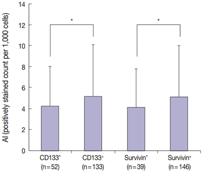
- 12,351 View
- 176 Download
- 20 Web of Science
- 19 Crossref
-
Abstract
PDF
- Background
Cancer stem cells have been investigated as new targets for colorectal cancer (CRC) treatment. We recently reported that CD133+ colon cancer cells showed chemoresistance to 5-fluorouracil through increased survivin expression and proposed the survivin inhibitor YM155 as an effective therapy for colon cancer in an in vitro study. Here, we investigate the relationship between survivin and CD133 expression in surgically resected CRC to identify whether the results obtained in our in vitro study are applicable to clinical samples.
Methods
We performed immunohistochemical staining for survivin and CD133 in surgically resected tissue from 187 stage II or III CRC patients. We also comparatively analyzed apoptosis according to survivin and CD133 expression using terminal deoxynucleotidyl transferase-mediated deoxyuridine triphosphate nick-end labeling.
Results
The results of the Mantel-Haenszel test established a linear association between nuclear survivin and CD133 expression (p = .018), although neither had prognostic significance, according to immunohistochemical expression level. No correlation was found between survivin expression and the following pathological parameters: invasion depth, lymph node metastasis, or histologic differentiation (p > .05). The mean apoptotic index in survivin+ and CD133+ tumors was higher than that in negative tumors: 5.116 ± 4.894 in survivin+ versus 4.103 ± 3.691 in survivin– (p = .044); 5.165 ± 4.961 in CD133+ versus 4.231 ± 3.812 in CD133– (p = .034).
Conclusions
As observed in our in vitro study, survivin expression is significantly related to CD133 expression. Survivin may be considered as a new therapeutic target for chemoresistant CRC. -
Citations
Citations to this article as recorded by- A comprehensive review and in silico analysis of the role of survivin (BIRC5) in hepatocellular carcinoma hallmarks: A step toward precision
Nermin M. Mohamed, Rania Hassan Mohamed, John F. Kennedy, Mahmoud M. Elhefnawi, Nadia M. Hamdy
International Journal of Biological Macromolecules.2025; 311: 143616. CrossRef - Genetic Variants in BIRC5 (rs8073069, rs17878467, and rs9904341) Are Associated with Susceptibility in Mexican Patients with Breast Cancer: Clinical Associations and Their Analysis In Silico
María Renee Jiménez-López, César de Jesús Tovar-Jácome, Alejandra Palacios-Ramírez, Martha Patricia Gallegos-Arreola, Teresa Giovanna María Aguilar-Macedo, Rubria Alicia González-Sánchez, Efraín Salas-González, José Elías García-Ortiz, Clara Ibet Juárez-V
Genes.2025; 16(7): 786. CrossRef - Upregulation of EMR1 (ADGRE1) by Tumor-Associated Macrophages Promotes Colon Cancer Progression by Activating the JAK2/STAT1,3 Signaling Pathway in Tumor Cells
Rokeya Akter, Rackhyun Park, Soo Kyung Lee, Eun ju Han, Kyu-Sang Park, Junsoo Park, Mee-Yon Cho
International Journal of Molecular Sciences.2024; 25(8): 4388. CrossRef - Antiapoptotic Gene Genotype and Allele Variations and the Risk of Lymphoma
Osama M. Al-Amer, Rashid Mir, Abdullah Hamadi, Mohammed I. Alasseiri, Malik A. Altayar, Waseem AlZamzami, Mamdoh Moawadh, Sael Alatawi, Hanan A. Niaz, Atif Abdulwahab A. Oyouni, Othman R. Alzahrani, Hanan E. Alatwi, Aishah E. Albalawi, Khalaf F. Alsharif,
Cancers.2023; 15(4): 1012. CrossRef - The Prognostic and Therapeutic Implications of the Chemoresistance Gene BIRC5 in Triple-Negative Breast Cancer
Getinet M. Adinew, Samia Messeha, Equar Taka, Karam F. A. Soliman
Cancers.2022; 14(21): 5180. CrossRef - EMR1/ADGRE1 Expression in Cancer Cells Upregulated by Tumor-Associated Macrophages Is Related to Poor Prognosis in Colorectal Cancer
Rokeya Akter, Kwangmin Kim, Hye Youn Kwon, Youngwan Kim, Young Woo Eom, Hye-mi Cho, Mee-Yon Cho
Biomedicines.2022; 10(12): 3121. CrossRef - Prognostic Significance of BIRC5/Survivin in Breast Cancer: Results from Three Independent Cohorts
Nina Oparina, Malin C. Erlandsson, Anna Fäldt Beding, Toshima Parris, Khalil Helou, Per Karlsson, Zakaria Einbeigi, Maria I. Bokarewa
Cancers.2021; 13(9): 2209. CrossRef - Obatoclax, a Pan-BCL-2 Inhibitor, Downregulates Survivin to Induce Apoptosis in Human Colorectal Carcinoma Cells Via Suppressing WNT/β-catenin Signaling
Chi-Hung R. Or, Chiao-Wen Huang, Ching-Chin Chang, You-Chen Lai, Yi-Ju Chen, Chia-Che Chang
International Journal of Molecular Sciences.2020; 21(5): 1773. CrossRef - M1 Macrophages Promote TRAIL Expression in Adipose Tissue-Derived Stem Cells, Which Suppresses Colitis-Associated Colon Cancer by Increasing Apoptosis of CD133+ Cancer Stem Cells and Decreasing M2 Macrophage Population
Young Woo Eom, Rokeya Akter, Wanlu Li, Suji Lee, Soonjae Hwang, Jiye Kim, Mee-Yon Cho
International Journal of Molecular Sciences.2020; 21(11): 3887. CrossRef - Emerging Importance of Survivin in Stem Cells and Cancer: the Development of New Cancer Therapeutics
Neerada Meenakshi Warrier, Prasoon Agarwal, Praveen Kumar
Stem Cell Reviews and Reports.2020; 16(5): 828. CrossRef - MMR-proficient and MMR-deficient colorectal cancer cells: 5-Fluorouracil treatment response and correlation to CD133 and MGMT expression
Jaime A. Oliver, Raúl Ortiz, Cristina Jiménez-Luna, Laura Cabeza, Gloria Perazzoli, Octavio Caba, Cristina Mesas, Consolación Melguizo, Jose Prados
Journal of Biosciences.2020;[Epub] CrossRef - Survivin rs9904341 polymorphism significantly increased the risk of cancer: evidence from an updated meta-analysis of case–control studies
Abdolkarim Moazeni-Roodi, Saeid Ghavami, Mohammad Hashemi
International Journal of Clinical Oncology.2019; 24(4): 335. CrossRef - CRISPR-Cas9 mediated CD133 knockout inhibits colon cancer invasion through reduced epithelial-mesenchymal transition
Wanlu Li, Mee-Yon Cho, Suji Lee, Mirae Jang, Junsoo Park, Rackhyun Park, Aamir Ahmad
PLOS ONE.2019; 14(8): e0220860. CrossRef - Diagnostic Approaches for Salivary Gland Tumors with Secretory and Microcystic Features
Ha Young Woo, Eun Chang Choi, Sun Och Yoon
Head and Neck Pathology.2018; 12(2): 237. CrossRef - MUC1‐ and Survivin‐based DNA Vaccine Combining Immunoadjuvants CpG and interleukin‐2 in a Bicistronic Expression Plasmid Generates Specific Immune Responses and Antitumour Effects in a Murine Colorectal Carcinoma Model
C. Liu, Y. Xie, B. Sun, F. Geng, F. Zhang, Q. Guo, H. Wu, B. Yu, J. Wu, X. Yu, W. Kong, H. Zhang
Scandinavian Journal of Immunology.2018; 87(2): 63. CrossRef - Activated STAT3 may participate in tumor progression through increasing CD133/survivin expression in early stage of colon cancer
Wanlu Li, Mi-Ra Lee, Taeyeong Kim, Young Wan Kim, Mee-Yon Cho
Biochemical and Biophysical Research Communications.2018; 497(1): 354. CrossRef - MiRNA-142-3p increases radiosensitivity in human umbilical cord blood mononuclear cells by inhibiting the expression of CD133
Fang Yuan, Lu Liu, Yonghong Lei, Yi Hu
Scientific Reports.2018;[Epub] CrossRef - MLH1 enhances the sensitivity of human endometrial carcinoma cells to cisplatin by activating the MLH1/c-Abl apoptosis signaling pathway
Yue Li, Shihong Zhang, Yuanjian Wang, Jin Peng, Fang Fang, Xingsheng Yang
BMC Cancer.2018;[Epub] CrossRef - Establishment of CMab-43, a Sensitive and Specific Anti-CD133 Monoclonal Antibody, for Immunohistochemistry
Shunsuke Itai, Yuki Fujii, Takuro Nakamura, Yao-Wen Chang, Miyuki Yanaka, Noriko Saidoh, Saori Handa, Hiroyoshi Suzuki, Hiroyuki Harada, Shinji Yamada, Mika K. Kaneko, Yukinari Kato
Monoclonal Antibodies in Immunodiagnosis & Immunotherapy.2017; 36(5): 231. CrossRef
- A comprehensive review and in silico analysis of the role of survivin (BIRC5) in hepatocellular carcinoma hallmarks: A step toward precision
- Meningeal Solitary Fibrous Tumors with Delayed Extracranial Metastasis
- Nayoung Han, Hannah Kim, Soo Kee Min, Sun-Ha Paek, Chul-Kee Park, Seung-Hong Choi, U-Ri Chae, Sung-Hye Park
- J Pathol Transl Med. 2016;50(2):113-121. Published online December 14, 2015
- DOI: https://doi.org/10.4132/jptm.2015.10.30
- 14,333 View
- 120 Download
- 26 Web of Science
- 23 Crossref
-
Abstract
PDF
- Background
The term solitary fibrous tumor (SFT) is preferred over meningeal hemangiopericytoma (HPC), because NAB2-STAT6 gene fusion has been observed in both intracranial and extracranial HPCs. HPCs are now considered cellular variants of SFTs. Methods: This study analyzes 19 patients with STAT6-confirmed SFTs, who were followed for over 11 years in a single institution. Ten patients (10/19, 56.2%) had extracranial metastases (metastatic group), while the remainder (9/19) did not (non-metastatic group). These two groups were compared clinicopathologically. Results: In the metastatic group, the primary metastatic sites were the lungs (n = 6), bone (n = 4), and liver (n = 3). There was a mean lag time of 14.2 years between the diagnosis of the initial meningeal tumor to that of systemic metastasis. The median age at initial tumor onset was 37.1 years in the metastatic group and 52.5 in the non-metastatic group. The 10-year survival rates of the metastatic- and non-metastatic groups were 100% and 33%, respectively. The significant prognostic factors for poor outcomes on univariate analysis included advanced age (≥45 years) and large initial tumor size (≥5 cm). In contrast, the patients with higher tumor grade, high mitotic rate (≥5/10 high-power fields), high Ki-67 index (≥5%), and the presence of necrosis or CD34 positivity showed tendency of poor prognosis but these parameters were not statistically significant poor prognostic markers. Conclusions: Among patients with SFTs, younger patients (<45 years) experienced longer survival times and paradoxically had more frequent extracranial metastases after long latent periods than did older patients. Therefore, young patients with SFTs require careful surveillance and follow-up for early detection of systemic metastases. -
Citations
Citations to this article as recorded by- Single-fraction stereotactic radiosurgery for residual, recurrent, or metastatic intracranial solitary fibrous tumors: An IRRF study toward management guidance
Salem M Tos, Ahmed Shaaban, Dawoud Hamdan, Georgios Mantziaris, Bardia Hajikarimloo, Mariam Ishaque, Yuki Shinya, Vanshika Lohia, Zhishuo Wei, Orbay Askeroglu, Christian Amezquita-Contreras, Andrea Becerril-Gaitan, Onam Verma, Keiss Douri, Nathalia Lora,
Neuro-Oncology.2026; 28(5): 1251. CrossRef - High-grade, metastatic disease, and adjuvant radiotherapy are independent prognostic factors for progression-free survival in patients with solitary fibrous tumors
Jan Paul Alker, Ramin Rahmanzade, Thomas Held, Christel Herold-Mende, Andreas Unterberg, Felix Sahm, Sandro Manuel Krieg, Gerhard Jungwirth
Neuro-Oncology Advances.2025;[Epub] CrossRef - Meningeal malignant solitary fibrous tumor with multiple recurrence, extracranial extension, cervical lymph node metastases: case report and review of the literature
Rong He, Peng Zhong, Juntao Hu, Guangkuo Guo, He Xiao, Lin Lei, Yun Liu, Mingying Geng, Jungang Ma
Discover Oncology.2025;[Epub] CrossRef - A Case of Intracranial Solitary Fibrous Tumor Followed by Distant Metastasis without Local Recurrence
Masafumi YOSHIDA, Koki MORIYOSHI, Kento DOI, Yukihiro YAMAO, Natsue KISHIDA, Hiroya UEMURA, Shunichi FUKUDA
NMC Case Report Journal.2025; 12: 181. CrossRef - The association between WHO grading and the long-term outcomes and radiotherapy efficacy of intracranial solitary fibrous tumors
Leihao Ren, Lingyang Hua, AO Feng, Jiaojiao Deng, Hiroaki Wakimoto, Tareq Juratli, Qing Xie, Ye Gong
Acta Neuropathologica Communications.2025;[Epub] CrossRef - Meningeal Solitary Fibrous Tumor: A Single-Center Retrospective Cohort Study
Siyer Roohani, Yasemin Alberti, Maximilian Mirwald, Felix Ehret, Carmen Stromberger, Soleiman Fabris Roohani, Katja Bender, Anne Flörcken, Sven Märdian, Daniel Zips, David Kaul, Manish Charan
Sarcoma.2024; 2024: 1. CrossRef - De-differentiation associated with drop metastasis of a recurrent intracranial solitary fibrous tumor: a case report and literature review
Chenhui Zhao, Xiran Fan, Wanwan Gao, Fan Zhang, Haijun Lv, Xiaochun Jiang, Guangfu Di
International Journal of Neuroscience.2022; 132(8): 843. CrossRef - Long-term extracranial metastatic relapse of an intraventricular solitary fibrous tumor: a case report
Tarek Assi, Elie Samaha, Hussein Nassereddine
Anti-Cancer Drugs.2022; 33(1): e764. CrossRef - Multidisciplinary Treatment of Liver Metastases from Intracranial SFTs/HPCs: A Report of Three Consecutive Cases
Felix J. Krendl, Franka Messner, Gregor Laimer, Angela Djanani, Andreas Seeber, Georg Oberhuber, Dietmar Öfner, Dominik Wolf, Stefan Schneeberger, Reto Bale, Christian Margreiter
Current Oncology.2022; 29(11): 8720. CrossRef - A review of solitary fibrous tumor/hemangiopericytoma tumor and a comparison of risk factors for recurrence, metastases, and death among patients with spinal and intracranial tumors.
Enrico Giordan, Elisabetta Marton, Alexandra M. Wennberg, Angela Guerriero, Giuseppe Canova
Neurosurgical Review.2021; 44(3): 1299. CrossRef - Intracranial Solitary Fibrous Tumor of the Skull Base: 2 Cases and Systematic Review of the Literature
Sricharan Gopakumar, Visish M. Srinivasan, Caroline C. Hadley, Adrish Anand, Marc Daou, Patrick J. Karas, Jacob Mandel, Shankar P. Gopinath, Akash J. Patel
World Neurosurgery.2021; 149: e345. CrossRef - Hemangiopericytoma/Solitary Fibrous Tumor in the central nervous system. Experience with surgery and radiotherapy as a complementary treatment: A 10-year analysis of a heterogeneous series in a single tertiary center
Pedro Miguel González-Vargas, José Luis Thenier-Villa, Pablo Sanromán Álvarez, Alexandre Serantes Combo, Lourdes Calero Félix, Raúl Alejandro Galárraga Campoverde, Eva Azevedo González, Álvaro Martín-Gallego, Rosa Martínez-Rolan, Adolfo de la Lama Zaragoz
Neurocirugía.2020; 31(1): 14. CrossRef - Hemangiopericytoma/Solitary Fibrous Tumor in the central nervous system. Experience with surgery and radiotherapy as a complementary treatment: A 10-year analysis of a heterogeneous series in a single tertiary center
Pedro Miguel González-Vargas, José Luis Thenier-Villa, Pablo Sanromán Álvarez, Alexandre Serantes Combo, Lourdes Calero Félix, Raúl Alejandro Galárraga Campoverde, Eva Azevedo González, Álvaro Martín-Gallego, Rosa Martínez-Rolan, Adolfo de la Lama Zaragoz
Neurocirugía (English Edition).2020; 31(1): 14. CrossRef - Solitary fibrous tumor/hemangiopericytoma: treatment results based on the 2016 WHO classification
Kyoung Su Sung, Ju Hyung Moon, Eui Hyun Kim, Seok-Gu Kang, Se Hoon Kim, Chang-Ok Suh, Sun Ho Kim, Kyu-Sung Lee, Won Seok Chang, Jong Hee Chang
Journal of Neurosurgery.2019; 130(2): 418. CrossRef - Grading of meningeal solitary fibrous tumors/hemangiopericytomas: analysis of the prognostic value of the Marseille Grading System in a cohort of 132 patients
Nicolas Macagno, Rob Vogels, Romain Appay, Carole Colin, Karima Mokhtari, Benno Küsters, Pieter Wesseling, Dominique Figarella‐Branger, Uta Flucke, Corinne Bouvier
Brain Pathology.2019; 29(1): 18. CrossRef - Solitary fibrous tumor of the pineal region with delayed ectopic intracranial metastasis: A case report and review of the literature
Yongjie Wang, Jingying Zhang, Qichang Liu, Fuyi Liu, Xiangdong Zhu, Jianmin Zhang
Medicine.2019; 98(21): e15737. CrossRef - Case report: neonatal giant forehead hemangiopericytoma with a 5-year follow-up
AiJun Peng, LiBing Zhang, Hai Zhao, LiangXue Zhou
Medicine.2019; 98(47): e17888. CrossRef - Liquid Biopsy in Rare Cancers: Lessons from Hemangiopericytoma
Chiara Nicolazzo, Luciano Colangelo, Alessandro Corsi, Guido Carpino, Angela Gradilone, Chiara Sonato, Cristina Raimondi, Eugenio Gaudio, Paola Gazzaniga, Walter Gianni
Analytical Cellular Pathology.2018; 2018: 1. CrossRef - Surveillance for metastatic hemangiopericytoma-solitary fibrous tumors-systematic literature review on incidence, predictors and diagnosis of extra-cranial disease
Tarini Ratneswaren, Florence Rosie Avila Hogg, Mathew Joseph Gallagher, Keyoumars Ashkan
Journal of Neuro-Oncology.2018; 138(3): 447. CrossRef - Intracranial Solitary Fibrous Tumor
Eveline Claus, Patrick Seynaeve, Jeroen Ceuppens, Alain Vanneste, Koenraad Verstraete
Journal of the Belgian Society of Radiology.2017;[Epub] CrossRef - Comparison and evaluation of risk factors for meningeal, pleural, and extrapleural solitary fibrous tumors: A clinicopathological study of 92 cases confirmed by STAT6 immunohistochemical staining
Ji Min Kim, Yoon-La Choi, Yu Jin Kim, Hyung Kyu Park
Pathology - Research and Practice.2017; 213(6): 619. CrossRef - Molecular Testing of Brain Tumor
Sung-Hye Park, Jaekyung Won, Seong-Ik Kim, Yujin Lee, Chul-Kee Park, Seung-Ki Kim, Seung-Hong Choi
Journal of Pathology and Translational Medicine.2017; 51(3): 205. CrossRef - Solitary fibrous tumour presenting with a single bone metastasis: report of six cases and literature review
Vittoria Colia, Salvatore Provenzano, Carlo Morosi, Paola Collini, Salvatore Lorenzo Renne, Paolo G. Dagrada, Claudia Sangalli, Angelo Paolo Dei Tos, Andrea Marrari, Paolo G. Casali, Silvia Stacchiotti
Clinical Sarcoma Research.2016;[Epub] CrossRef
- Single-fraction stereotactic radiosurgery for residual, recurrent, or metastatic intracranial solitary fibrous tumors: An IRRF study toward management guidance
- Immunohistochemical Expression and Clinical Significance of Suggested Stem Cell Markers in Hepatocellular Carcinoma
- Jong Jin Sung, Sang Jae Noh, Jun Sang Bae, Ho Sung Park, Kyu Yun Jang, Myoung Ja Chung, Woo Sung Moon
- J Pathol Transl Med. 2016;50(1):52-57. Published online November 18, 2015
- DOI: https://doi.org/10.4132/jptm.2015.10.09
- 12,962 View
- 78 Download
- 21 Web of Science
- 19 Crossref
-
Abstract
PDF
- Background
Increasing evidence has shown that tumor initiation and growth are nourished by a small subpopulation of cancer stem cells (CSCs) within the tumor mass. CSCs are posited to be responsible for tumor maintenance, growth, distant metastasis, and relapse after curative operation. We examined the expression of CSC markers in paraffin-embedded tissue sections of hepatocellular carcinoma (HCC) and correlated the results with clinicopathologic characteristics. Methods: Immunohistochemical staining for the markers believed to be expressed in the CSCs, including epithelial cell adhesion molecule (EpCAM), keratin 19 (K19), CD133, and CD56, was performed in 82 HCC specimens. Results: EpCAM expression was observed in 56% of the HCCs (46/82) and K19 in 6% (5/82). EpCAM expression in HCC significantly correlated with elevated α-fetoprotein level, microvessel invasion of tumor cells, and high histologic grade. In addition, Ep- CAM expression significantly correlated with K19 expression. The overall survival and relapsefree survival rates in patients with EpCAM-expressing HCC were relatively lower than those in patients with EpCAM-negative HCC. All but two of the 82 HCCs were negative for CD133 and CD56, respectively. Conclusions: Our results suggest that HCCs expressing EpCAM are associated with unfavorable prognostic factors and have a more aggressive clinical course than those not expressing EpCAM. Further, the expression of either CD133 or CD56 in paraffin-embedded HCC tissues appears to be rare. -
Citations
Citations to this article as recorded by- Spatial immune scoring system predicts hepatocellular carcinoma recurrence
Gengjie Jia, Peiqi He, Tianli Dai, Denise Goh, Jiabei Wang, Mengyuan Sun, Felicia Wee, Fuling Li, Jeffrey Chun Tatt Lim, Shuxia Hao, Yao Liu, Tony Kiat Hon Lim, Nye-Thane Ngo, Qingping Tao, Wei Wang, Ahitsham Umar, Björn Nashan, Yongchang Zhang, Chen Ding
Nature.2025; 640(8060): 1031. CrossRef - Evolving Landscape of Systemic Therapy for Hepatocellular Carcinoma in 2025
Karan Kumar, Vivek A. Saraswat
Journal of Clinical and Experimental Hepatology.2025; 15(5): 102547. CrossRef - Recent Progress in Systemic Therapy for Advanced Hepatocellular Carcinoma
Narayanan Sadagopan, Aiwu Ruth He
International Journal of Molecular Sciences.2024; 25(2): 1259. CrossRef - Diagnostic value of expressions of cancer stem cell markers for adverse outcomes of hepatocellular carcinoma and their associations with prognosis: A Bayesian network meta‑analysis
Zhengrong Ou, Shoushuo Fu, Jian Yi, Jingxuan Huang, Weidong Zhu
Oncology Letters.2024;[Epub] CrossRef - Clinicopathological and prognostic value of epithelial cell adhesion molecule in solid tumours: a meta-analysis
Peiwen Ding, Panyu Chen, Jiqi Ouyang, Qiang Li, Shijie Li
Frontiers in Oncology.2023;[Epub] CrossRef - PD-L1 Downregulation and DNA Methylation Inhibition for Molecular Therapy against Cancer Stem Cells in Hepatocellular Carcinoma
Caecilia Sukowati, Loraine Kay D. Cabral, Beatrice Anfuso, Francesco Dituri, Roberto Negro, Gianluigi Giannelli, Claudio Tiribelli
International Journal of Molecular Sciences.2023; 24(17): 13357. CrossRef - EpCAM, Ki67, and ESM1 Predict Hepatocellular Carcinoma Recurrence After Liver Transplantation
Aiat Shaban Hemida, Doha Maher Taie, Moshira Mohamed Abd El-Wahed, Mohammed Ibrahim Shabaan, Mona Saeed Tantawy, Nermine Ahmed Ehsan
Applied Immunohistochemistry & Molecular Morphology.2023; 31(9): 596. CrossRef - The clinical, prognostic and therapeutic significance of liver cancer stem cells and their markers
Izabela Zarębska, Arkadiusz Gzil, Justyna Durślewicz, Damian Jaworski, Paulina Antosik, Navid Ahmadi, Marta Smolińska-Świtała, Dariusz Grzanka, Łukasz Szylberg
Clinics and Research in Hepatology and Gastroenterology.2021; 45(3): 101664. CrossRef - Detection of oncogenic mutations in paired circulating tumor DNA and circulating tumor cells in patients with hepatocellular carcinoma
Zhouhong Ge, Jean C.A. Helmijr, Maurice P.H.M. Jansen, Patrick P.C. Boor, Lisanne Noordam, Maikel Peppelenbosch, Jaap Kwekkeboom, Jaco Kraan, Dave Sprengers
Translational Oncology.2021; 14(7): 101073. CrossRef - Hepatocellular Carcinoma Score and Subclassification Into Aggressive Subtypes Using Immunohistochemical Expression of p53, β-Catenin, CD133, and Ki-67
Asmaa G. Abdou, Nanis S. Holah, Dina S. Elazab, Walaa G. El-Gendy, Mohammed T. Badr, Dalia R. Al-Sharaky
Applied Immunohistochemistry & Molecular Morphology.2021; 29(1): 20. CrossRef - The prognostic significance of neuroendocrine markers and somatostatin receptor 2 in hepatocellular carcinoma
Keigo Murakami, Hiroyuki Kumata, Shigehito Miyagi, Takashi Kamei, Hironobu Sasano
Pathology International.2021; 71(10): 682. CrossRef - Predictors of recurrence and survival of hepatocellular carcinoma: A prospective study including transient elastography and cancer stem cell markers
Hend Ibrahim Shousha, Rabab Fouad, Tamer Mahmoud Elbaz, Dina Sabry, Mohamed Mahmoud Nabeel, Ahmed Hosni Abdelmaksoud, Aisha Mahmoud Elsharkawy, Zeinab Abdellatif Soliman, Ghada Habib, Ashraf Omar Abdelaziz
Arab Journal of Gastroenterology.2020; 21(2): 95. CrossRef - Napabucasin Reduces Cancer Stem Cell Characteristics in Hepatocellular Carcinoma
Ya Li, Qiuju Han, Huajun Zhao, Quanjuan Guo, Jian Zhang
Frontiers in Pharmacology.2020;[Epub] CrossRef - The mRNA Distribution of Cancer Stem Cell Marker CD90/Thy-1 Is Comparable in Hepatocellular Carcinoma of Eastern and Western Populations
An B. Luong, Huy Q. Do, Paola Tarchi, Deborah Bonazza, Cristina Bottin, Loraine Kay D. Cabral, Long D. C. Tran, Thao P. T. Doan, Lory S. Crocè, Hoa L. T. Pham, Claudio Tiribelli, Caecilia H. C. Sukowati
Cells.2020; 9(12): 2672. CrossRef - The prognostic relationship between histopathological and immunohistochemical features of hepatocellular carcinoma, intrahepatic cholangiocarcinoma and mixed type
Fatmagül Kuşku Çabuk, Nuray Başsüllü, İlknur Türkmen, Murat Dayangaç, Murat Akyıldız, Yıldıray Yüzer, Yaman Tokat, Gülen Bülbül Doğusoy
Polish Journal of Pathology.2020; 71(2): 79. CrossRef - Histological architectural classification determines recurrence pattern and prognosis after curative hepatectomy in patients with hepatocellular carcinoma
Hirohisa Okabe, Tomoharu Yoshizumi, Yo-ichi Yamashita, Katsunori Imai, Hiromitsu Hayashi, Shigeki Nakagawa, Shinji Itoh, Norifumi Harimoto, Toru Ikegami, Hideaki Uchiyama, Toru Beppu, Shinichi Aishima, Ken Shirabe, Hideo Baba, Yoshihiko Maehara, Motoyuki
PLOS ONE.2018; 13(9): e0203856. CrossRef - Overexpression of epithelial cell adhesion molecule as a predictor of poor outcome in patients with hepatocellular carcinoma
Chih‑Jan Ko, Chia‑Jung Li, Meng‑Yu Wu, Pei‑Yi Chu
Experimental and Therapeutic Medicine.2018;[Epub] CrossRef - Clinicopathologic Significance of Survivin Expression in Relation to CD133 Expression in Surgically Resected Stage II or III Colorectal Cancer
Wanlu Li, Mi-Ra Lee, EunHee Choi, Mee-Yon Cho
Journal of Pathology and Translational Medicine.2017; 51(1): 17. CrossRef - PIN1 in hepatocellular carcinoma is associated with TP53 gene status
Jun Sang Bae, Sang Jae Noh, Kyoung Min Kim, Kyu Yun Jang, Ho Sung Park, Myoung Ja Chung, Byung-Hyun Park, Woo Sung Moon
Oncology Reports.2016; 36(4): 2405. CrossRef
- Spatial immune scoring system predicts hepatocellular carcinoma recurrence
- The Continuing Value of Ultrastructural Observation in Central Nervous System Neoplasms in Children
- Na Rae Kim, Sung-Hye Park
- J Pathol Transl Med. 2015;49(6):427-437. Published online October 13, 2015
- DOI: https://doi.org/10.4132/jptm.2015.09.19
- 10,987 View
- 78 Download
-
Abstract
PDF
- Central nervous system (CNS) neoplasms are the second most common childhood malignancy after leukemia and the most common solid organ neoplasm in children. Diagnostic dilemmas with small specimens from CNS neoplasms are often the result of multifactorial etiologies such as frozen or fixation artifact, biopsy size, or lack of knowledge about rare or unfamiliar entities. Since the late 1950s, ultrastructural examination has been used in the diagnosis of CNS neoplasms, though it has largely been replaced by immunohistochemical and molecular cytogenetic studies. Nowadays, pathologic diagnosis of CNS neoplasms is achieved through intraoperative cytology, light microscopy, immunohistochemistry, and molecular cytogenetic results. However, the utility of electron microscopy (EM) in the final diagnosis of CNS neoplasms and investigation of its pathogenetic origin remains critical. Here, we reviewed the distinguishing ultrastructural features of pediatric CNS neoplasms and emphasize the continuing value of EM in the diagnosis of CNS neoplasms.
- Current Issues and Clinical Evidence in Tumor-Infiltrating Lymphocytes in Breast Cancer
- Sung Gwe Ahn, Joon Jeong, SoonWon Hong, Woo Hee Jung
- J Pathol Transl Med. 2015;49(5):355-363. Published online August 17, 2015
- DOI: https://doi.org/10.4132/jptm.2015.07.29
- 17,843 View
- 244 Download
- 45 Web of Science
- 39 Crossref
-
Abstract
PDF
- With the advance in personalized therapeutic strategies in patients with breast cancer, there is an increasing need for biomarker-guided therapy. Although the immunogenicity of breast cancer has not been strongly considered in research or practice, tumor-infiltrating lymphocytes (TILs) are emerging as biomarkers mediating tumor response to treatments. Earlier studies have provided evidence that the level of TILs has prognostic value and the potential for predictive value, particularly in triple-negative and human epidermal growth factor receptor 2–positive breast cancer. Moreover, the level of TILs has been associated with treatment outcome in patients undergoing neoadjuvant chemotherapy. To date, no standardized methodology for measuring TILs has been established. In this article, we review current issues and clinical evidence for the use of TILs in breast cancer.
-
Citations
Citations to this article as recorded by- Prognostic relevance of specific TIL (CD4+, CD8+, and FOXP3 + T-cell infiltrates) in triple-negative breast cancer: short- and long-term outcomes
Olga Caramelo, Vânia Almeida, Ana Fidalgo, Augusta Cipriano, Teresa Almeida-Santos
Breast Cancer.2026; 33(2): 386. CrossRef - Cytokine Networks in Triple-Negative Breast Cancer: Mechanisms, Therapeutic Targets, and Emerging Strategies
María Rosado-Sanz, Nuria Martínez-Alarcón, Adrián Abellán-Soriano, Raúl Golfe, Eva M. Trinidad, Jaime Font de Mora
Biomedicines.2025; 13(8): 1945. CrossRef - Unveiling Metabolic Signatures as Potential Biomarkers in Common Cancers: Insights from Lung, Breast, Colorectal, Liver, and Gastric Tumours
Kha Wai Hon, Rakesh Naidu
Biomolecules.2025; 15(10): 1376. CrossRef - Systemic chemotherapy in metastatic TNBC polarizes effector T cell differentiation
Hye-Yeon Ju, Hyundong Yoon, Seungpil Jung, Serk In Park, Tae Kon Kim, Mi-Ryung Han
Cancer Cell International.2025;[Epub] CrossRef - Immune characteristics and clinical significance of peripheral blood lymphocytes in breast cancer
Hongyu Gao, Dengjie Ouyang, Xinyu Guan, Jiachi Xu, Qitong Chen, Liyun Zeng, Jian Pang, Qiongyan Zou, Ke Qian, Wenjun Yi
BMC Cancer.2024;[Epub] CrossRef - Precision unveiled: Synergistic genomic landscapes in breast cancer—Integrating single‐cell analysis and decoding drug toxicity for elite prognostication and tailored therapeutics
Chenglu Jiang, Shengke Zhang, Lai Jiang, Zipei Chen, Haiqing Chen, Jinbang Huang, Jingyi Tang, Xiufang Luo, Guanhu Yang, Jie Liu, Hao Chi
Environmental Toxicology.2024; 39(6): 3448. CrossRef - Exploring histological predictive biomarkers for immune checkpoint inhibitor therapy response in non–small cell lung cancer
Uiju Cho, Soyoung Im, Hyung Soon Park
Journal of Pathology and Translational Medicine.2024; 58(2): 49. CrossRef - Review: Merging from traditional to potential novel breast cancer biomarkers
Hanan Alismail
Journal of King Saud University - Science.2024; 36(11): 103551. CrossRef -
A non-inferiority, phase III trial of gemcitabine plus capecitabine
versus
gemcitabine plus carboplatin as first-line therapy and tumor-infiltrating lymphocytes as a prognostic biomarker in patients with advanced tripl
Xiaodong Liu, Weipeng Zhao, Yongsheng Jia, Yehui Shi, Xu Wang, Shufen Li, Pin Zhang, Chen Wang, Chunfang Hao, Zhongsheng Tong
Therapeutic Advances in Medical Oncology.2024;[Epub] CrossRef - Predictive and Prognostic Role of Tumor‐Infiltrating Lymphocytes in Patients with Advanced Breast Cancer Treated with Primary Systemic Therapy
Gaurav Agarwal, K. M. M. Vishvak Chanthar, Shweta Katiyar, Niraj Kumari, Narendra Krishnani, M. Sabaretnam, Gyan Chand, Anjali Mishra, Punita Lal
World Journal of Surgery.2023; 47(5): 1238. CrossRef - The role of tumor-infiltrating lymphocytes in triple-negative breast cancer and the research progress of adoptive cell therapy
Ruonan Li, Lili Cao
Frontiers in Immunology.2023;[Epub] CrossRef - Using Self-Assembling ADDomer Platform to Display B and T Epitopes of Type O Foot-and-Mouth Disease Virus
Chaowei Luo, Quanhui Yan, Juncong Huang, Jiameng Liu, Yuwan Li, Keke Wu, Bingke Li, Mingqiu Zhao, Shuangqi Fan, Hongxing Ding, Jinding Chen
Viruses.2022; 14(8): 1810. CrossRef - Modification of the Tumor Microenvironment Enhances Anti-PD-1 Immunotherapy in Metastatic Melanoma
Guilan Shi, Megan Scott, Cathryn G. Mangiamele, Richard Heller
Pharmaceutics.2022; 14(11): 2429. CrossRef - Assessment of Immune Status in Dynamics for Patients with Cancer Undergoing Immunotherapy
Bacinschi Xenia Elena, Laurentia Nicoleta Gales, Anca Florina Zgura, Laura Iliescu, Rodica Maricela Anghel, Bogdan Haineala, Xiaosheng Wang
Journal of Oncology.2021; 2021: 1. CrossRef - Differences in immune-related gene expressions and tumor-infiltrating lymphocytes according to chemotherapeutic response in ovarian high-grade serous carcinoma
Kyung Un Choi, Ahrong Kim, Jee Yeon Kim, Ki Hyung Kim, Chungsu Hwang, So Jung Lee, Won Young Park, Sejin Jung, Hye Jeong Choi, Kyungbin Kim
Journal of Ovarian Research.2020;[Epub] CrossRef Predictors of Neoadjuvant Chemotherapy Response in Breast Cancer: A Review
Weilin Xu, Xiu Chen, Fei Deng, Jian Zhang, Wei Zhang, Jinhai Tang
OncoTargets and Therapy.2020; Volume 13: 5887. CrossRef- Immune phenotype of patients with stage IV metastatic inflammatory breast cancer
Sandra V. Fernandez, Alexander W. MacFarlane, Mowafaq Jillab, Maria F. Arisi, Jennifer Yearley, Lakshmanan Annamalai, Yulan Gong, Kathy Q. Cai, R. Katherine Alpaugh, Massimo Cristofanilli, Kerry S. Campbell
Breast Cancer Research.2020;[Epub] CrossRef - Infiltrating stromal immune cells in inflammatory breast cancer are associated with an improved outcome and increased PD-L1 expression
C. Van Berckelaer, C. Rypens, P. van Dam, L. Pouillon, M. Parizel, K. A. Schats, M. Kockx, W. A. A. Tjalma, P. Vermeulen, S. van Laere, F. Bertucci, C. Colpaert, L. Dirix
Breast Cancer Research.2019;[Epub] CrossRef - The composition of T cell infiltrates varies in primary invasive breast cancer of different molecular subtypes as well as according to tumor size and nodal status
Anna Glajcar, Joanna Szpor, Diana Hodorowicz-Zaniewska, Katarzyna Ewa Tyrak, Krzysztof Okoń
Virchows Archiv.2019; 475(1): 13. CrossRef - Retinoid X receptor agonist LG100268 modulates the immune microenvironment in preclinical breast cancer models
Ana S. Leal, Kayla Zydeck, Sarah Carapellucci, Lyndsey A. Reich, Di Zhang, Jessica A. Moerland, Michael B. Sporn, Karen T. Liby
npj Breast Cancer.2019;[Epub] CrossRef - Stromal tumor‑infiltrating lymphocytes evaluated on H&E‑stained slides are an independent prognostic factor in epithelial ovarian cancer and ovarian serous carcinoma
Chungsu Hwang, So Lee, Jung Lee, Ki Kim, Dong Suh, Byung‑Su Kwon, Kyung Choi
Oncology Letters.2019;[Epub] CrossRef - The therapeutic candidate for immune checkpoint inhibitors elucidated by the status of tumor-infiltrating lymphocytes (TILs) and programmed death ligand 1 (PD-L1) expression in triple negative breast cancer (TNBC)
Nobumoto Tomioka, Manabu Azuma, Mayuko Ikarashi, Mitsugu Yamamoto, Masako Sato, Ken-ichi Watanabe, Katsushige Yamashiro, Masato Takahashi
Breast Cancer.2018; 25(1): 34. CrossRef - Triple negative breast cancer – prognostic role of immune-related factors: a systematic review
Elisabeth Specht Stovgaard, Dorte Nielsen, Estrid Hogdall, Eva Balslev
Acta Oncologica.2018; 57(1): 74. CrossRef - Lack of effective translational regulation of PLD expression and exosome biogenesis in triple-negative breast cancer cells
Julian Gomez-Cambronero
Cancer and Metastasis Reviews.2018; 37(2-3): 491. CrossRef - Lymphoid environment in molecular subtypes of breast cancer
Anna Glajcar, Joanna Szpor, Katarzyna Ewa Tyrak, Joanna Streb, Diana Hodorowicz-Zaniewska, Krzysztof Okoń
Polish Journal of Pathology.2018; 69(2): 169. CrossRef - Expression of Myxovirus Resistance A (MxA) Is Associated with Tumor-Infiltrating Lymphocytes in Human Epidermal Growth Factor Receptor 2 (HER2)–Positive Breast Cancers
So Jeong Lee, Cheong-Soo Hwang, Young-Keum Kim, Hyun Jung Lee, Sang-Jeong Ahn, Nari Shin, Jung Hee Lee, Dong Hoon Shin, Kyung Un Choi, Do Youn Park, Chang Hun Lee, Gi Young Huh, Mi Young Sol, Hee Jin Lee, Gyungyub Gong, Jee Yeon Kim, Ahrong Kim
Cancer Research and Treatment.2017; 49(2): 313. CrossRef - Glutaminase expression is a poor prognostic factor in node-positive triple-negative breast cancer patients with a high level of tumor-infiltrating lymphocytes
Joo Young Kim, Sun-Hee Heo, Seul Ki Choi, In Hye Song, In Ah Park, Young-Ae Kim, Hye Seon Park, Suk Young Park, Won Seon Bang, Gyungyub Gong, Hee Jin Lee
Virchows Archiv.2017; 470(4): 381. CrossRef - Whole Slide Imaging for Analytical Anatomic Pathology and Telepathology: Practical Applications Today, Promises, and Perils
Alton Brad Farris, Cynthia Cohen, Thomas E. Rogers, Geoffrey H. Smith
Archives of Pathology & Laboratory Medicine.2017; 141(4): 542. CrossRef - Tumor-infiltrating lymphocytes and breast cancer: Beyond the prognostic and predictive utility
Andrea Ravelli, Giandomenico Roviello, Daniele Cretella, Andrea Cavazzoni, Alessandra Biondi, Maria Rosa Cappelletti, Laura Zanotti, Giuseppina Ferrero, Marco Ungari, Fabrizio Zanconati, Alberto Bottini, Roberta Alfieri, Pier Giorgio Petronini, Daniele Ge
Tumor Biology.2017; 39(4): 101042831769502. CrossRef - Predictive Value of Tertiary Lymphoid Structures Assessed by High Endothelial Venule Counts in the Neoadjuvant Setting of Triple-Negative Breast Cancer
In Hye Song, Sun-Hee Heo, Won Seon Bang, Hye Seon Park, In Ah Park, Young-Ae Kim, Suk Young Park, Jin Roh, Gyungyub Gong, Hee Jin Lee
Cancer Research and Treatment.2017; 49(2): 399. CrossRef - Lymphocyte count or percentage: which can better predict the prognosis of advanced cancer patients following palliative care?
Weiwei Zhao, Peng Wang, Huixun Jia, Menglei Chen, Xiaoli Gu, Minghui Liu, Zhe Zhang, Wenwu Cheng, Zhenyu Wu
BMC Cancer.2017;[Epub] CrossRef - Programmed death-ligand 1 (PD-L1) expression in tumour cell and tumour infiltrating lymphocytes of HER2-positive breast cancer and its prognostic value
Ahrong Kim, So Jeong Lee, Young Keum Kim, Won Young Park, Do Youn Park, Jee Yeon Kim, Chang Hun Lee, Gyungyub Gong, Gi Yeong Huh, Kyung Un Choi
Scientific Reports.2017;[Epub] CrossRef - Negative association between GATA3 and fascin could predict relapse-free and overall survival in patients with breast cancer
Kyueng-Whan Min, Dong-Hoon Kim, Sung-Im Do, Seoung Wan Chae, Kyungeun Kim, Jin Hee Sohn, Jung-Soo Pyo, Hyun Joo Lee, Dong Hyun Kim, Sukjoong Oh, Seon Hyeong Choi, Yong Lai Park, Chan Heun Park, Eun-Kyung Kim, Mi Jung Kwon, Jinwon Seo, Kyoung Min Moon
Virchows Archiv.2016; 468(4): 409. CrossRef - Cytoplasmic expression of high mobility group B1 (HMGB1) is associated with tumor‐infiltrating lymphocytes (TILs) in breast cancer
Hee Jin Lee, Ahrong Kim, In Hye Song, In Ah Park, Jong Han Yu, Jin Hee Ahn, Gyungyub Gong
Pathology International.2016; 66(4): 202. CrossRef - Clinical pharmacologic aspects of immune checkpoint inhibitors in cancer therapy
Kyu-pyo Kim, Hun Jung
Translational and Clinical Pharmacology.2016; 24(1): 7. CrossRef - The next level of 3D tumour models: immunocompetence
Agata Nyga, Joana Neves, Katerina Stamati, Marilena Loizidou, Mark Emberton, Umber Cheema
Drug Discovery Today.2016; 21(9): 1421. CrossRef - An Examination of the Local Cellular Immune Response to Examples of Both Ductal Carcinoma In Situ (DCIS) of the Breast and DCIS With Microinvasion, With Emphasis on Tertiary Lymphoid Structures and Tumor Infiltrating Lymphoctytes
Ahrong Kim, Sun-Hee Heo, Young-Ae Kim, Gyungyub Gong, Hee Jin Lee
American Journal of Clinical Pathology.2016; 146(1): 137. CrossRef - High Ki67/BCL2 index is associated with worse outcome in early stage breast cancer
Kyueng-Whan Min, Dong-Hoon Kim, Sung-Im Do, Jung-Soo Pyo, Seoung Wan Chae, Jin Hee Sohn, Kyungeun Kim, Hyun Joo Lee, Dong Hyun Kim, Sukjoong Oh, Seon Hyeong Choi, Yong Lai Park, Chan Heun Park, Mi Jung Kwon, Kyoung Min Moon
Postgraduate Medical Journal.2016; 92(1094): 707. CrossRef - Expression of T-Lymphocyte Markers in Human Epidermal Growth Factor Receptor 2-Positive Breast Cancer
Changro Lee, Seho Park, Joo Heung Kim, Sung Mook Lim, Hyung Seok Park, Seung Il Kim, Byeong-Woo Park
Journal of Breast Cancer.2016; 19(4): 385. CrossRef
- Prognostic relevance of specific TIL (CD4+, CD8+, and FOXP3 + T-cell infiltrates) in triple-negative breast cancer: short- and long-term outcomes
- Expression of CD99 in Multiple Myeloma: A Clinicopathologic and Immunohistochemical Study of 170 Cases
- Su-Jin Shin, Hyangsin Lee, Geunyoung Jung, Minchan Gil, Hosub Park, Young Soo Park, Dok Hyun Yoon, Cheolwon Suh, Chan-Jeoung Park, Jooryung Huh, Chan-Sik Park
- Korean J Pathol. 2014;48(3):209-216. Published online June 26, 2014
- DOI: https://doi.org/10.4132/KoreanJPathol.2014.48.3.209
- 10,513 View
- 82 Download
- 9 Crossref
-
Abstract
PDF
Background Multiple myeloma (MM) is a heterogeneous and ultimately fatal disease. Risk stratification using prognostic biomarkers is crucial to individualize treatments. We sought to investigate the role of CD99, a transmembrane protein highly expressed in many hematopoietic cells including subpopulations of normal and neoplastic plasma cells, for MM risk stratification.
Methods CD99 expression was measured in paraffin samples of bone marrow and extramedullary biopsies of 170 patients with MM. Patients were divided into those with high score (moderately and strongly positive) and low score (negative and weakly positive), with all staining being cytoplasmic and/or membranous.
Results High anti-CD99 immunostaining was observed in 72 of 136 (52.9%) bone marrow biopsies and 24 of 87 (27.6%) extramedullary biopsies in MM. High CD99 expression of extramedullary specimens was associated with significantly longer overall survival (OS; p=.016). High CD99 expression of extramedullary specimens was also associated with better prognosis in the nonautologous stem cell transplantation group of MM patients (p=.044). In multivariate analysis, International Staging System stage was an independent prognostic factor, whereas CD99 expression was no longer statistically significant.
Conclusions Expression of CD99 in extramedullary specimens was correlated with longer OS, suggesting that CD99 may be a helpful immunohistochemical marker for risk stratification.
-
Citations
Citations to this article as recorded by- Cell Adhesion Molecule CD99 in Cancer Immunotherapy
Feng Yu, Guodong Liu, Hailing Zhang, Xiaoyan Wang, Zhi Wu, Qinggang Xu, Yan Wu, Dongfeng Chen
Current Molecular Medicine.2023; 23(10): 1028. CrossRef - Detection of Circulating Tumor Plasma Cells in Monoclonal Gammopathies: Methods, Pathogenic Role, and Clinical Implications
Luzalba Sanoja-Flores, Juan Flores-Montero, Martín Pérez-Andrés, Noemí Puig, Alberto Orfao
Cancers.2020; 12(6): 1499. CrossRef - Tumor suppressor CD99 is downregulated in plasma cell neoplasms lacking CCND1 translocation and distinguishes neoplastic from normal plasma cells and B-cell lymphomas with plasmacytic differentiation from primary plasma cell neoplasms
Qi Gao, Venkata Yellapantula, Maly Fenelus, Janine Pichardo, Lu Wang, Ola Landgren, Ahmet Dogan, Mikhail Roshal
Modern Pathology.2018; 31(6): 881. CrossRef - EWSR1 fusion proteins mediate PAX7 expression in Ewing sarcoma
Gregory W Charville, Wei-Lien Wang, Davis R Ingram, Angshumoy Roy, Dafydd Thomas, Rajiv M Patel, Jason L Hornick, Matt van de Rijn, Alexander J Lazar
Modern Pathology.2017; 30(9): 1312. CrossRef - Activation of the polycomb repressive complex pathway in the bone marrow resident cells of diffuse large B-cell lymphoma patients
Eun Ji Oh, Eun Kyung Kim, Woo Ick Yang, Sun Och Yoon
Leukemia & Lymphoma.2016; 57(8): 1921. CrossRef - CD99 Is Strongly Expressed in Basal Cells of the Normal Adult Epidermis and Some Subpopulations of Appendages: Comparison with Developing Fetal Skin
Gawon Choi, Jin Roh, Chan-Sik Park
Journal of Pathology and Translational Medicine.2016; 50(5): 361. CrossRef - Towards Stratified Medicine in Plasma Cell Myeloma
Philip Egan, Stephen Drain, Caroline Conway, Anthony Bjourson, H. Alexander
International Journal of Molecular Sciences.2016; 17(10): 1760. CrossRef - Human Myeloma Cell Lines Induce Osteoblast Downregulation of CD99 Which Is Involved in Osteoblast Formation and Activity
Angela Oranger, Giacomina Brunetti, Claudia Carbone, Graziana Colaianni, Teresa Mongelli, Isabella Gigante, Roberto Tamma, Giorgio Mori, Adriana Di Benedetto, Marika Sciandra, Selena Ventura, Katia Scotlandi, Silvia Colucci, Maria Grano
Journal of Immunology Research.2015; 2015: 1. CrossRef - CD99 regulates CXCL12-induced chemotaxis of human plasma cells
Minchan Gil, Hyo-Kyung Pak, A-Neum Lee, Seo-Jung Park, Yoonkyung Lee, Jin Roh, Hyunji Lee, Yoo-Sam Chung, Chan-Sik Park
Immunology Letters.2015; 168(2): 329. CrossRef
- Cell Adhesion Molecule CD99 in Cancer Immunotherapy
- Evaluation of Protein Expression in Housekeeping Genes across Multiple Tissues in Rats
- Hye Jeong Kim, Jong In Na, Byung Woo Min, Joo Young Na, Kyung Hwa Lee, Jae Hyuk Lee, Young Jik Lee, Hyung Seok Kim, Jong Tae Park
- Korean J Pathol. 2014;48(3):193-200. Published online June 26, 2014
- DOI: https://doi.org/10.4132/KoreanJPathol.2014.48.3.193
- 19,007 View
- 168 Download
- 22 Crossref
-
Abstract
PDF
Background Housekeeping genes, which show constant protein expression patterns between different tissue types, are very important in molecular biological studies as an internal control for protein research.
Methods The protein expression profiles of seven housekeeping genes (
HPRT1 ,PPIA ,GYS1 ,TBP ,YWHAZ ,GAPDH andACTB ) in various rat tissues (cerebrum, cerebellum, cardiac ventricle and atrium, psoas muscle, femoral muscle, liver, spleen, kidney, and aorta) were analyzed by Western blot and compared by coefficient of variation (CV).Results HPRT1 was stably expressed (CV≤10%) in six tissues (cerebrum, cerebellum, ventricle, femoral muscle, spleen, and kidney),PPIA was stably expressed in five tissues (cerebrum, cerebellum, ventricle, spleen and kidney),YWHAZ was stably expressed in three tissues (cerebrum, cerebellum, and kidney), andGAPDH was stably expressed in four tissues (cerebrum, ventricle, psoas muscle, and kidney). In comparison,GYS1 ,TBP , andACTB were found to have CV values over 10% in all tissues. Of the seven genes examined, four (HPRT1 ,PPIA ,YWHAZ , andGAPDH ) were found to be stably expressed across multiple organs, with low CV values (≤10%).Conclusions These results will provide fundamental information regarding internal controls for protein expression studies and can be used for analysis of postmortem protein degradation patterns in forensic medicine.
-
Citations
Citations to this article as recorded by- scCompass: An Integrated Multi‐Species scRNA‐seq Database for AI‐Ready
Pengfei Wang, Wenhao Liu, Jiajia Wang, Yana Liu, Pengjiang Li, Ping Xu, Wentao Cui, Ran Zhang, Qingqing Long, Zhilong Hu, Chen Fang, Jingxi Dong, Chunyang Zhang, Yan Chen, Chengrui Wang, Guole Liu, Hanyu Xie, Yiyang Zhang, Meng Xiao, Shubai Chen, Haiping
Advanced Science.2025;[Epub] CrossRef - Evolutionary Insights into the Length Variation of DNA Damage Response Proteins Across Eukaryotes
Dominic Wiredu-Boakye, Laurence Higgins, Ondřej Gahura, Anzhelika Butenko, Guy Leonard, Mark A Freeman, Árni Kristmundsson, Karen Moore, Jamie W Harrison, Shani Mac Donald, Vyacheslav Yurchenko, Bryony A P Williams, Richard Chahwan, Courtney Stairs
Genome Biology and Evolution.2025;[Epub] CrossRef - Tissue-Specific Reference Genes in Rats: Insights into Cardiac, Muscular, and Skeletal Systems
Emre Can Günaydın, Beyza Göncü
International Journal of Life Sciences and Biotechnology.2025; 8(3): 219. CrossRef -
Reference Genes for Gene Expression Profiling in Mouse Models of
Listeria Monocytogenes
Infection
Lethicia Souza Tavares, Roberta Lane Oliveira-Silva, Marcelo Tigre Moura, Jéssica Barboza da Silva, Ana Maria Benko-Iseppon, José Vitor Lima-Filho
BioTechniques.2024; 76(3): 104. CrossRef - Sarcolipin relates to fattening, but not sarco/endoplasmic reticulum Ca2+-ATPase uncoupling, in captive migratory gray catbirds
Cory R. Elowe, Maria Stager, Alexander R. Gerson
Journal of Experimental Biology.2024;[Epub] CrossRef - Assessment of the alpha 7 nicotinic acetylcholine receptor as an imaging marker of cardiac repair-associated processes using NS14490
Victoria J. M. Reid, Wesley K. X. McLoughlin, Kalyani Pandya, Holly Stott, Monika Iškauskienė, Algirdas Šačkus, Judit A. Marti, Dominic Kurian, Thomas M. Wishart, Christophe Lucatelli, Dan Peters, Gillian A. Gray, Andrew H. Baker, David E. Newby, Patrick
EJNMMI Research.2024;[Epub] CrossRef - The interaction effect of high intensity interval swimming and gallic acid consumption on pain tolerance mechanisms in an animal model of Parkinson's disease
Raheleh Rastegarian, Mehrzad Moghadasi, Zahra Mosalanezhad, Reza Zeinalebadi
The Neuroscience Journal of Shefaye Khatam.2024; 12(4): 10. CrossRef - Identification of differentially expressed genes at different post-natal development stages of longissimus dorsi muscle in Tianzhu white yak
Bingang Shi, Xuehong Shi, Zhi Zuo, Shijie Zhao, Zhidong Zhao, Jiqing Wang, Huitong Zhou, Yuzhu Luo, Jiang Hu, Jon G.H. Hickford
Gene.2022; 823: 146356. CrossRef - Specialized androgen synthesis in skeletal muscles that actuate elaborate social displays
Eric R. Schuppe, Daniel Tobiansky, Franz Goller, Matthew J. Fuxjager
Journal of Experimental Biology.2022;[Epub] CrossRef - Deep-ultraviolet laser ablation sampling for proteomic analysis of tissue
Remilekun O. Lawal, Luke T. Richardson, Chao Dong, Fabrizio Donnarumma, Touradj Solouki, Kermit K. Murray
Analytica Chimica Acta.2021; 1184: 339021. CrossRef - Protective effects of grape seed proanthocyanidins against iron overload-induced renal oxidative damage in rats
Shaojun Yun, Dongyang Chu, Xingshuai He, Wenfang Zhang, Cuiping Feng
Journal of Trace Elements in Medicine and Biology.2020; 57: 126407. CrossRef - Gene Expression Variation of Candidate Endogenous Control Genes Across Latitudinal Populations of the Bank Vole (Clethrionomys glareolus)
Lucie Němcová, Silvia Marková, Petr Kotlík
Frontiers in Ecology and Evolution.2020;[Epub] CrossRef - High-Fat Diet Alters the Expression of Reference Genes in Male Mice
Xiuqin Fan, Hongyang Yao, Xuanyi Liu, Qiaoyu Shi, Liang Lv, Ping Li, Rui Wang, Tiantian Tang, Kemin Qi
Frontiers in Nutrition.2020;[Epub] CrossRef - The applicability of forensic time since death estimation methods for buried bodies in advanced decomposition stages
Stefan Pittner, Valentina Bugelli, M. Eric Benbow, Bianca Ehrenfellner, Angela Zissler, Carlo P. Campobasso, Roelof-Jan Oostra, Maurice C. G. Aalders, Richard Zehner, Lena Lutz, Fabio C. Monticelli, Christian Staufer, Katharina Helm, Vilma Pinchi, Joseph
PLOS ONE.2020; 15(12): e0243395. CrossRef - Doublecortin-like Kinase 1 Regulates α-Synuclein Levels and Toxicity
Gabriel E. Vázquez-Vélez, Kristyn A. Gonzales, Jean-Pierre Revelli, Carolyn J. Adamski, Fatemeh Alavi Naini, Aleksandar Bajić, Evelyn Craigen, Ronald Richman, Sabrina M. Heman-Ackah, Matthew J.A. Wood, Maxime W.C. Rousseaux, Huda Y. Zoghbi
The Journal of Neuroscience.2020; 40(2): 459. CrossRef - The role of extracellular matrix stiffness in regulating cytoskeletal remodeling via vinculin in synthetic smooth muscle cells
Kai Shen, Harshavardhan Kenche, Hua Zhao, Jinping Li, Jasimine Stone
Biochemical and Biophysical Research Communications.2019; 508(1): 302. CrossRef - Combined proteomic and miRNome analyses of mouse testis exposed to an endocrine disruptors chemicals mixture reveals altered toxicological pathways involved in male infertility
Julio Buñay, Eduardo Larriba, Daniel Patiño-Garcia, Paulina Urriola-Muñoz, Ricardo D Moreno, Jesús del Mazo
MHR: Basic science of reproductive medicine.2019; 25(3): 156. CrossRef - Postmortem proteomics to discover biomarkers for forensic PMI estimation
Kyoung-Min Choi, Angela Zissler, Eunjung Kim, Bianca Ehrenfellner, Eunji Cho, Se-in Lee, Peter Steinbacher, Ki Na Yun, Jong Hwan Shin, Jin Young Kim, Walter Stoiber, Heesun Chung, Fabio Carlo Monticelli, Jae-Young Kim, Stefan Pittner
International Journal of Legal Medicine.2019; 133(3): 899. CrossRef - Aging‐associated changes in hippocampal glycogen metabolism in mice. Evidence for and against astrocyte‐to‐neuron lactate shuttle
Dominika Drulis‐Fajdasz, Agnieszka Gizak, Tomasz Wójtowicz, Jacek R. Wiśniewski, Dariusz Rakus
Glia.2018; 66(7): 1481. CrossRef - New insights into the distribution, protein abundance and subcellular localisation of the endogenous peroxisomal biogenesis proteins PEX3 and PEX19 in different organs and cell types of the adult mouse
Claudia Colasante, Jiangping Chen, Barbara Ahlemeyer, Rocio Bonilla-Martinez, Srikanth Karnati, Eveline Baumgart-Vogt, Stephan N. Witt
PLOS ONE.2017; 12(8): e0183150. CrossRef - Identification of valid endogenous control genes for determining gene expression in C6 glioma cell line treated with conditioned medium from adipose-derived stem cell
I.C. Iser, R.P. de Campos, A.P.S. Bertoni, M.R. Wink
Biomedicine & Pharmacotherapy.2015; 75: 75. CrossRef - Sclerosing Mucoepidermoid Carcinoma in the Parotid Gland: Literature Review
Chang-Ki Woo, Bae-Hyun Kim, Byung-Joo Lee, Jin-Choon Lee
Korean Journal of Otorhinolaryngology-Head and Neck Surgery.2012; 55(8): 508. CrossRef
- scCompass: An Integrated Multi‐Species scRNA‐seq Database for AI‐Ready
- Diagnostic Accuracy of Cerebrospinal Fluid (CSF) Cytology in Metastatic Tumors: An Analysis of Consecutive CSF Samples
- Yoon Sung Bae, June-Won Cheong, Won Seok Chang, Sewha Kim, Eun Ji Oh, Se Hoon Kim
- Korean J Pathol. 2013;47(6):563-568. Published online December 24, 2013
- DOI: https://doi.org/10.4132/KoreanJPathol.2013.47.6.563
- 11,507 View
- 78 Download
- 18 Crossref
-
Abstract
PDF
Background Cerebrospinal fluid (CSF) examination can be used to verify the presence of primary malignancies as well as cases of central nervous system (CNS) metastasis. Because of its importance, there have been several studies concerning the sensitivity of CSF cytology. To determine the practical use and reproducibility of diagnoses based on CSF cytology, we evaluated this test by analyzing cytology results from consecutive CSF samples.
Methods Between July 2010 and June 2013, 385 CSF cytology samples from 42 patients were collected. The samples were gathered using a ventricular catheter and reservoir. CSF cytology of all patients was examined more than two times with immunocytochemistry for cytokeratin.
Results Primary neoplastic sites and histologic types of patients' metastatic cancer were diverse. The overall sensitivity for detecting malignancy was 41.3%. Even within short-term intervals, diagnoses frequently changed.
Conclusions Our results were inconsistent, with low sensitivity, when compared to the results of previous studies. However, CSF evaluation can still provide valuable diagnostic and prognostic information because adjuvant treatments are now routinely performed in patients with CNS metastasis. Negative CSF cytology results should not be ignored, and continuous CSF follow-up is essential for following the clinical course of patients with metastatic cancer involving the CNS.
-
Citations
Citations to this article as recorded by- Application of the International System for Serous Fluid Cytopathology in cerebrospinal fluid cytology: Overall survival analysis associated with diagnostic categories
Isil Z. Yildiz Aktas, Baidarbhi Chakraborty, Jia Meng Zhang, Yuan Wang, Jocelyn Chandler, Carlos Mauricio Mejia Arbelaez, Maximilian Wegener, Travis Blake Fenlon, Abdol Aziz Ould Ismail, Jonathan D. Marotti, Zhihui Zhang, Xiaoying Liu, He Wang
Cancer Cytopathology.2026;[Epub] CrossRef - Analytical validation of the Belay Vantage™ assay for evaluation of MGMT promoter methylation using enzymatically converted tumorDNA from cerebrospinal fluid
Kala F Schilter, Qian Nie, Jennifer N Adams, Rakshitha Jagadish, Anthony Acevedo, Alexandra Larson, Samantha A Vo, Brett A Domagala, Kyle M Hernandez, Christopher Douville, Yuxuan Wang, Brian Coe, Chetan Bettegowda, Honey V Reddi
Cancer Genetics.2025; 294-295: 94. CrossRef - Analytical Validation and Clinical Sensitivity of the Belay Summit Assay for the Detection of DNA Variants in Cerebrospinal Fluid of Primary and Metastatic Central Nervous System Cancer
Qian Nie, Kala F. Schilter, Kyle M. Hernandez, Jennifer N. Adams, Rakshitha Jagadish, Anthony Acevedo, Alexandra Larson, Brett A. Domagala, Samantha A. Vo, Sakshi Khurana, Kathleen Mitchell, Dean Ellis, Baymuhammet Muhammedov, Yuxuan Wang, Christopher Dou
The Journal of Molecular Diagnostics.2025; 27(7): 615. CrossRef - Demonstrating the clinical utility of genomic profiling using cerebrospinal fluid to inform management of central nervous system tumors – a meta analysis of the literature
Sakshi Khurana, Qian Nie, Kala F. Schilter, Honey V. Reddi
The Journal of Liquid Biopsy.2025; 9: 100317. CrossRef - Application of the International System for Serous Fluid Cytopathology in Cerebrospinal Fluid Cytology
Ioannina Vidali, Konstantinos Christofidis, Georgia Bairaktari, Maria Sevastiadou, Alexandros Pergaris, Aglaia Dimitrakopoulou, Panagiota Keramari, Panagiota Mikou
Cytopathology.2025; 36(6): 589. CrossRef - The Spectrum of Malignant Diagnoses in Cerebrospinal Fluid (CSF) Cytology in Both Pediatric and Adult Populations: A Single‐Institutional Retrospective Review
Nida Babar, Asif Loya, Sajid Mushtaq, Maryam Hameed, Usman Hassan, Mudassar Hussain
Diagnostic Cytopathology.2025; 53(12): 620. CrossRef - Molecular Analysis of Cerebrospinal Fluid Tumor-Derived DNA to Aid in the Diagnosis and Targeted Treatment of Breast Cancer Brain Metastasis
Michael Youssef, Alexandra Larson, Vindhya Udhane, Viriya Keo, Kala F. Schilter, Qian Nie, Honey V. Reddi
Diseases.2025; 13(10): 336. CrossRef - Numb cheek syndrome in breast cancer: a case report
Zhibin Tan, Si Ying Tan
Frontiers in Oncology.2024;[Epub] CrossRef - Utility and performance of cell blocks in cerebrospinal fluid cytology: Experience at two teaching hospitals
Hyeji Yoon, Constance V. Chen, Vimal Krishnan, Jill Grochowski, Gioia Iezza, Poonam Vohra, Ronald Balassanian, Nancy Y. Greenland
Cancer Cytopathology.2024; 132(10): 621. CrossRef - Liquid biopsy for evaluating mutations and chromosomal aberrations in cerebrospinal fluid from patients with primary or metastatic CNS tumors
Ahmad Charifa, Sally Agersborg, Arash Mohtashamian, Andrew Ip, Andre Goy, Maher Albitar
The Journal of Liquid Biopsy.2024; 6: 100281. CrossRef - Body fluids
Shyam H. Nemade, Meherbano M. Kamal
Indian Journal of Pathology and Microbiology.2023; 66(1): 75. CrossRef - Standardizing a volume benchmark for cerebrospinal fluids for optimal diagnostic accuracy
David Kim, Susan A. Alperstein, Momin T. Siddiqui
Diagnostic Cytopathology.2021; 49(2): 258. CrossRef - Evaluating Infectious, Neoplastic, Immunological, and Degenerative Diseases of the Central Nervous System with Cerebrospinal Fluid-Based Next-Generation Sequencing
Konstantinos I. Tsamis, Hercules Sakkas, Alexandros Giannakis, Han Suk Ryu, Constantina Gartzonika, Ilias P. Nikas
Molecular Diagnosis & Therapy.2021; 25(2): 207. CrossRef - Imaging of Intraspinal Tumors
Luke N. Ledbetter, John D. Leever
Radiologic Clinics of North America.2019; 57(2): 341. CrossRef - Isolated leptomeningeal carcinomatosis and possible fungal meningitis as late sequelae of oesophageal adenocarcinoma
Richard Dumbill, Sanja Thompson, Heiko Peschl, GDH Turner, Charles Woodrow
BMJ Case Reports.2019; 12(11): e230117. CrossRef - Cytomorphological and immunocytochemical examinations of cerebrospinal fluid in primary and metastatic brain lesions
M. V. Savostikova, L. Ya. Fomina, E. S. Fedoseeva, E. Yu. Furminskaya
Onkologiya. Zhurnal imeni P.A.Gertsena.2018; 7(1): 28. CrossRef - Metastatic Breast Carcinoma in Cerebrospinal Fluid: A Cytopathological Review of 15 Cases
Rema Rao, Syed A. Hoda, Alan Marcus, Rana S. Hoda
The Breast Journal.2017; 23(4): 456. CrossRef - Clinicocytological analysis of cases with positive cerebrospinal fluid in our hospital
Nozomi IWAMOTO, Mitsuaki ISHIDA, Akiko KAGOTANI, Nozomi KASUGA, Muneo IWAI, Yuji HAYASHI, Namie ARITA, Yoshimitsu MIYAHIRA, Ryoji KUSHIMA
The Journal of the Japanese Society of Clinical Cytology.2016; 55(5): 291. CrossRef
- Application of the International System for Serous Fluid Cytopathology in cerebrospinal fluid cytology: Overall survival analysis associated with diagnostic categories
- Endogenous Gastric-Resident Mesenchymal Stem Cells Contribute to Formation of Cancer Stroma and Progression of Gastric Cancer
- Eun-Kyung Kim, Hye-Jung Kim, Young-Il Yang, Jong Tae Kim, Min-Young Choi, Chang Soo Choi, Kwang-Hee Kim, Jeong-Han Lee, Won-Hee Jang, Soon-Ho Cheong
- Korean J Pathol. 2013;47(6):507-518. Published online December 24, 2013
- DOI: https://doi.org/10.4132/KoreanJPathol.2013.47.6.507
- 11,392 View
- 55 Download
- 18 Crossref
-
Abstract
PDF
Background Carcinoma-associated fibroblasts (CAFs) contribute to carcinogenesis and cancer progression, although their origin and role remain unclear. We recently identified and investigated the
in situ identity and implications of gastric submucosa-resident mesenchymal stem cells (GS-MSCs) in the progression of gastric carcinogenesis.Methods We isolated GS-MSCs from gastric submucosa using hydrogel-supported organ culture and defined their identity. Isolated cells were assessed
in vitro by immunophenotype and mesengenic multipotency. Reciprocal interactions between GS-MSCs and gastric cancer cells were evaluated. To determine the role of GS-MSCs, xenografts were constructed of gastric cancer cells admixed with or without GS-MSCs.Results Isolated cells fulfilled MSCs requirements in regard to plastic adherence, stromal cell immunophenotype, and multipotency. We demonstrated a paracrine loop that gastric cancer cells enhanced the migration, proliferation, and differentiation of GS-MSCs; additionally, GS-MSCs promoted the proliferation of gastric cancer cell
in vitro . Xenograft experiments showed that GS-MSCs significantly promoted cancer growth and angiogenesis. GS-MSCs that integrated into gastric cancer became not only CAFs but also rarely endothelial cells which contributed to the formation of cellular and vascular cancer stroma.Conclusions Endogenous GS-MSCs play an important role in gastric cancer progression.
-
Citations
Citations to this article as recorded by- Tumor microenvironment dynamics in gastric cancer pathogenesis and therapeutic resistance
Zhenhua Lu, Qinnan Zhang, Jing Han, Jiafu Ji, Xiaofang Xing
Molecular Cancer.2026;[Epub] CrossRef - The therapeutic potential of mesenchymal stem cells in intestinal diseases: from mechanisms to clinical translation
Jia-Zhi Yang, Li-Yan He, Xian-Zhou Lu
Stem Cell Research & Therapy.2025;[Epub] CrossRef - Gastric cancer and mesenchymal stem cell-derived exosomes: from pro-tumorigenic effects to anti-cancer vehicles
Maryam Dolatshahi, Ahmad Reza Bahrami, Qaiser Iftikhar Sheikh, Mohsen Ghanbari, Maryam M. Matin
Archives of Pharmacal Research.2024; 47(1): 1. CrossRef - Extracellular vesicles derived from SARS-CoV-2 M-protein-induced triple negative breast cancer cells promoted the ability of tissue stem cells supporting cancer progression
Hoai-Nga Thi Nguyen, Cat-Khanh Vuong, Mizuho Fukushige, Momoko Usuda, Liora Kaho Takagi, Toshiharu Yamashita, Mana Obata-Yasuoka, Hiromi Hamada, Motoo Osaka, Toru Tsukada, Yuji Hiramatsu, Osamu Ohneda
Frontiers in Oncology.2024;[Epub] CrossRef - Revisiting the role of mesenchymal stromal cells in cancer initiation, metastasis and immunosuppression
Yanyan Zhang, Charles Wang, Jian Jian Li
Experimental Hematology & Oncology.2024;[Epub] CrossRef - Mesenchymal stem cells and their potential therapeutic benefits and challenges in the treatment and pathogenesis of gastric cancer
Asma'a H. Mohamed, Alaa Shafie, Waleed Al Abdulmonem, Hassan Swed Alzahrani, Amal Adnan Ashour, Ahmed Hjazi, Azfar Jamal, Allolo D. Aldreiwish, Mohammad Azhar Kamal, Fuzail Ahmad, Nazia Khan
Pathology - Research and Practice.2024; 260: 155422. CrossRef - Cancer-associated fibroblasts promote proliferation, angiogenesis, metastasis and immunosuppression in gastric cancer
Peiyuan Li, Huan Zhang, Tao Chen, Yajing Zhou, Jiaoyang Yang, Jin Zhou
Matrix Biology.2024; 132: 59. CrossRef - Mesenchymal Stem Cell-Derived Exosomes Modulate Angiogenesis in Gastric Cancer
Fawzy Akad, Veronica Mocanu, Sorin Nicolae Peiu, Viorel Scripcariu, Bogdan Filip, Daniel Timofte, Florin Zugun-Eloae, Magdalena Cuciureanu, Monica Hancianu, Teodor Oboroceanu, Laura Condur, Radu Florin Popa
Biomedicines.2023; 11(4): 1031. CrossRef - LRRC15 expression indicates high level of stemness regulated by TWIST1 in mesenchymal stem cells
Kensuke Toriumi, Yuta Onodera, Toshiyuki Takehara, Tatsufumi Mori, Joe Hasei, Kanae Shigi, Natsumi Iwawaki, Toshifumi Ozaki, Masao Akagi, Mahito Nakanishi, Takeshi Teramura
iScience.2023; 26(7): 106946. CrossRef - Rat Adipose-Derived Stromal Cells (ADSCs) Increases the Glioblastoma Growth and Decreases the Animal Survival
Isabele Cristiana Iser, Liziane Raquel Beckenkamp, Juliana Hofstatter Azambuja, Francine Luciano Rahmeier, Paula Andreghetto Bracco, Ana Paula Santin Bertoni, Rita de Cássia Sant’Anna Alves, Elizandra Braganhol, Léder Leal Xavier, Marilda da Cruz Fernande
Stem Cell Reviews and Reports.2022; 18(4): 1495. CrossRef - Mesenchymal/stromal stem cells: necessary factors in tumour progression
Xinyu Li, Qing Fan, Xueqiang Peng, Shuo Yang, Shibo Wei, Jingang Liu, Liang Yang, Hangyu Li
Cell Death Discovery.2022;[Epub] CrossRef - Mesenchymal stromal cells in cancer: a review of their immunomodulatory functions and dual effects on tumor progression
Sabine Galland, Ivan Stamenkovic
The Journal of Pathology.2020; 250(5): 555. CrossRef - A highly efficient non-viral process for programming mesenchymal stem cells for gene directed enzyme prodrug cancer therapy
Yoon Khei Ho, Jun Yung Woo, Geraldine Xue En Tu, Lih-Wen Deng, Heng-Phon Too
Scientific Reports.2020;[Epub] CrossRef - Fruit-Derived Polysaccharides and Terpenoids: Recent Update on the Gastroprotective Effects and Mechanisms
Mohammed Safwan Ali Khan, Syeda Umme Kulsoom Khundmiri, Syeda Rukhaiya Khundmiri, Mohammad M. Al-Sanea, Pooi Ling Mok
Frontiers in Pharmacology.2018;[Epub] CrossRef - Gastric cancer mesenchymal stem cells derived IL-8 induces PD-L1 expression in gastric cancer cells via STAT3/mTOR-c-Myc signal axis
Li Sun, Qianqian Wang, Bin Chen, Yuanyuan Zhao, Bo Shen, Hua Wang, Jing Xu, Miaolin Zhu, Xiangdong Zhao, Changgen Xu, Zhihong Chen, Mei Wang, Wenrong Xu, Wei Zhu
Cell Death & Disease.2018;[Epub] CrossRef - Mesenchymal stem cells: key players in cancer progression
Sarah M. Ridge, Francis J. Sullivan, Sharon A. Glynn
Molecular Cancer.2017;[Epub] CrossRef - Gastric cancer-derived mesenchymal stem cells prompt gastric cancer progression through secretion of interleukin-8
Wei Li, Ying Zhou, Jin Yang, Xu Zhang, Huanhuan Zhang, Ting Zhang, Shaolin Zhao, Ping Zheng, Juan Huo, Huiyi Wu
Journal of Experimental & Clinical Cancer Research.2015;[Epub] CrossRef - Mesenchymal stem cells – A new hope for radiotherapy-induced tissue damage?
Nils H. Nicolay, Ramon Lopez Perez, Juergen Debus, Peter E. Huber
Cancer Letters.2015; 366(2): 133. CrossRef
- Tumor microenvironment dynamics in gastric cancer pathogenesis and therapeutic resistance
- Proposal for a Standardized Pathology Report of Gastroenteropancreatic Neuroendocrine Tumors: Prognostic Significance of Pathological Parameters
- Mee-Yon Cho, Jin Hee Sohn, So Young Jin, Hyunki Kim, Eun Sun Jung, Mi-Jung Kim, Kyoung-Mee Kim, Woo Ho Kim, Joon Mee Kim, Yun Kyung Kang, Joon Hyuk Choi, Dae Young Kang, Youn Wha Kim, Eun Hee Choi
- Korean J Pathol. 2013;47(3):227-237. Published online June 25, 2013
- DOI: https://doi.org/10.4132/KoreanJPathol.2013.47.3.227
- 16,618 View
- 148 Download
- 12 Crossref
-
Abstract
PDF
Background There is confusion in the diagnosis and biological behaviors of gastroenteropancreatic neuroendocrine tumors (GEP-NETs), because of independently proposed nomenclatures and classifications. A standardized form of pathology report is required for the proper management of patients.
Methods We discussed the proper pathological evaluation of GEP-NET at the consensus conference of the subcommittee meeting for the Gastrointestinal Pathology Study Group of the Korean Society of Pathologists. We then verified the prognostic significance of pathological parameters from our previous nationwide collection of pathological data from 28 hospitals in Korea to determine the essential data set for a pathology report.
Results Histological classification, grading (mitosis and/or Ki-67 labeling index), T staging (extent, size), lymph node metastasis, and lymphovascular and perineural invasion were significant prognostic factors and essential for the pathology report of GEP-NET, while immunostaining such as synaptophysin and chromogranin may be optional. Furthermore, the staging system, either that of the 2010 American Joint Cancer Committee (AJCC) or the European Neuroendocrine Tumor Society (ENETS), should be specified, especially for pancreatic neuroendocrine neoplasms.
Conclusions A standardized pathology report is crucial for the proper management and prediction of prognosis of patients with GEP-NET.
-
Citations
Citations to this article as recorded by- Analysis of Prognostic Risk Factors of Endoscopic Submucosal Dissection (ESD) and Curative Resection of Gastrointestinal Neuroendocrine Neoplasms
Yuan Si, ChaoKang Huang, JingBin Yuan, XianHui Zhang, QingQiang He, ZhiJin Lin, Ling He, ZhongXin Liu, Yuvaraja Teekaraman
Contrast Media & Molecular Imaging.2022;[Epub] CrossRef - Standardization of the pathologic diagnosis of appendiceal mucinous neoplasms
Dong-Wook Kang, Baek-hui Kim, Joon Mee Kim, Jihun Kim, Hee Jin Chang, Mee Soo Chang, Jin-Hee Sohn, Mee-Yon Cho, So-Young Jin, Hee Kyung Chang, Hye Seung Han, Jung Yeon Kim, Hee Sung Kim, Do Youn Park, Ha Young Park, So Jeong Lee, Wonae Lee, Hye Seung Lee,
Journal of Pathology and Translational Medicine.2021; 55(4): 247. CrossRef - Preoperative diagnosis of well‐differentiated neuroendocrine tumor in common hepatic duct by brush cytology: A case report
Jiwoon Choi, Kyong Joo Lee, Sung Hoon Kim, Mee‐Yon Cho
Diagnostic Cytopathology.2019; 47(7): 720. CrossRef - Primary renal well-differentiated neuroendocrine tumors: report of six cases with an emphasis on the Ki-67 index and mitosis
Bohyun Kim, Han-Seong Kim, Kyung Chul Moon
Diagnostic Pathology.2019;[Epub] CrossRef - Primary low‐grade neuroendocrine carcinoma of the skin: An exceedingly rare entity
Tiffany Y. Chen, Annie O. Morrison, Joe Susa, Clay J. Cockerell
Journal of Cutaneous Pathology.2017; 44(11): 978. CrossRef - Prognostic Validity of the American Joint Committee on Cancer and the European Neuroendocrine Tumors Staging Classifications for Pancreatic Neuroendocrine Tumors
Jae Hee Cho, Ji Kon Ryu, Si Young Song, Jin-Hyeok Hwang, Dong Ki Lee, Sang Myung Woo, Young-Eun Joo, Seok Jeong, Seung-Ok Lee, Byung Kyu Park, Young Koog Cheon, Jimin Han, Tae Nyeun Kim, Jun Kyu Lee, Sung-Hoon Moon, Hyunjin Kim, Eun Taek Park, Jae Chul Hw
Pancreas.2016; 45(7): 941. CrossRef - Early diagnosis and treatment of gastrointestinal neuroendocrine tumors
Hong Shen, Zhuo Yu, Jing Zhao, Xiu-Zhen Li, Wen-Sheng Pan
Oncology Letters.2016; 12(5): 3385. CrossRef - Recent Updates on Neuroendocrine Tumors From the Gastrointestinal and Pancreatobiliary Tracts
Joo Young Kim, Seung-Mo Hong
Archives of Pathology & Laboratory Medicine.2016; 140(5): 437. CrossRef - Pancreatic neuroendocrine tumors: Correlation between the contrast-enhanced computed tomography features and the pathological tumor grade
Koji Takumi, Yoshihiko Fukukura, Michiyo Higashi, Junnichi Ideue, Tomokazu Umanodan, Hiroto Hakamada, Ichiro Kanetsuki, Takashi Yoshiura
European Journal of Radiology.2015; 84(8): 1436. CrossRef - Tumeurs neuroendocrines du tube digestif et du pancréas : ce que le pathologiste doit savoir et doit faire en 2014
Jean-Yves Scoazec, Anne Couvelard
Annales de Pathologie.2014; 34(1): 40. CrossRef - Spectrum of Gastroenteropancreatic NENs in Routine Histological Examinations of Bioptic and Surgical Specimen: A Study of 161 Cases Collected from 17 Departments of Pathology in the Czech Republic
Václav Mandys, Tomáš Jirásek
Gastroenterology Research and Practice.2014; 2014: 1. CrossRef - p27 Loss Is Associated with Poor Prognosis in Gastroenteropancreatic Neuroendocrine Tumors
Hee Sung Kim, Hye Seung Lee, Kyung Han Nam, Jiwoon Choi, Woo Ho Kim
Cancer Research and Treatment.2014; 46(4): 383. CrossRef
- Analysis of Prognostic Risk Factors of Endoscopic Submucosal Dissection (ESD) and Curative Resection of Gastrointestinal Neuroendocrine Neoplasms
- Intracranial Fibromatosis: A Case Report.
- Jeong Ju Lee, Jeoung Hun Kim, Shin Kwang Khang, Kyung Ja Cho, Jihun Kim
- Korean J Pathol. 2011;45:S89-S92.
- DOI: https://doi.org/10.4132/KoreanJPathol.2011.45.S1.S89
- 4,812 View
- 30 Download
- 1 Crossref
-
Abstract
PDF
- Fibromatosis can occur at various sites, but intracranial fibromatosis is exceptionally rare. Here, we report a case of intracranial fibromatosis arising in the suprasellar area of a 52-year-old woman who had undergone a surgery at that site. A computed tomography scan revealed a heavily calcified, highly enhancing, poorly demarcated mass in the left sellar area that extended into the left suprasellar, parasellar areas, and orbital apex and completely encased the left distal inferior cerebral artery. Histologic and immunohistochemical features were compatible with those of fibromatosis, although the cellularity was focally higher than usual. The etiology of extra-abdominal fibromatosis is unknown, but physical injuries such as trauma and irradiation have been reported to be associated with its occurrence. Although fibromatosis is rare in the intracranial area, it should be considered as a differential diagnosis when an intracranial mass occurs at a previously injured site.
-
Citations
Citations to this article as recorded by- Infantile Intracranial Aggressive Fibromatosis: Report of Two Cases with a Review of the Literature
Baocheng Wang, Jie Ma, Huiming Jin
Pediatric Neurosurgery.2012; 48(3): 181. CrossRef
- Infantile Intracranial Aggressive Fibromatosis: Report of Two Cases with a Review of the Literature
- The Significance of Ventricular Volume in the Evaluation of Secondary Cardiomyopathy at Autopsy.
- Joo Young Na, Byung Woo Min, Yeong Hui Kim, Seung Hyun Chung, Young Jik Lee, Hyung Seok Kim, Jong Tae Park
- Korean J Pathol. 2011;45(4):336-347.
- DOI: https://doi.org/10.4132/KoreanJPathol.2011.45.4.336
- 4,026 View
- 21 Download
-
Abstract
PDF
- BACKGROUND
The weight, shape and consistency of the heart, and the thickness of the ventricular wall are used as parameters for evaluating postmortem heart and diagnosing cardiomyopathy at autopsy.
METHODS
The weight and volume of the ventricles and the thickness of the left ventricular wall of 58 hearts were measured and analyzed.
RESULTS
In the group of dilated hearts, the ventricular weight, ventricular volume, ventricular volume/ventricular weight, and left ventricular volume/right ventricular volume increased, whereas ventricular wall thickness decreased. In the group of hypertrophied hearts, the ventricular weight, ventricular volume, and thickness of the ventricular wall increased but ventricular volume/ventricular weight and left ventricular volume/right ventricular volume did not change significantly. In the group of undetermined hearts, it was later found that four of the cases should have been included in the dilated heart group and another two cases in the hypertrophied heart group.
CONCLUSIONS
In addition to conventional methods, the measuring ventricular volume is useful for evaluating a postmortem heart and may suggest postmortem differential diagnoses of dilated or hypertrophied forms of secondary cardiomyopathies.
- A Consideration of MGMT Gene Promotor Methylation Analysis for Glioblastoma Using Methylation-Specific Polymerase Chain Reaction and Pyrosequencing.
- Sang Hwa Lee, Tae Sook Hwang, Young Cho Koh, Wook Youn Kim, Hye Seung Han, Wan Seop Kim, Young Sin Ko, So Dug Lim
- Korean J Pathol. 2011;45(1):21-29.
- DOI: https://doi.org/10.4132/KoreanJPathol.2011.45.1.21
- 5,836 View
- 56 Download
- 3 Crossref
-
Abstract
PDF
- BACKGROUND
O6-methylguanine-DNA methyltransferase (MGMT) gene promoter methylation is currently the most promising predictive marker for the outcome and benefit from temozolomide treatment in patients with glioblastoma, but there is no consensus on the analysis method for assessing the methylation status in the molecular diagnostic field. The objective of this study was to evaluate methylation-specific polymerase chain reaction (MSP) and pyrosequencing methods for assessing MGMT gene promotor methylation of glioblastoma as well as assessing the MGMT protein expression by immunohistochemistry.
METHODS
Twenty-seven cases of glioblastoma from the archives at the Department of Pathology Konkuk University Hospital were selected. MGMT promoter methylation was evaluated by MSP and the pyrosequencing methods. The MGMT expression was also measured at the protein level by immunohistochemistry.
RESULTS
Overall, MGMT hypermethylation was observed in 44.4% (12/27 cases) of the case of glioblastoma using either MSP or pyrosequencing. The concordant rate was 70.3% (19/27 cases) between MSP and pyrosequencing for MGMT methylation. There was no correlation between MGMT methylation and the protein expression. No significant differences in progression free survival and overall survival were seen between the methylated group and the unmethylated group by using either MSP or pyrosequencing. The status of the MGMT protein expression was correlated with progression free survival (p=0.026).
CONCLUSIONS
In this study the concordance rate between MSP and the pyrosequencing methods for assessing MGMT gene promotor methylation was relatively low for the cases of glioblastoma. This suggests that more reliable techniques for routine MGMT methylation study of glioblastoma remain to be developed because of quality control and assurance issues. -
Citations
Citations to this article as recorded by- Prognostic Role of Methylation Status of theMGMTPromoter Determined Quantitatively by Pyrosequencing in Glioblastoma Patients
Dae Cheol Kim, Ki Uk Kim, Young Zoon Kim
Journal of Korean Neurosurgical Society.2016; 59(1): 26. CrossRef - Distinct genetic alterations in pediatric glioblastomas
Sun-ju Byeon, Jae Kyung Myung, Se Hoon Kim, Seung-Ki Kim, Ji Hoon Phi, Sung-Hye Park
Child's Nervous System.2012; 28(7): 1025. CrossRef - MGMTGene Promoter Methylation Analysis by Pyrosequencing of Brain Tumour
Young Zoon Kim, Young Jin Song, Ki Uk Kim, Dae Cheol Kim
The Korean Journal of Pathology.2011; 45(5): 455. CrossRef
- Prognostic Role of Methylation Status of theMGMTPromoter Determined Quantitatively by Pyrosequencing in Glioblastoma Patients
- Clinicopathological Significance of Invasive Ductal Carcinoma with High Prevalence of CD44(+)/CD24(-/low) Tumor Cells in Breast Cancer.
- Ji Youn Sung, Gou Young Kim, Yong Koo Park, Juhie Lee, Youn Wha Kim, Sung Jig Lim
- Korean J Pathol. 2010;44(4):390-396.
- DOI: https://doi.org/10.4132/KoreanJPathol.2010.44.4.390
- 5,051 View
- 36 Download
- 3 Crossref
-
Abstract
PDF
- BACKGROUND
Epithelial tumor cells with a CD44(+)/CD24(-/low) immunoprofile may have the ability to cause breast cancer. We studied these cells and their clinicopathological significance.
METHODS
The clinicopathologic findings of 100 invasive ductal carcinoma (IDC) cases and 45 ductal carcinoma in situ (DCIS) cases were reviewed. CD44(+)/CD24(-/low) tumor cells were identified by immunohistochemistry, and their clinicopathological implications in IDC and DCIS were analyzed.
RESULTS
IDC with a high prevalence of CD44(+)/CD24(-/low) tumor cells was significantly associated with larger mass, higher grade, estrogen receptor (ER) negativity, and tumor cells with a higher frequency of metastasis. The proportion of CD44(+)/CD24(-/low) tumor cells in IDC, and its DCIS components was not significantly different, whereas the proportion of CD44(+)/CD24(-/low) tumor cells was higher in DCIS than in the DCIS component of IDC (p < 0.001).
CONCLUSIONS
IDC with a high prevalence of CD44(+)/CD24(-/low) tumor cells might correlate with aggressive features, such as ER and higher grades. Moreover, the proportion of CD44(+)/CD24(-/low) tumor cells in the DCIS components of IDC and DCIS might harbor different biology, which may lead to differences in cancer progression and early carcinogenesis. -
Citations
Citations to this article as recorded by- CD44 Marks Dormant Tumor Cells After HER2 Inhibition in Breast Cancer Cells
Carla Vargas, Adam Aguirre-Ducler, Karina Cereceda, Sebastián Quijada, Nicolás Escobar-Gómez, Rodrigo L. Castillo, Matías Escobar-Aguirre
International Journal of Molecular Sciences.2025; 26(10): 4907. CrossRef - Clinicopathologic Characteristics of Breast Cancer Stem Cells Identified on the Basis of Aldehyde Dehydrogenase 1 Expression
Yoon Seok Kim, Min Jung Jung, Dong Won Ryu, Chung Han Lee
Journal of Breast Cancer.2014; 17(2): 121. CrossRef - CD44/CD24 as potential prognostic markers in node-positive invasive ductal breast cancer patients treated with adjuvant chemotherapy
Agnieszka Adamczyk, Joanna A. Niemiec, Aleksandra Ambicka, Anna Mucha-Małecka, Jerzy Mituś, Janusz Ryś
Journal of Molecular Histology.2014; 45(1): 35. CrossRef
- CD44 Marks Dormant Tumor Cells After HER2 Inhibition in Breast Cancer Cells
- A Case of Gastric Carcinosarcoma with Neuroendocrine and Smooth Muscle Differentiation.
- Se Min Jang, Si Hyong Jang, Kyueng Whan Min, Woong Na, Young Jin Jun, Seung Sam Paik
- Korean J Pathol. 2010;44(1):87-91.
- DOI: https://doi.org/10.4132/KoreanJPathol.2010.44.1.87
- 5,011 View
- 33 Download
- 6 Crossref
-
Abstract
PDF
- Carcinosarcoma of the stomach is a rarely occurring malignant biphasic tumor that consists of both carcinomatous and sarcomatous components simultaneously in a single tumor. The common carcinoma component is tubular or papillary adenocarcinoma and the mesenchymal sarcomatous components are variable and these include leiomyosarcoma, rhabdomyosarcoma, osteosarcoma and chondrosarcoma. However, neuroendocrine carcinomatous differentiation in the carcinomatous component is extremely rare. We present here a rare gastric carcinosarcoma that demonstrated neuroendocrine carcinomatous and leiomyosarcomatous differentiation in a 47-year-old man.
-
Citations
Citations to this article as recorded by- Asymptomatic giant gastric carcinosarcoma
Hitesh Sarda, Anshuman Pandey, Abhishek Gatuam, Hareesh Shanthappa Nellikoppad
BMJ Case Reports.2023; 16(10): e255680. CrossRef - Histological heterogeneity and distributional difference of gastric carcinosarcoma: report of 4 cases and literature review
Ling Nie, Xiaoli Zhou, Lijun Peng, Yao Fu, Yuqing Cheng, Qin Huang
Polish Journal of Pathology.2019; 69(4): 366. CrossRef - Carcinosarcoma of Stomach Confined to the Mucosa
Hirohiko Kuroda, Hiroaki Saito, Yusuke Kono, Yuki Murakami, Yuji Shishido, Tomoyuki Matsunaga, Yoji Fukumoto, Tomohiro Osaki, Keigo Ashida, Kanae Nosaka, Yoshihisa Umekita, Yoshiyuki Fujiwara
Yonago Acta Medica.2017; 60(4): 246. CrossRef - A Rare Case of Gastric Carcinosarcoma with Neuroendocrine Differentiation
Hae Jin Shin, Jong Seok Ju, Hee Seok Moon, Sun Hyung Kang, Jae Kyu Sung, Hyun Yong Jeong, Kyu Sang Song
The Korean Journal of Helicobacter and Upper Gastrointestinal Research.2014; 14(2): 121. CrossRef - Gastric Sarcomatoid Carcinoma
Fabio Carboni, Giovanni Battista Levi Sandri, Mario Valle, Renato Covello, Alfredo Garofalo
Journal of Gastrointestinal Surgery.2013; 17(11): 2025. CrossRef - Gastric carcinosarcoma: A case report and review of the literature
ROBERTO CIROCCHI, STEFANO TRASTULLI, JACOPO DESIDERIO, VERONICA GRASSI, IVAN BARILLARO, ALBERTO SANTORO, CARLO BOSELLI, NICOLA AVENIA, GIUSEPPE NOYA, ANGELO DE SOL
Oncology Letters.2012; 4(1): 53. CrossRef
- Asymptomatic giant gastric carcinosarcoma
- Graft-Versus-Host Disease of the Lung after Allogeneic Hematopoietic Stem Cell Transplantation: A Report of Two Cases.
- Ji Hyeon Roh, Joungho Han, Keon Hee Yoo, Kang Mo Ahn, Jihye Kim
- Korean J Pathol. 2009;43(4):378-381.
- DOI: https://doi.org/10.4132/KoreanJPathol.2009.43.4.378
- 3,976 View
- 71 Download
-
Abstract
PDF
- Herein, we describe cases of pulmonary acute graft-versus-host disease (aGVHD) in two patients occurring after allogeneic hematopoietic stem cell transplantation (HSCT) due to precursor B-cell acute lymphoblastic leukemia in a 6-year-old patient and in acute myeloid leukemia in a 14-year-old boy. In both cases, chest CT revealed confluent ground-glass attenuation along the bronchovascular bundles, as well as some bronchial dilatation. Microscopically, in both cases we noted a characteristic bronchiolocentric pattern and bronchiolar epithelial changes, which included denudation of the bronchiolar epithelium, regenerating atypical cells, and wall thickening with subepithelial or transmural fibroblast proliferation, along with some lymphocytic infiltration. One patient died on day 86 after allogeneic HSCT due to sudden acute respiratory distress syndrome, and the other patient currently remains alive without active aGVHD. The authors' experiences in these two cases demonstrate that intense awareness of the pathologic findings of GVHD is mandatory after allogeneic HSCT.
- Computerization of Surgical Pathology Reporting and Data Storage by Automatic Coding System using Personal Computer.
- Woo Ho Kim, Jeong Wook Seo, Yong Il Kim
- Korean J Pathol. 1989;23(4):410-415.
- 2,078 View
- 14 Download
-
Abstract
PDF
- The authors developed a computer program for use in report printing as well as data storage and retrieval system at the surgical pathology and its efficacy was evaluated at the Department of Pathology, Seoul National University Hospital. This program used IBM PC XT and was written in DBASE III plus language. The main features of the program included an automatic coding and decoding of the diagnosis, automatic searching of the previous biopsy during gross dictation, powerful word processing function and flexibility of the program. The data storage was carried out during the typewriting of the report, so that the typist's workload became markedly reduced. Two kinds of data files wer stored in the hard disk ; the temporary file contained full informations and the permanent file contained the core data only. Searching of a specific case was performed by pathology accession number, chart number, patient's name or by SNOMED code within a second. All the cases were arranged by copied to the diskette during the daily service automatically, with which data were easily restored in case of hard disk failure. The advantages of this program using a persosnal computer were discussed with comparison to those of larger computer system. Based on the experience of 8 months usage in Seoul National University, we assume that this program gives a sufficient solution to the surgical pathology service of many institutes where a large computer system with well designed software is not available yet.
- Grading System for Gastric Epithelial Proliferative Diseases Standardized Guidelines proposed by Korean Study Group for Pathology of Digestive Diseases.
- Hoguen Kim, So Young Jin, Ja June Jang, Woo Ho Kim, Sang Yong Song, Kyu Rae Kim, Eun Sil Yu, Hyung Sik Shin, Han Kyeom Kim, Jin Hee Sohn, Eun Kyung Hong, Youn Wha Kim, Jin Sook Jeong, Chang Jin Kim, Shin Eun Choi, In Suh Park, Chan Il Park, Yong Il Kim
- Korean J Pathol. 1997;31(5):389-400.
- 2,422 View
- 45 Download
-
Abstract
PDF
- The assessment of epithelial changes in gastric mucosal biopsies has been one of the major problems caused by inconsistencies in and disagreements about nomenclature and interpretation. To resolve these issues, members of the Study Group for Pathology of Digestive Diseases reviewed microslides of 50 gastric lesions showing varying degrees of mucosal abnormality and reached the following consensus; 1) the proliferating gastric epithelium can be divided into hyperplastic and neoplastic; 2) the term "dysplasia" is reserved for the microscopic epithelial changes that are unequivocally neoplastic; 3) Biopsy specimens can be categorized as regenerative(negative for dysplasia), indefinite(questionable dysplasia), positive(positive for dysplasia) and overt carcinoma; 4) The positive category can be divided into two subgroups, high grade dysplasia and low grade dysplasia. Criteria for each grade are presented and discussed. We offer these guidelines for establishing the correct diagnosis of the gastric mucosal biopsy specimens and for prospective studies.
- Cytologic Features and Distribution of Primary Sites of Malignant Cells in Cerebrospinal Fluid .
- Yeon Mee Kim, Mi Yeong Jeon, Je Geun Chi
- J Pathol Transl Med. 2000;11(2):65-73.
- 2,275 View
- 16 Download
-
Abstract
PDF
- Cytologic evaluation of cerebrospinal fluid(CSF) is an effective mean for diagnosing many disorders involving the central nervous systems(CNS). One of the most important reasons for cytologic examination of CSF is to detect metastatic or primary neoplasms of the CNS. We did a retrospective study of 1,438 CSF specimens obtained between 1992 and 1996. A total of 1,205 adult and 233 pediatric CSF specimens from 947 patients were accessed at the Department of Pathology of Seoul National University Hospital and Children's Hospital, respectively. Among 1,438 CSF cytology specimens, 169 cases(11.8%, 77 patients) including 135 adult cases(59 patients) and 34 pediatric cases(18 patients) were positive for malignant cells. Diagnoses included 60 metastatic carcinomas(adult, 60; pediatric, 0); 46 malignant lymphomas(adult, 44; pediatric, 2); 21 leukemias(adult, 20; pediatric, 1); 4 retinoblastomas(adult, 0; pediatric 4); 2 rhabdomyosarcomas(adult, 0; pediatric, 2); 1 multiple myeloma(adult, 1; pediatric, 0), and 35 primary CNS neoplasms(adult, 10; pediatric, 25). The most commonly identified metastatic carcinomas in adults were adenocarcinoma. Their primary sites were the lung, gastrointestinal tract, and breast in order of frequency. The most common primary CNS neoplasm in children was medulloblastoma.
- An Analysis of 342 Forensic Autopsies (1987~1989).
- Yoon Seong Lee
- Korean J Pathol. 1993;27(3):249-255.
- 1,823 View
- 16 Download
-
Abstract
PDF
- The post-mortem investigation (PI) system in a jurisdiction varies greatly with its lagal and social system and culture. Korean PI system considred as "judical autopsy prime" is not well astablished. To develop our own system, it is neccessary to overview the forensic autopsis performed in Korea. Three hundred forty two forensic autopsies were performed prospectively, every 7 to 15 days among the cases of National Institute of Scientific Investigation (NSI), Ministry of Internal Affairs, during the period from October, 1987 to March, 1989. Cases were those registered to NISI on the author's duty day, not selected or rejected. For each case, autopsy protocols, autopsy reports and police's investigation reports were reviewed. The postmortem hours prior to autopsy were about 60 hours in average, mainly due to the defective PI system. The commonest cause of death(COD) among the natural deaths was the circulatory diseases, followed by diseases f the digestive system. As to the manners of death(MOD), the proportion of the natural deaths was now, comparing to those reported in America and Japan, partly because of our PI system where the public prosecutors determine whether autopsy is necessary on the particular area or not. For the proper application of medical knowledge to the practice of law, the independent institute and qualified specialist for the forensic medicine are mandatory.
- The Bethedsa System 2001 Workshop Report.
- Eun Kyung Hong, Jong Hee Nam, Moon Hyang Park
- J Pathol Transl Med. 2001;12(1):1-15.
- 2,105 View
- 15 Download
-
Abstract
PDF
- The Bethesda System (TBS) was first developed in 1988 for the need to enhance the communication of the cytopathologic findings to the referring physician in unambiguous diagnostic terms. The terminology used in this reporting system should reflect current understanding of the pathogenesis of cervical/vaginal disease, so the framework of the reporting system should be flexible enough to accommodate advances in medicine, including virology, molecular biology, and pathology. Three years after the introduction of TBS, the second Bethesda workshop was held to set or amend diagnostic criteria for each categories of TBS. TBS 1991 is now widely used. The third Bethesda workshop, The Bethesda System 2001 Workshop, was held in National Cancer Institute, Bethesda, Maryland from April 30 to May 2, 2001. Again, the goals of this workshop were to promote effective communication and to clarify in reporting cervical cytopathology results to clinicians and to provide with the information to make appropriate decisions about diagnosis and treatment. Nine forum groups were made and there were Web-based bulletin board discussions between October, 2000 and the first week of April, 2001. On the basis of bulletin board comments and discussions, the forum moderators recommended revised terminologies in the Workshop. Hot discussions were followed after the presentation by forum moderators during the workshop. Terminologies confusing clinicians and providing no additional informations regarding patient management were deleted in the workshop to clarify the cervicovaginal cytology results. Any informations related to the patient management were encouraged to add. So 'Satisfactory for evaluation but limited by' of 'Specimen Adequacy' catergory was deleted. Terminology of 'Unsatisfactory' was further specified as 'Specimen rejected' and 'Specimen processed and examined, but unsatisfactory'. Terminologies of 'Benign Cellular Change' and 'Within Normal Limits' were combined and terminology was changed to 'Negative for intraepithelial lesion
- The Spontaneously Occurred Apoptosis in Squamous Carcinoams of the Uterine Cervix.
- Chan Hwan Kim, Kwan Kyu Park, Kun Young Kwon, Sang Sook Lee, Eun Sook Chang
- Korean J Pathol. 1990;24(3):254-266.
- 2,067 View
- 12 Download
-
Abstract
PDF
- The apoptosis, a distinctive type of individual cell necrosis, has been considered to play a complementary but opposite role to mitosis in the regulation of animal cell populations. It can be initiated or inhibited by a variety of environmental stimuli, physiologically and pathologically. Apoptosis seems to appear in either non-neoplastic or neoplastic tissues, even malignant tumors in the state of untreatment or irradiation. This study was carried out to investigate the spontaneous occurrence of apoptosis in squamous carcinomas of the uterine cervix and its mechanisms. Light microscopically, noted were the condensation and fragmentation of individual tumor cells with formation of apoptotic bodies that were frequently phagocytosed by nearby intact tumor cells. They were commonly seen in the neighbourhood of coagulative necrosis. Electron microscopically (TEM and SEM), noted were nuclear condensation, margination toward the nuclear membrane and fragmentation of membrane-bounded apoptotic bodies that were well preserved. The intracellular apoptotic bodies were phagosomes and reduced to electron-dense lysosomal residual bodies. The conclusion obtained was as follow: Apoptosis was found in all cases of squamous carcinoma of the uterine cervix, of which the frequency was higher in tumors of poor differentiation than those of well to moderate differentiation. The process of the apoptosis is considered to pass through the step of formation of the apoptotic bodies, phagocytosis by adjoining tumor cells or histiocytes, and then degradation as lysosmal residual bodies.
- Qualification of Atypical Squamous Cells of Undetermined Significance - "ASCUS, R/O HSIL": Cytologic Features and Histologic Correlation.
- Hye Sun Kim, Bock Man Kim, Yee Jung Kim, Hy Sook Kim
- J Pathol Transl Med. 2002;13(1):14-20.
- 2,831 View
- 26 Download
-
Abstract
PDF
- Cytologic and histopathologic features and human papillomavirus (HPV) DNA detection associated with 101 cervicovaginal smears which are classified as 'atypical squamous cells of undetermined significance, rule out high grade squamous intraepithelial lesion(ASCUS, R/O HSIL)' were reviewed and compared to 89 smears of 'ASCUS, not otherwise specified(NOS)' . Cytologic fieatures of ASCUS, R/O HSIL included atypical single small cells(36.6%), hyperchromatic tissue fragments(35.6%), atypical metaplastic cells(18.8%), endometrial cell-like clusters(5.9%), and atypical parakeratotic cells(3.0%). A final diagnosis of HSIL on biopsy was assigned to 47(54.0%) of 87 women with ASCUS, R/O HSIL and to 13(14.6%) of 89 women with ASCUS, NOS ( p=0.000). There was no difference in HPV DNA detection rate between ASCUS, R/O HSIL and ASCUS, NOS smears. These data suggest that subclassification of ASCUS is helpful to manage patients because ASCUS, R/O HSIL is more often associated with an underlying HSIL on biopsy. Therefore, women with ASCUS, rule out HSIL should be actively managed with colposcopic examination.
- Pathologic Analysis of 71 Cases of Cerebral Cortical Dysplasia.
- Sang Pyo Kim, Seung Che Cho
- Korean J Pathol. 1997;31(9):815-822.
- 2,281 View
- 18 Download
-
Abstract
PDF
- Cortical dysplasia (CD) is considered to be a malformative lesion of the neocortex which exhibits a spectrum of pathologic changes reflecting a disturbance in the process of its development. CD is recently recognized as a major cause of intractable epilepsy with non-neoplastic lesions. Mischel et al. proposed that CD can be graded mild, moderate and severe with regard to nine specific microscopic abnormalities: mild CD consists of 1) cortical laminar disorganization, 2) single heterotopic white matter neurons, 3) neurons in the cortical molecular layer, 4) persistent remnants of the subpial granular cell layer, and 5) marginal glioneuronal heterotopia; moderate CD displays 6) polymicrogyria and 7) white matter neuronal heterotopia; severe CD phows 8) neuronal cytomegaly with associated cytoskeletal abnormalities and 9) balloon cell change. We reassessed 71 cases of cortical dysplasia to elucidate the proportion and histologic features of each group, using Mischel's grading system. CD was most frequently found in the temporal lobe with 50 cases (70%). Mild CD was predominently seen and was noted in 61 cases (86%) Cortical laminar disorganization and single heterotopic white matter neurons were identified in all mild CD cases. Neurons in the cortical molecular layer, persistent subpial granular cell layer, and marginal glioneuronal heterotopia were also noted in case numbers 40, 3, and 1 of mild CD, respectively. Moderate CD was composed of 2 cases with polymicrogyria, and the remaining 8 cases had severe CD. All moderate and severe CD were associated with the various histological features of mild CD. Thirty eight cases (51%) of CD showed dual pathology, composed of both CD and hippocampal sclerosis, and 5 cases of dysembryoplastic neuroepithelial tumor also had CD. Neurofilament immunostain revealed disarray of abnormally beaded axons in CD. We believe that the grading system of CD is very important to the evaluation and classification of CD.
- The Pattern of Cell Proliferation and Apoptosis in Human Embryonic and Fetal Brain.
- Suk Jin Choi, Jung Ran Kim
- Korean J Pathol. 2002;36(1):38-44.
- 2,378 View
- 16 Download
-
Abstract
PDF
- BACKGROUND
Cell proliferation and apoptosis account for the major morphogenetic mechanisms during development of the central nervous system. We investigated these processes in developing human brains.
METHODS
We examined human embryonic and fetal brains. Cell proliferation was analysed by classical histology and MIB-1 immunohistochemistry; cell death was investigated by the TdT-mediated dUTP-biotin nick end labelling method.
RESULTS
Most proliferating cells were observed in the ventricular zone (VZ) in the 3rd-10th week of gestational age (GA), and in both the VZ and the subventricular zone (SV) in the 19-24th week of GA. The proliferation index of the VZ was highest in the 8th week of GA and then decreased as the GA advanced. Apoptotic cells were observed in the VZ as early as the 5th week of GA. They were also observed in the intermediate zone in the 19-24th week of GA, although they were significantly lower in amount compared to that in the VZ and SV.
CONCLUSIONS
These results suggest that apoptosis occurring early in the embryonic period is related to a cellular mechanism which selects and determines the cells that are committed to migration and differentiation during the development of the human brain.
- Computerization of 100,000 cases of Surgical Pathology Data at SNUH by Automatic Coding System using Personal Computer.
- Woo Ho Kim, Ghee Young Choe, Jeong Wook Seo, Yong Il Kim
- Korean J Pathol. 1990;24(4):509-512.
- 2,107 View
- 21 Download
-
Abstract
PDF
- A computer program using automatic coding of the diagnosis has been used for report printing as well as data storage and retrieval system at the Department of Pathology, Seoul National University Hospital. Previous surgical pathology files were also computerized by the automatic coding system using personal computer, and 100,000 cases of surgical pathology data during 7 years were computerized at present. The cpmputerized surgical files were counted and listed according to topograph and morphologic diagnosis. It is available to print out the list of a specific diagnosis or to copy the records to a floppy diskette. Collection of cases in surgical pathology files using cpmputerized automatic coding system becomes much convenient and accurate than using stored file cards or log books. In addition, previous biopsy records of the patient are automatically searched during the routine work so that understanding of a patient as a whole is possible through the informations about previous pathologic diagnosis. We confirm that automatic coding methods is the most practical and economic method for computerization of the surgical pathology records.
- Sensitivity of AutoPap Primary Screening System with Location-Guided Screening in Uterine Cervical Cytology.
- Jong Sun Choi, Hoi Sook Jang, Hy Sook Kim, Yi Kyeong Chun, Hye Sun Kim, Ji Young Park, In Sou Park, Sung Ran Hong
- J Pathol Transl Med. 2003;14(2):60-65.
- 2,309 View
- 15 Download
-
Abstract
PDF
- OBJECTIVE: The sensitivity of the AutoPap Primary Screening System with Location-Guided Screening (AutoPap LGS) for identifying atypical cells in cervicovaginal smears was evaluated. METHODS: Two hundred forty one slides with atypical cervical cytology randomly sampled were rescreened both manually and by the AutoPap LGS. The AutoPap LGS localized the atypical cells as 15 fields of view(FOVs), which were reexamined by manual review. The sensitivity was also evaluated in accordance with the cellularity of the smears. RESULTS: The AutoPap LGS successfully processed 232 out of 241 slides. The sensitivity of the AutoPap LGS identifying the atypical cells in successfully processed slides was 97.4%(226/232). The false negative rate was 2.6%(6/232). There was no false negative case in high grade squamous intraepithelial lesion (HSIL) or squamous cell carcinoma(SCC) smears in the AutoPap LGS. The FOVs localized the diagnostic-atypical cells in 97.8%(221/226). The number of diagnostic-atypical FOVs was increased in higher-degree of atypical cytology. The AutoPap LGS localized the atypical cells in 100% of adequately cellular smears and in 92.5% even in low cellular smears. CONCLUSION: The AutoPap LGS showed relatively good sensitivity to detect atypical cells. It can be a valuable system to localize atypical cells, especially in HSIL or cancer slides, even in smears with low cellularity.
- Ras Gene Mutations and Expression of ERK1 and ERK2 Proteins in Stomach Cancer.
- Jinyoung Yoo, Seok Jin Kang, Byung Kee Kim, Chang Suk Kang
- Korean J Pathol. 2002;36(2):77-83.
- 2,319 View
- 14 Download
-
Abstract
PDF
- BACKGROUND
We investigated stomach cancers for ras abnormalities and expression of ERK1 and ERK2 to determine their significance in the tumor development and/or progression and to evaluate their potential correlation with clinicopathologic parameters.
METHODS
Seventy gastric adenocarcinomas were studied immunohistochemically in paraffin-embedded tissue sections for the expression of ERK1 and ERK2 proteins. All tumors were further analyzed with the use of a polymerase chain reaction technique and a direct sequence analysis procedure for the presence of the mutated ras gene.
RESULTS
ERK1 and/or ERK2 was expressed in 65.7% (46/70) of the tumors; overexpression of ERK1 was observed in 38 (54.3%) tumors, whereas ERK2 was detected in 29 (41.4%). Nine (12.8%) samples demonstrated multations in the ras gene: 4 in H-ras and 5 in K-ras. Seven of the 9 (77.8%) mutated tumors were of the intestinal type. No association was established between the ras abnormalities and the overexpression of ERK1 and/or ERK2. However, the correlation between ERK2 and progression (early vs. advanced) was statistically significant (p<0.05).
CONCLUSIONS
These data indicate that ras abnormalities are uncommon events in gastric adenocarcinomas. The majority of ras mutations, however, occurred in intestinal-type tumors, supporting the notion of different molecular mechanisms involved between the intestinal-and diffuse-type lesions. Enhanced ERK2 activity may provide assistance in the determination of tumor penetration in these tumors.
- Cytology of Crush Preparation in Central Nervous System Lesion.
- Young Il Yang, Sul Mi Park, Young Joo Kim, Shin Kwang Khang
- J Pathol Transl Med. 1994;5(2):79-89.
- 2,329 View
- 19 Download
-
Abstract
PDF
- This study was performed in order to evaluate the accuracy and the usefulness of the cytology of crush preparation in central nervous system(CNS) lesions. Forty four intraoperative biopsies were performed at the time of craniotomy including 34 benign and 10 malignant lesions. Crush preparations were prepared from tiny tissue fragments of craiotomy products. All cases were stained with toluidine blue. Intraoperative diagnoses made on cytologic examination were compared with the final paraffin section diagnoses. Comparison between the results of the cytologic and histologic findings revealed an overall diagnostic accuracy of 88.6%. This study attests to the diagnostic accuracy of cytologic examination in CNS lesions. The detailed cytologic features are described and important criteria for the cytodiagnosis of CNS lesions are discussed.
- Liquid-Based Cytology Using MonoPrep2(TM) System in Cervicovaginal Cytology: Comparative Study with Conventional Pap Smear and Histology.
- Yoon Kyung Jeon, Ok Ran Kim, Ki Wha Park, Soon Beom Kang, In Ae Park
- J Pathol Transl Med. 2004;15(1):33-39.
- 2,364 View
- 22 Download
-
Abstract
PDF
- We compared the diagnostic accuracy of liquid-based cervicovaginal cytology using MonoPrep2(TM) system (Monogen, Herndon, Virginia, USA), a manual system based on membrane filtration method, with conventional Pap smear. Study population included 92 patients visiting the gynecologic department under the suspicion of uterine cervical disease. In thirty of them, surgical biopsy was performed. MonoPrep2(TM) system provided well-preserved monolayer specimen with good nuclear morphology. However, about 19% of specimens were inadequate to interpret due to low cellularity. The detection rate of abnormal cells more than ASCUS (atypical squamous cells of unknown significance) was 23.9% and higher than 19.4 % of conventional Pap smear. Diagnostic concordance rate with conventional Pap smear was 81%, and severe discordance rate influencing on the management of patient was 7.6 %. Among these seven cases, MonoPrep2(TM) system was more diagnostic only in four. In comparison with histology, the sensitivity of diagnosis of MonoPrep2(TM) system was 78.9% and slightly higher than 73.5% of conventional Pap smear. However, the specificity was 81.1% and lower than 90.9% of Pap smear. In conclusion, MonoPrep2(TM) system provided diagnostic accuracies similar to the conventional Pap smear. The inexpertness of slide preparation and the low cellularity were considered to endow a limitation in more accurate evaluation.
- Non-neoplastic Lesions in Temporal Lobe Epilepsy: A Pathologic Review of 64 cases.
- Sang Pyo Kim, Kun Young Kwon, Eun Sook Chang, Kwan Kyu Park, Sang Do Yi, Eun Ik Son
- Korean J Pathol. 1996;30(4):281-292.
- 2,450 View
- 20 Download
-
Abstract
PDF
- Temporal lobe epilepsy is characterized by complex partial seizures with either primary intracranial neoplasms or other non-neoplastic lesions. We reviewed 64 cases of surgically resected temporal lobes and amygdalo-hippocampal regions for temporal lobe epilepsy ansed by non-neoplastic lesions to elucidate the incidence and histologic features of each histologic group for a period of 2 years. The patient's age ranged from 12 to 49 years and the ratio of male to female was 42:22. There were 37 cases(57.8%) with single pathology and an additional 20 cases(31.3%) with dual pathology. The emaining 7 cases(10.9%) had no structural alternations. The most common temporal lobe pathology was hippocampal sclerosis in 41 cases(64.1%), diagnosed alone in 21 cases and as dual lesions in 20 cases. The hippocampal neuron loss was most pro,omemt in CA1, followed by CA4, CA3, and CA2. Amygdaloid sclerosis was present in 28 cases(43.8%), lases had 13 dual lesions, 25 cases also had hippocampal sclerosis. The 20 dual lesions showed that 6 cortical dysplasia, 10 microdysgenesis, 1 chronic non-specific inflammatory lesion, and 3 cysticercosis were associated with the various degree of mesial temporal sclerosis. Neuronoglial malformative lesions were identified in 21 cases(32.8%) including 16 dual lesion cases, which composed of 15 microdysgenesis and 6 cortical dysplasia. Neurofilament immunostain for cortical dysplasia revealed abnormally beaded disarray of axons in dysplastic pyramidal cells. The remaining pathologic lesions observed were 1 cysticercosis, 1 chronic non-specific inflammatory lesion, 3 arteriovenous malformation, 2 fibrous nodule, and 1 fibrous adhesions of the arachnoid.
- Ultrastructural Feature of Proximal Convoluted Tubular Cells of Rat Induced by Gentamicin.
- Byoung Yuk Lee, Tae Jung Shon, Jong Min Chae
- Korean J Pathol. 1998;32(1):43-50.
- 2,084 View
- 14 Download
-
Abstract
PDF
- Myeloid body formation is an ultrastructural feature of gentamicin induced nephrotoxicity in human being and experimental animals. The origin of the myeloid body is not satisfactorily understood and morphological verification of the developing process of this structure is not fully accomplished. We injected 100 mg/kg/12 hour of gentamicin in 20 Spraque-Dawley rats and examined the ultrastructural feature of the proximal convoluted tubular cells of the kidney every 30 minutes in the first 4 hours, and in 5 hours, 6 hours, 12 hours, 24 hours and 48 hours after injection of gentamicin, with a TEM and a SEM. Myeloid bodies were noted as concentric layers of membranous structures of degenerated endoplasmic reticulum and mitochondria in the lysosome. The number and size of the myeloid body containing lysosomes were increased with time. We can deduce from this observation that injured cell organelles by diffusible gentamicin within the cells are autophagocytosed by lysosomes which were also injured by the drug from pinocytotic vesicles, and incompletely digested organellar remnants are retained in the lysosomes as myeloid bodies. So we think that the myeloid body formation is a result of an exaggerated and a pathologic autophagocytic process due to cell injury induced by gentamicin.
- Pathologic Analysis of 39 Cases of Epilepsy Surgery.
- Young Mee Cho, Joong Koo Kang, Youn Mee Hwang, Jung Kyo Lee, Ghee Young Choe
- Korean J Pathol. 1996;30(5):388-395.
- 2,144 View
- 17 Download
-
Abstract
PDF
- Pharmacologic therapy is still the primary management for epilpsy; however, surgical treatment is a reasonable therapeutic option for patients suffering from medically intractable seizures, especially temporal lobe epilepsy having a documented unilateral epileptogenic area. Thirty nine patients with pharmaco-resistant complex partial seizures underwent anterior temporal lobectomy and hippocampectomy in 38 cases and frontal cortisectomy in one case. On pathological examination, hippocampal sclerosis was a predominent pathologic finding and was identified in 18 cases. Other non-neoplastic lesions consisted of 5 cases of vascular lesions(2 cavernous angiomas, 2 arteriovenous malformations and 1 angiomatosis), 3 cases of fibrous nodule, 2 cases of cicatrical changes of cerebral cortex, and 1 case of parasitic infection. Neoplasms including two cases of oligodendroglioma and one case of anaplastic astrocytoma were also noted. In seven cases, there was no detectable lesion on gross and microscopic examination. On post-operative follow-up, seizures were completely terminated in most cases(31 cases, 79%). The rest of the patients also displayed marked alleviation of symptoms. The seizures tended to recur more aften among the patients with neoplasm or no pathologically detectable lesion. In order to detect any minute pathological lesion, thorough gross and microcsopic examinations are considered to be essential.
 First
First Prev
Prev


