 E-submission
Search
- Page Path
- HOME > Search
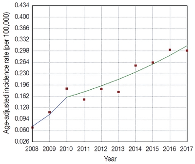
- Upward trend in follicular lymphoma among the Korean population: 10-year experience at a large tertiary institution
- Meejeong Kim, Hee Sang Hwang, Hyungwoo Cho, Dok Hyun Yoon, Cheolwon Suh, Chan Sik Park, Heounjeong Go, Jooryung Huh
- J Pathol Transl Med. 2021;55(5):330-337. Published online September 2, 2021
- DOI: https://doi.org/10.4132/jptm.2021.07.25
- 8,189 View
- 129 Download
- 4 Web of Science
- 5 Crossref
-
 Abstract
 PDF
PDF Supplementary Material
 - Background
Follicular lymphoma (FL) is the second most common non-Hodgkin lymphoma (NHL) in Western countries. However, it is relatively rare in Asia. This study examined epidemiologic characteristics of FL in South Korea, with an emphasis on recent trends of increase in cases.
Methods
We retrospectively examined 239 cases of newly diagnosed FL at a large tertiary institution in Korea (Asan Medical Center, Seoul, Republic of Korea) between 2008 and 2017. Age-adjusted incidence rates and clinicopathological variables were analyzed, and joinpoint regression analysis was used to identify the changes.
Results
The age-adjusted incidence of FL significantly increased during the study period (p = .034), and the ratio of (relative incidence) patients with FL to patients with NHL increased from 4.28% to 9.35% in the same period. Over the 10-year study assessment duration, the proportion of patients with stage III/IV FL (p = .035) and expression of BCL2 (p = .022) or BCL6 (p = .039) significantly increased. From 2013–2017, the proportion of patients with highrisk Follicular Lymphoma International Prognostic Index (FLIPI) score increased (21.5% to 28.7%), whereas that of low-risk FLIPI decreased (55.4% to 38.6%), although those results were not statistically significant (p = .066).
Conclusions
We found an increasing incidence of FL, with a disproportionate increase in the incidence of high-stage disease and recent changes in the clinicopathologic features of the Korean patient population. -
Citations
Citations to this article as recorded by
- Incidence Trend of Follicular Lymphoma in Taiwan Compared to Japan and Korea, 2001–2019
Liang-Chun Chiu, Chih-Wen Lin, Hung-Ju Li, Jian-Han Chen, Fu-Cheng Chuang, Sheng-Fung Lin, Yu Chang, Yu-Chieh Su
Journal of Clinical Medicine.2023; 12(4): 1417. CrossRef - A Case Report on the Complete Response of a Patient with Recurrent Follicular Lymphoma Treated with Integrative Medicine
Kyung-dug Park, Jisoo Kim, Yoona Oh, Beom-Jin Jeong, Yu-jin Jung, Sunhwi Bang
The Journal of Internal Korean Medicine.2023; 44(3): 585. CrossRef - Recent Updates on Diagnosis and Treatment of Follicular Lymphoma
Ga-Young Song, Deok-Hwan Yang
The Korean Journal of Medicine.2023; 98(5): 231. CrossRef - Classical Hodgkin lymphoma following follicular lymphoma: a case report
Bomi Kim
Journal of Yeungnam Medical Science.2023; 40(Suppl): S113. CrossRef - Incidence, clinicopathological features and genetics of in‐situ follicular neoplasia: a comprehensive screening study in a Japanese cohort
Naoki Oishi, Takahiro Segawa, Kunio Miyake, Kunio Mochizuki, Tetsuo Kondo
Histopathology.2022; 80(5): 820. CrossRef
- Incidence Trend of Follicular Lymphoma in Taiwan Compared to Japan and Korea, 2001–2019
- Human Papillomavirus Serologic Profiles of Selected Filipinos with Head and Neck Squamous Cell Carcinoma
- Pia Marie Albano, Christianne Salvador, Jose Orosa, Sheryl Racelis, Modesty Leaño, Angelika Michel, John Donnie Ramos, Dana Holzinger, Michael Pawlita
- J Pathol Transl Med. 2019;53(5):273-279. Published online May 30, 2019
- DOI: https://doi.org/10.4132/jptm.2019.05.12
- 9,641 View
- 196 Download
- 1 Web of Science
- 1 Crossref
-
Abstract
PDF
- Background
The low prevalence of human papillomavirus (HPV) DNA and mRNA in biopsy samples of Filipinos with head and neck squamous cell carcinoma (HNSCC) has been reported previously. Here, the HPV serologic profiles of HNSCC cases were analyzed and associated with life-style and sexual practices.
Methods
Serum samples were collected between May 2012 and September 2013 from HNSCC patients (n = 22) in the northwest region of the Philippines, and age- and sex-matched clinically healthy controls. Antibodies to capsid and early oncoproteins of HPV16, 18, 31, 33, 45, 52, 58, 6, and 11 were analyzed using multiplex serology.
Results
Most of the cases were males with tumors of the oral cavity or larynx. Two of the cases tested positive for at least one of the early oncoproteins (E6, E7, E1, and/or E2) of HPV16, and 11 did not display reactivity to any HPV early or late oncoproteins. Of the controls, four tested positive for at least one of the HPV16 early oncoproteins, and 10 were non-reactive to all HPV types. Titers to HPV16 E6 or E7 of the seropositive cases and controls were considerably lower than those typically observed in economically developed countries.
Conclusions
The low HPV titers seen here are consistent with the results of molecular analyses for this population. Hence, the seropositivity of some of the HNSCC cases is likely an indication of prior exposure to the virus and not the presence of HPV-driven tumors. -
Citations
Citations to this article as recorded by- Social determinants of sex disparities in cancer in Southeast Asia
Ma. Veronica Pia N. Arevalo, Ethan Angelo S. Maslog, Katherine Donatela Manlongat, Eric David B. Ornos, Imjai Chitapanarux, Michelle Ann B. Eala, Edward Christopher Dee
iScience.2023; 26(7): 107110. CrossRef
- Social determinants of sex disparities in cancer in Southeast Asia
- Acute Atherosis of the Uterine Spiral Arteries: Clinicopathologic Implications
- Joo-Yeon Kim, Yeon Mee Kim
- J Pathol Transl Med. 2015;49(6):462-471. Published online November 4, 2015
- DOI: https://doi.org/10.4132/jptm.2015.10.23
- 22,585 View
- 246 Download
- 36 Web of Science
- 40 Crossref
-
Abstract
PDF
- Acute atherosis is unique vascular changes of the placenta associated with poor placentation. It is characterized by subendothelial lipid-filled foam cells, fibrinoid necrosis of the arterial wall, perivascular lymphocytic infiltration, and it is histologically similar to early-stage atherosclerosis. Acute atherosis is rare in normal pregnancies, but is frequently observed in non- transformed spiral arteries in abnormal pregnancies, such as preeclampsia, small for gestational age (SGA), fetal death, spontaneous preterm labor and preterm premature rupture of membranes. In preeclampsia, spiral arteries fail to develop physiologic transformation and retain thick walls and a narrow lumen. Failure of physiologic transformation of spiral arteries is believed to be the main cause of uteroplacental ischemia, which can lead to the production of anti-angiogenic factors and induce endothelial dysfunction and eventually predispose the pregnancy to preeclampsia. Acute atherosis is more frequently observed in the spiral arteries of the decidua of the placenta (parietalis or basalis) than in the decidual or myometrial segments of the placental bed. The presence and deeper location of acute atherosis is associated with poorer pregnancy outcomes, more severe disease, earlier onset of preeclampsia, and a greater frequency of SGA neonates in patients with preeclampsia. Moreover, the idea that the presence of acute atherosis in the placenta may increase the risk of future cardiovascular disease in women with a history of preeclampsia is of growing concern. Therefore, placental examination is crucial for retrospective investigation of pregnancy complications and outcomes, and accurate placental pathology based on universal diagnostic criteria in patients with abnormal pregnancies is essential for clinicopathologic correlation.
-
Citations
Citations to this article as recorded by- Placental vascular remodeling in preeclampsia: A three-dimensional analysis of microvascular alterations across disease severity
Mingqun Li, Xiaoqiang Han, Yao Peng, Yang He, Qiangqiang You, Jiaqi Zhang
Placenta.2026; 174: 96. CrossRef - Placental aberrant inflammation and spatial-specific lipid metabolism contribute to hypertensive disorder of pregnancy susceptibility in preeclampsia offspring
Pei-ran Hu, Jing-hui Xu, Yan Shi, Ying Zhu, Gao-chen Zhang, Jie-ru Yang, Yue Xu, Ming-hao Li, Xian-hua Lin, Yu Zhang, He-feng Huang
Biochimica et Biophysica Acta (BBA) - Molecular Basis of Disease.2026; 1872(4): 168168. CrossRef - Cardiac Implications of Preeclampsia: A Review
Beani J. Forst, Linda R. Chambliss, David S. Majdalany
Journal of Personalized Medicine.2026; 16(5): 265. CrossRef - Circulating vascular endothelial growth factor receptor‐3, a pro‐lymphangiogenic and pro‐angiogenic mediator, is decreased in pre‐eclampsia
Ana C. Palei, Julyane N. S. Kaihara, Ricardo C. Cavalli, Valeria C. Sandrim
International Journal of Gynecology & Obstetrics.2025; 168(1): 210. CrossRef - ECHS1 as a Lipid Metabolism Biomarker for Pediatric Focal Segmental Glomerulosclerosis
Chao He, Wei Peng, Sheng Li, Can Xu, Xiuping Chen, Yuanhan Qin, Nasar Alwahaibi
PLOS ONE.2025; 20(3): e0319049. CrossRef - PlacEntal Acute atherosis RefLecting Subclinical systemic atherosclerosis in women up to 20 years after pre-eclampsia (PEARLS): research protocol for a cohort study
Gwyneth Jansen, Robert-Jan Alers, Emma BNJ Janssen, Laura M Jorissen, Eri Morina - Shijaku, Carmen Severens-Rijvers, Arnoud van ’t Hof, J van Drongelen, Ralph R Scholten, Salwan Al-Nasiry, Droima Stevens, Wessel Ganzevoort, Sanne Gordijn, Jérôme Cornette,
BMJ Open.2025; 15(5): e100542. CrossRef - Understanding Preeclampsia: Cardiovascular Pathophysiology, Histopathological Insights and Molecular Biomarkers
Kaltrina Kutllovci Hasani, Nurxhan Ajeti, Nandu Goswami
Medical Sciences.2025; 13(3): 154. CrossRef - Evidence that atherosis of the spiral artery represents atherosclerotic lesions similar to those of native and transplant-induced atherosclerosis: implications for understanding the pathophysiology of obstetrical syndromes and long-term cardiovascular ris
Carlos A. Labarrere, Roberto Romero, Hector L. DiCarlo, James W. Hardin, Yeon Mee Kim, Arun Meyyazhagan, Offer Erez, Piya Chaemsaithong, Liliana Voto, Awoniyi Awonuga, Tinnakorn Chaiworapongsa, Ghassan S. Kassab
American Journal of Obstetrics and Gynecology.2025;[Epub] CrossRef - Human Placenta and Evolving Insights into Pathological Changes of Preeclampsia: A Comprehensive Review of the Last Decade
Diana Maria Chiorean, Esra Cobankent Aytekin, Melinda-Ildiko Mitranovici, Sabin Gligore Turdean, Mirpooya Salehi Moharer, Ovidiu Simion Cotoi, Havva Serap Toru
Fetal and Pediatric Pathology.2024; 43(1): 33. CrossRef - Effects of hypertensive disorders of pregnancy on the complications in very low birth weight neonates
Baoquan Zhang, Xiujuan Chen, Changyi Yang, Huiying Shi, Wenlong Xiu
Hypertension in Pregnancy.2024;[Epub] CrossRef - Prevention of Pregnancy Complications Using a Multimodal Lifestyle, Screening, and Medical Model
Jim Parker, Pierre Hofstee, Shaun Brennecke
Journal of Clinical Medicine.2024; 13(15): 4344. CrossRef - Placental growth factor mediates pathological uterine angiogenesis by activating the NFAT5-SGK1 signaling axis in the endometrium: implications for preeclampsia development
Janet P. Raja Xavier, Toshiyuki Okumura, Melina Apweiler, Nirzari A. Chacko, Yogesh Singh, Sara Y Brucker, Satoru Takeda, Florian Lang, Madhuri S Salker
Biological Research.2024;[Epub] CrossRef - Genome-wide DNA methylation and gene expression in human placentas derived from assisted reproductive technology
Pauliina Auvinen, Jussi Vehviläinen, Karita Rämö, Ida Laukkanen, Heidi Marjonen-Lindblad, Essi Wallén, Viveca Söderström-Anttila, Hanna Kahila, Christel Hydén-Granskog, Timo Tuuri, Aila Tiitinen, Nina Kaminen-Ahola
Communications Medicine.2024;[Epub] CrossRef - Missing links in preeclampsia cell model systems of endothelial dysfunction
Sarah Viana-Mattioli, Miriam Helena Fonseca-Alaniz, Iguaracy Pinheiro-de-Sousa, José Eduardo Krieger, Valéria Cristina Sandrim
Trends in Molecular Medicine.2023; 29(7): 541. CrossRef - Roles of maternal HDL during pregnancy
Laura A. Woollett, Janet M. Catov, Helen N. Jones
Biochimica et Biophysica Acta (BBA) - Molecular and Cell Biology of Lipids.2022; 1867(3): 159106. CrossRef - The role of the placenta in spontaneous preterm labor and delivery with intact membranes
Sunil Jaiman, Roberto Romero, Gaurav Bhatti, Eunjung Jung, Francesca Gotsch, Manaphat Suksai, Dahiana M. Gallo, Tinnakorn Chaiworapongsa, Nicholas Kadar
Journal of Perinatal Medicine.2022; 50(5): 553. CrossRef - Gestational Antibodies to C. pneumoniae, H. pylori and CMV in Women with Preeclampsia and in Matched Controls
Abdul Wajid, David Todem, Mark R. Schleiss, David F. Colombo, Nigel S. Paneth
Maternal and Child Health Journal.2022; 26(10): 2040. CrossRef - Toward a new taxonomy of obstetrical disease: improved performance of maternal blood biomarkers for the great obstetrical syndromes when classified according to placental pathology
Roberto Romero, Eunjung Jung, Tinnakorn Chaiworapongsa, Offer Erez, Dereje W. Gudicha, Yeon Mee Kim, Jung-Sun Kim, Bomi Kim, Juan Pedro Kusanovic, Francesca Gotsch, Andreea B. Taran, Bo Hyun Yoon, Sonia S. Hassan, Chaur-Dong Hsu, Piya Chaemsaithong, Nardh
American Journal of Obstetrics and Gynecology.2022; 227(4): 615.e1. CrossRef - Preeclampsia and Fetal Growth Restriction as Risk Factors of Future Maternal Cardiovascular Disease—A Review
Sylwia Sławek-Szmyt, Katarzyna Kawka-Paciorkowska, Aleksandra Ciepłucha, Maciej Lesiak, Mariola Ropacka-Lesiak
Journal of Clinical Medicine.2022; 11(20): 6048. CrossRef - The Role of NF-κB in Uterine Spiral Arteries Remodeling, Insight into the Cornerstone of Preeclampsia
Maciej W. Socha, Bartosz Malinowski, Oskar Puk, Mateusz Wartęga, Martyna Stankiewicz, Anita Kazdepka-Ziemińska, Michał Wiciński
International Journal of Molecular Sciences.2021; 22(2): 704. CrossRef - Pathogenesis of uteroplacental acute atherosis: An update on current research
Shu Li, Yan‐Wei Hu
American Journal of Reproductive Immunology.2021;[Epub] CrossRef - Disorders of placental villous maturation are present in one-third of cases with spontaneous preterm labor
Sunil Jaiman, Roberto Romero, Percy Pacora, Offer Erez, Eunjung Jung, Adi L. Tarca, Gaurav Bhatti, Lami Yeo, Yeon Mee Kim, Chong Jai Kim, Jung-Sun Kim, Faisal Qureshi, Suzanne M. Jacques, Nardhy Gomez-Lopez, Chaur-Dong Hsu
Journal of Perinatal Medicine.2021; 49(4): 412. CrossRef - The COVID-19 Pandemic: an Appraisal of its Impact on Human Immunodeficiency Virus Infection and Pre-Eclampsia
Rowen Govender, Jagidesa Moodley, Thajasvarie Naicker
Current Hypertension Reports.2021;[Epub] CrossRef - Acute Atherosis Lesions at the Fetal-Maternal Border: Current Knowledge and Implications for Maternal Cardiovascular Health
Daniel Pitz Jacobsen, Heidi Elisabeth Fjeldstad, Guro Mørk Johnsen, Ingrid Knutsdotter Fosheim, Kjartan Moe, Patji Alnæs-Katjavivi, Ralf Dechend, Meryam Sugulle, Anne Cathrine Staff
Frontiers in Immunology.2021;[Epub] CrossRef - Aetiology, prophylaxis and management of preeclampsia
Karolina Gronkowska
Acta Universitatis Lodziensis. Folia Biologica et Oecologica.2021; 17: 111. CrossRef - HMOX1 is partly responsible for phenotypic and functional abnormalities in mesenchymal stem cells/stromal cells from placenta of preeclampsia (PE) patients
Yasser S. Basmaeil, Dana Algudiri, Reem Alenzi, Abdullah Al Subayyil, Ayodele Alaiya, Tanvir Khatlani
Stem Cell Research & Therapy.2020;[Epub] CrossRef - Analyzing Preeclampsia as the Tip of the Iceberg Represented by Women with Long-Term Cardiovascular Disease, Atherosclerosis, and Inflammation
Angélica Lemos Debs Diniz, Maria Marta Bini Martins Paes, Aline Debs Diniz
Current Atherosclerosis Reports.2020;[Epub] CrossRef - Lipids in preeclampsia: pathogenic parallels to atherosclerosis
V. I. Shcherbakov, Ya. V. Polonskaya, E. V. Kashtanova, A. V. Shirinskaya
"Arterial’naya Gipertenziya" ("Arterial Hypertension").2020; 26(2): 163. CrossRef - Transthyretin increases migration and invasion of rat placental trophoblast cells
Xiao‐Peng Ma, Chong‐Dong Liu, Guang‐Ming Cao, Zhen‐Yu Zhang
FEBS Open Bio.2020; 10(8): 1568. CrossRef - Early Onset Preeclampsia Is Associated With Glycocalyx Degradation and Reduced Microvascular Perfusion
Tracey L. Weissgerber, Oscar Garcia‐Valencia, Natasa M. Milic, Elizabeth Codsi, Hajrunisa Cubro, Meryl C. Nath, Wendy M. White, Karl A. Nath, Vesna D. Garovic
Journal of the American Heart Association.2019;[Epub] CrossRef - The immunophenotype of decidual macrophages in acute atherosis
Navleen Gill, Yaozhu Leng, Roberto Romero, Yi Xu, Bogdan Panaitescu, Derek Miller, Afrah Arif, Salma Mumuni, Faisal Qureshi, Chaur‐Dong Hsu, Sonia S. Hassan, Anne Cathrine Staff, Nardhy Gomez‐Lopez
American Journal of Reproductive Immunology.2019;[Epub] CrossRef - The potential effects of pomegranate peel extract and bee venom in improving the diabetes induced damaging of spiral artery
HIH El-Sayyad, HA El-Ghawet, AMA El-Sayed
Studies on Stem Cells Research and Therapy.2019; 5(1): 007. CrossRef - Race and risk of maternal vascular malperfusion lesions in the placenta
Vanessa Assibey-Mensah, W. Tony Parks, Alison D. Gernand, Janet M. Catov
Placenta.2018; 69: 102. CrossRef - Preclinical atherosclerosis at the time of pre‐eclamptic pregnancy and up to 10 years postpartum: systematic review and meta‐analysis
N. M. Milic, J. Milin‐Lazovic, T. L. Weissgerber, G. Trajkovic, W. M. White, V. D. Garovic
Ultrasound in Obstetrics & Gynecology.2017; 49(1): 110. CrossRef - Is an episode of suspected preterm labor that subsequently leads to a term delivery benign?
Roberto Romero, Offer Erez, Eli Maymon, Percy Pacora
American Journal of Obstetrics and Gynecology.2017; 216(2): 89. CrossRef - Establishment of the Human Uteroplacental Circulation: A Historical Perspective
Kenna Degner, Ronald R. Magness, Dinesh M. Shah
Reproductive Sciences.2017; 24(5): 753. CrossRef - Preeclampsia and coronary plaque erosion: Manifestations of endothelial dysfunction resulting in cardiovascular events in women
Saskia C.A. de Jager, John A.L. Meeuwsen, Freeke M. van Pijpen, Gerbrand A. Zoet, Arjan D. Barendrecht, Arie Franx, Gerard Pasterkamp, Bas B. van Rijn, Marie-José Goumans, Hester M. den Ruijter
European Journal of Pharmacology.2017; 816: 129. CrossRef - Placental histopathology lesions and pregnancy outcome in pregnancies complicated with symptomatic vs. non-symptomatic placenta previa
Eran Weiner, Hadas Miremberg, Ehud Grinstein, Letizia Schreiber, Shimon Ginath, Jacob Bar, Michal Kovo
Early Human Development.2016; 101: 85. CrossRef - Porphyromonas gingivalis within Placental Villous Mesenchyme and Umbilical Cord Stroma Is Associated with Adverse Pregnancy Outcome
Sizzle F. Vanterpool, Jasper V. Been, Michiel L. Houben, Peter G. J. Nikkels, Ronald R. De Krijger, Luc J. I. Zimmermann, Boris W. Kramer, Ann Progulske-Fox, Leticia Reyes, Motohiro Komaki
PLOS ONE.2016; 11(1): e0146157. CrossRef - Pregnant women with heart disease: Placental characteristics and their association with fetal adverse events
Fabio V. Lima, Paraskevi Koutrolou-Sotiropoulou, Puja B. Parikh, Cecilia Avila, Javed Butler, Kathleen Stergiopoulos
Acute Cardiac Care.2016; 18(3): 56. CrossRef
- Placental vascular remodeling in preeclampsia: A three-dimensional analysis of microvascular alterations across disease severity
- Oncocytic Lipoadenoma: A Rare Case of Parotid Gland Tumor and Review of the Literature
- Chen-lin Chi, Tseng-tong Kuo, Li-yu Lee
- J Pathol Transl Med. 2015;49(2):144-147. Published online March 12, 2015
- DOI: https://doi.org/10.4132/jptm.2014.02.10
- 12,213 View
- 70 Download
- 9 Web of Science
- 9 Crossref
-
Abstract
PDF
- Oncocytic lipoadenoma is a rare tumor, with only 18 cases having been reported since the first in 1998. We encountered a case of oncocytic lipoadenoma presenting as a slowly growing parotid mass in a 71-year-old man. This tumor is characteristically comprised of a mixture of oncocytes and adipocytes. The present case is one of five reported cases of oncocytic lipoadenoma showing sebaceous differentiation. The results of immunohistochemical study with DOG1 antibody supported the origination of this tumor in the striated duct.
-
Citations
Citations to this article as recorded by- Multimodal Imaging of Oncocytic Lipoadenoma Arising from the Parotid Deep Lobe with Medial Extension into the Parapharyngeal Space: A Case Report with Histopathologic Findings and Literature Review
Jong-Uk Lee, Hye Jin Baek, Kwang Ho Choi, Eun Cho, Hyo Jung An
Diagnostics.2026; 16(9): 1366. CrossRef - Oncocytic lipoadenoma of the parotid gland: a case report and a review of the literature
Jood K Alotaibi, Turki Mohammed Almuhaimid, Ghada Abdallah Moumneh
Journal of Surgical Case Reports.2024;[Epub] CrossRef - Oncocytic sialolipoma of parotid gland: Case report and literature review
VenuPatel Sureja, KoyyeRavindranath Tagore
Indian Journal of Pathology and Microbiology.2023; 66(3): 591. CrossRef - Complex Component of Oncocytic and Non-Oncocytic Lipoadenomas in the Parotid Gland: A Case Report
Fuyuki Sato, Takashi Nakajima, Takashi Sugino
Diagnostics.2021; 11(8): 1478. CrossRef - Oncocyitic lipoadenoma of the parotid gland
Renato PIANTANIDA, Alberto CARANTI, Adele CHIESA, Jessica BARIZZI, Ulrike PERRIARD, Filippo BARUCCA, Antonio PELLANDA
Otorinolaringologia.2021;[Epub] CrossRef - A case of oncocytic lipoadenoma of the submandibular gland and its diagnostic cytology challenges
Khaled A. Murshed, Ammar Khalafalla, Belal Alani, Hanan Farghaly, Moustafa Alkhalil
Diagnostic Cytopathology.2020; 48(4): 364. CrossRef - An extremely rare case of giant oncocytic adenolipoma of the parotid gland
Dipesh Shakya, Ajit Nepal
Clinical Case Reports.2020; 8(12): 2390. CrossRef - A rare cause of primary hyperparathyroidism: Parathyroid lipoadenoma
Sabri Özden, Servet Güreşci, Barış Saylam, Gül Dağlar
Auris Nasus Larynx.2018; 45(6): 1245. CrossRef - Oncocytic osteolipoadenoma of the submandibular gland
Domenico Corradi, Rodolfo Monaco, Giulia D'Angelo, Paola Bini, Teore Ferri, Enrico M Silini
Histopathology.2016; 69(1): 148. CrossRef
- Multimodal Imaging of Oncocytic Lipoadenoma Arising from the Parotid Deep Lobe with Medial Extension into the Parapharyngeal Space: A Case Report with Histopathologic Findings and Literature Review
- A Case of Mixed Adenoneuroendocrine Carcinoma of the Common Bile Duct: Initially Diagnosed as Cholangiocarcinoma
- Soon Wook Lee, In Seok Lee, Yu Kyung Cho, Jae Myung Park, Sang Woo Kim, Myung-Gyu Choi, Kyu Yong Choi, Myung Ah Lee, Tae Ho Hong, Young Kyoung You, Eun-Sun Jung
- Korean J Pathol. 2014;48(6):445-448. Published online December 31, 2014
- DOI: https://doi.org/10.4132/KoreanJPathol.2014.48.6.445
- 11,078 View
- 44 Download
- 12 Crossref
-
PDF
-
Citations
Citations to this article as recorded by- Long-term survival after chemotherapy combined immunotherapy for recurrent mixed neuroendocrine–non-neuroendocrine neoplasms of the common bile duct
Jia Li, Chunyan Yuan, Yulin Pan, Yuanyuan Yang, Nannan Lai, Xia Sheng
Clinical Journal of Gastroenterology.2025; 18(4): 653. CrossRef - Neuroendocrine carcinoma of the common hepatic duct coexisting with distal cholangiocarcinoma: A case report and review of literature
Fei Chen, Wei-Wei Li, Juan-Fen Mo, Min-Jie Chen, Su-Hang Wang, Shu-Ying Yang, Zheng-Wei Song
World Journal of Gastrointestinal Surgery.2024; 16(5): 1449. CrossRef - Comparison of Metastatic Patterns Among Neuroendocrine Tumors, Neuroendocrine Carcinomas, and Nonneuroendocrine Carcinomas of Various Primary Organs
Hyung Kyu Park, Ghee Young Kwon
Journal of Korean Medical Science.2023;[Epub] CrossRef - Mixed adenoneuroendocrine carcinoma of the distal bile duct: a case report
Takashi Maeda, Kyohei Yugawa, Nao Kinjo, Hiroto Kayashima, Daisuke Imai, Koto Kawata, Shinichiro Ikeda, Keitaro Edahiro, Kazuki Takeishi, Tomohiro Iguchi, Noboru Harada, Mizuki Ninomiya, Shohei Yamaguchi, Kozo Konishi, Shinichi Tsutsui, Hiroyuki Matsuda
Surgical Case Reports.2020;[Epub] CrossRef - The clinical profiles, management, and prognostic factors of biliary mixed neuroendocrine nonneuroendocrine neoplasms
Li-Jia Wen, Jun-Hong Chen, Hong-Ji Xu, Qiong Yu, Yu Deng, Kai Liu
Medicine.2020; 99(50): e23271. CrossRef - Rapidly progressed neuroendocrine carcinoma in the extrahepatic bile duct: a case report and review of the literature
Mariko Kamiya, Naoto Yamamoto, Yuto Kamioka, Hirohide Inoue, Hirokazu Yotsumoto, Masaaki Murakawa, Toru Aoyama, Kota Washimi, Kae Kawachi, Takashi Oshima, Makoto Ueno, Norio Yukawa, Yasushi Rino, Munetaka Masuda, Soichiro Morinaga
Surgical Case Reports.2020;[Epub] CrossRef - Mixed adenoneuroendocrine carcinoma of the hepatic bile duct: a case report and review of the literature
Sulai Liu, Zhendong Zhong, Meng Xiao, Yinghui Song, Youye Zhu, Bo Hu, Zengpeng Sun, Weimin Yi, Chuang Peng
BMC Gastroenterology.2020;[Epub] CrossRef - Mixed adenoendocrine carcinoma in the extrahepatic biliary tract: A case report and literature review
Liang Zhang, Zhengtao Yang, Qing Chen, Mengxia Li, Xiaolu Zhu, Dalong Wan, Haiyang Xie, Shusen Zheng
Oncology Letters.2019;[Epub] CrossRef - Large Cell Neuroendocrine Carcinoma Coexisting with Adenocarcinoma in the Extrahepatic Bile Duct
Masami Yuda, Teruyuki Usuba, Shin Hagiwara, Masahisa Okuma, Hisatoshi Asano, Hitoshi Sakuda, Hiroaki Katagi, Yoshiyuki Furukawa
The Japanese Journal of Gastroenterological Surgery.2018; 51(3): 187. CrossRef - Mixed Adenoneuroendocrine Carcinoma of the Distal Bile Duct
Chiaki Uchida, Yoshikazu Toyoki, Keinosuke Ishido, Daisuke Kudo, Norihisa Kimura, Shinji Tsutsumi, Takuji Kagiya, Toshiro Kimura, Kenichi Hakamada
The Japanese Journal of Gastroenterological Surgery.2017; 50(1): 43. CrossRef - Mixed adenoneuroendocrine carcinoma of the distal bile duct: A case report
Toshiaki Komo, Toshihiko Kohashi, Akira Nakashima, Ichiro Ohmori, Jun Hihara, Hidenori Mukaida, Mayumi Kaneko, Naoki Hirabayashi
International Journal of Surgery Case Reports.2017; 39: 203. CrossRef - Common Hepatic Duct Mixed Adenoneuroendocrine Carcinoma Masquerading as Cholangiocarcinoma
Sali Priyanka Akhilesh, Yadav Kamal Sunder, Tampi Chandralekha, Parikh Samir, Wagle Prasad Kashinath
Case Reports in Gastrointestinal Medicine.2016; 2016: 1. CrossRef
- Long-term survival after chemotherapy combined immunotherapy for recurrent mixed neuroendocrine–non-neuroendocrine neoplasms of the common bile duct
- Myoepithelial Carcinoma of Soft Tissue: A Case Report and Review of the Literature
- Chang Hwan Choi, Young Chae Chu, Lucia Kim, Suk Jin Choi, In Suh Park, Jee Young Han, Joon Mee Kim
- Korean J Pathol. 2014;48(6):413-417. Published online December 31, 2014
- DOI: https://doi.org/10.4132/KoreanJPathol.2014.48.6.413
- 15,284 View
- 134 Download
- 9 Crossref
-
PDF
-
Citations
Citations to this article as recorded by- Hyalinizing Clear Cell Carcinoma of the Lung With Uncommon Distant Cutaneous Metastasis and Aggressive Clinical Course
David M. Gustafson, Sansar Tiwari, Catherine G. Chung
Journal of Cutaneous Pathology.2026;[Epub] CrossRef - Myoepithelial Carcinoma Mimicking Basal Cell Carcinoma: A Case Report
Farlin Asharaff, Neena Nayak, Roger Webb, Karwan Moutasim, Soogan Lalla
Cureus.2025;[Epub] CrossRef - Fine‐needle aspiration cytology of retroperitoneal myoepithelial carcinoma: A rare encounter with diagnostic dilemmas
Aadya Kerkar, Ajay Savlania, Reetu Kundu, Suvradeep Mitra, Manish Rohilla, Harmandeep Singh, Harish Bhujade
Diagnostic Cytopathology.2024;[Epub] CrossRef - EWSR1::NR4A3 gene fusion in a cutaneous atypical myoepithelial neoplasm
Ashley Rose Scholl, Evelyna Kliassov, Diana M. Cardona, Rex Bentley, Rami N. Al‐Rohil
Journal of Cutaneous Pathology.2023; 50(7): 601. CrossRef - Abdominal myoepithelial carcinoma: A rare abdominal wall entity of an uncommon tumor
Daania Shoaib, Saqib Raza Khan, Yasmin Abdul Rashid, Muhammad Nauman Zahir
International Journal of Surgery Case Reports.2022; 99: 107618. CrossRef - Adult soft tissue myoepithelial carcinoma: treatment outcomes and efficacy of chemotherapy
Florence Chamberlain, Elena Cojocaru, Mariana Scaranti, Jonathan Noujaim, Anastasia Constantinou, Khin Thway, Cyril Fisher, Christina Messiou, Dirk C. Strauss, Aisha Miah, Shane Zaidi, Charlotte Benson, Spyridon Gennatas, Robin L. Jones
Medical Oncology.2020;[Epub] CrossRef - Foot plantar soft tissue malignant myoepithelioma tumor: Case report and review of the literature
Manuel Trevino, Chetan Moorthy, Lisa Kafchinski, Daniel Bustamante
Clinical Imaging.2020; 61: 90. CrossRef - Presumed choroidal metastasis from soft tissue myoepithelial carcinoma
Michelle M. Hui, Rohan Merani, Fiona Bonar, Angela M. Hong, Adrian T. Fung
American Journal of Ophthalmology Case Reports.2019; 14: 55. CrossRef - Myoepithelial carcinoma of the elbow diagnosed by immunohistochemistry: Case report of an uncommon neoplasm with metastatic recurrence
Madhura Mahapatra, Travis Lambert, Abdal Rahman El-Mallah, Andressa Balbi, Mohamad Aziz
Case Reports International.2019; 8(2): 1. CrossRef
- Hyalinizing Clear Cell Carcinoma of the Lung With Uncommon Distant Cutaneous Metastasis and Aggressive Clinical Course
- Myxoid Liposarcoma with Cartilaginous Differentiation: A Case Study with Cytogenetical Analysis
- Hyunchul Kim, Won Hwangbo, Sangjeong Ahn, Suhjin Kim, Insun Kim, Chul Hwan Kim
- Korean J Pathol. 2013;47(3):284-288. Published online June 25, 2013
- DOI: https://doi.org/10.4132/KoreanJPathol.2013.47.3.284
- 10,510 View
- 43 Download
- 3 Crossref
-
Abstract
PDF
Myxoid liposarcoma is a subtype of liposarcoma. This specific subtype can be identified based on its characteristic histological and cytogenetical features. The tumor has a fusion transcript of the
CHOP andTLS genes, which is caused by t(12;16)(q13;p11). Most of the fusion transcripts that have been identified fall into three categories, specifically type I (exons 7-2), type II (exons 5-2), and type III (exons 8-2). A total of seven myxoid liposarcomas associated with the rare phenomenon of cartilaginous differentiation have been documented in the literature. Currently, only one of these cases has been cytogenetically analyzed, and the analysis indicated that it was a type IITLS-CHOP fusion transcript in both the typical myxoid liposarcoma and cartilaginous areas. This study presents a second report of myxoid liposarcoma with cartilaginous differentiation, and includes a cytogenetical analysis of both the myxoid and cartilaginous areas.-
Citations
Citations to this article as recorded by- Myxoid liposarcoma with nuclear pleomorphism: a clinicopathological and molecular study
Naoki Kojima, Takashi Kubo, Taisuke Mori, Kaishi Satomi, Yuko Matsushita, Shintaro Iwata, Yasushi Yatabe, Koichi Ichimura, Akira Kawai, Hitoshi Ichikawa, Akihiko Yoshida
Virchows Archiv.2024; 484(1): 71. CrossRef - The Conundrum of Dedifferentiation in a Liposarcoma at a Peculiar Location: A Case Report and Literature Review
Ana-Maria Ciongariu, Adrian-Vasile Dumitru, Cătălin Cîrstoiu, Bogdan Crețu, Maria Sajin, Dana-Antonia Țăpoi, Aminia-Diana Ciobănoiu, Adrian Bejenariu, Andrei Marin, Mariana Costache
Medicina.2023; 59(5): 967. CrossRef - Myxoid liposarcoma with cartilaginous differentiation showing DDIT3 rearrangement
Kayo Suzuki, Taketoshi Yasuda, Kenta Watanabe, Takeshi Hori, Masahiko Kanamori, Tomoatsu Kimura
Oncology Letters.2017;[Epub] CrossRef
- Myxoid liposarcoma with nuclear pleomorphism: a clinicopathological and molecular study
- Adenolipoma of the Skin Arising at Neck Region
- Hyun Seung Lee, Yoon Sang Song
- Korean J Pathol. 2012;46(6):587-589. Published online December 26, 2012
- DOI: https://doi.org/10.4132/KoreanJPathol.2012.46.6.587
- 11,678 View
- 66 Download
- 1 Crossref
-
Abstract
PDF
We report here a case of adenolipoma of the skin, an unusual variant of lipoma, arising on the neck. A 56-year-old man visited our hospital due to an anterior neck mass. An excisional biopsy was performed. The mass revealed a tan-yellow soft cut surface. We could not find any difference from other lipoma on gross inspection. Microscopically, the mass showed proliferation of mature adipocytes admixed with several eccrine units. The eccrine units were demonstrated by periodic acid-Schiff-positive granules in the secretory portions and by positivity of smooth muscle actin in the myoepithelial cells surrounding the eccrine glands. The tumor was completely excised, and the patient has been followed up without any evidence of recurrence so far.
-
Citations
Citations to this article as recorded by- Adenolipoma of the Skin: A Report of 11 Cases
Rawan Amir, Salwa Sheikh
Case Reports in Dermatology.2018; 10(1): 76. CrossRef
- Adenolipoma of the Skin: A Report of 11 Cases
- Primary Pulmonary Myxoid Liposarcoma with Translocation t(12;16)(q13;p11) in a Young Female Patient: A Brief Case Report
- Choonhee Son, Phil Jo Choi, Mee Sook Roh
- Korean J Pathol. 2012;46(4):392-394. Published online August 23, 2012
- DOI: https://doi.org/10.4132/KoreanJPathol.2012.46.4.392
- 8,943 View
- 50 Download
- 4 Crossref
-
Abstract
PDF
Primary liposarcoma of the lung is an extremely rare disease. To date, only 14 cases have been reported in the literature. We experienced a case of myxoid liposarcoma of the lung treated by surgery. The tumor was well-defined, solid, lobulated mass measuring 3.5×2 cm, involving the bronchus of the left lower lobe. Microscopically, myxoid liposarcoma was identified. The fluorescence
in situ hybridization confirmed the presence of a reciprocal translocation involving DNA damage-inducible transcript 3 (DDIT3 ) and fused in sarcoma (FUS ) genes. The patient is still alive with no recurrence or metastasis at the time of writing this report (on 20 months postoperatively). To our knowledge, this is the first cytogenetic case report of pulmonary myxoid liposarcoma.-
Citations
Citations to this article as recorded by- Primary Liposarcoma of the Spleen: Case Report With Review of the Literature
Elisa M. Wächtershäuser, Gabriele Köhler, Verena Böhmer, Alexander Marx, Achim Hellinger
International Journal of Surgical Pathology.2025; 33(4): 928. CrossRef - Primary intrathoracic liposarcomas: A clinicopathologic and molecular study of 43 cases in one of the largest medical centers of China
You Xie, Wenyi Jing, Wei Zhao, Ran Peng, Min Chen, Ting Lan, Heng Peng, Xin He, Huijiao Chen, Zhang Zhang, Hongying Zhang
Frontiers in Oncology.2022;[Epub] CrossRef - Primary Pulmonary Liposarcoma with Pancreatic Metastasis: A Rarest of Rare Intrathoracic Malignancy
Anirban Halder, Rituparna Biswas, Sujit Shukla, Nisha Rana, Vikas Yadav, Jaspreet Kaur
Indian Journal of Medical and Paediatric Oncology.2020; 41(04): 605. CrossRef - Primary dedifferentiated liposarcoma of the lung with rhabdomyoblastic and chrondroblastic differentiation
Anthony Longano, Alexandra DuGuesclin, Catherine Mitchell
Histopathology.2015; 67(6): 923. CrossRef
- Primary Liposarcoma of the Spleen: Case Report With Review of the Literature
- Diagnostic Value of
MDM2 andDDIT3 FluorescenceIn Situ Hybridization in Liposarcoma Classification: A Single-Institution Experience - Junhun Cho, Seung Eun Lee, Yoon-La Choi
- Korean J Pathol. 2012;46(2):115-122. Published online April 25, 2012
- DOI: https://doi.org/10.4132/KoreanJPathol.2012.46.2.115
- 12,292 View
- 105 Download
- 10 Crossref
-
Abstract
PDF
Background The amplification of murine double minutes (
MDM2 ) is the primary feature of well-differentiated liposarcomas (WDLPS) and dedifferentiated liposarcomas (DDLPS), whileDDIT3 rearrangement is the main one of myxoid liposarcomas (MLPS). Our aim was to evaluate the added value ofMDM2 amplification andDDIT3 rearrangement in making a diagnosis and classifying lipogenic tumors.Methods Eighty-two cases of liposarcoma and 60 lipomas diagnosed between 1995 and 2010 were analysed for
MDM2 amplification andDDIT3 rearrangement using a fluorescencein situ hybridization (FISH). The subtypes of liposarcoma were reclassified according to the molecular results, whose results were reviewed with an analysis of the relevant histologic and immunohistochemical findings.Results One case of lipoma (1.67%) was reclassified as a WDLPS. Of the liposarcomas, 13.4% (16/82) were reclassified after the molecular testing. Five cases of MLPS were reclassified as four cases of DDLPS and one case of myxoid lipoma. Two cases of WDLPS were reclassified as one case of spindle cell lipoma and another case of myxofibrosarcoma. Four cases of DDLPS were reclassified as two cases of leiomyosarcoma, one case of angiomyolipoma and another case of fibroinflammatory lesion. Of the six cases of pleomorphic liposarcoma, five were reclassified as DDLPS.
Conclusions In our series, a critical revision of diagnosis was found at a rate of 3.5% (5/142) after a review of the lipomatous lesions. The uses of molecular testing by
MDM2 andDDIT3 FISH were valuable to make an accurate subtyping of liposarcomas as well as to differentiate WDLPS from benign lipomatous tumor.-
Citations
Citations to this article as recorded by- Synchronous Presentation of Intraperitoneal and Retroperitoneal Dedifferentiated Liposarcoma: A Case Report
Yousra Mhande, Laila Merghat, Amal Hajri, Driss Erguibi, Saad Rifki El Jai
Cureus.2026;[Epub] CrossRef - Myxoid liposarcoma with nuclear pleomorphism: a clinicopathological and molecular study
Naoki Kojima, Takashi Kubo, Taisuke Mori, Kaishi Satomi, Yuko Matsushita, Shintaro Iwata, Yasushi Yatabe, Koichi Ichimura, Akira Kawai, Hitoshi Ichikawa, Akihiko Yoshida
Virchows Archiv.2024; 484(1): 71. CrossRef - FISH Diagnostic Assessment of MDM2 Amplification in Liposarcoma: Potential Pitfalls and Troubleshooting Recommendations
Alessandro Gambella, Luca Bertero, Milena Rondón-Lagos, Ludovica Verdun Di Cantogno, Nelson Rangel, Chiara Pitino, Alessia Andrea Ricci, Luca Mangherini, Isabella Castellano, Paola Cassoni
International Journal of Molecular Sciences.2023; 24(2): 1342. CrossRef - Expression of CTAG1B clone EPR13780 versus DDIT3 gene rearrangement distinguishes myxoid liposarcoma from its mimics with detection of novel DDIT3 gene copy number variations
Marwa M. Abdelaziz, Hanan Y. Tayel, Amany Abdel-Bary, Omnia M. Badawy
Journal of Histotechnology.2022; 45(2): 56. CrossRef - Musculoskeletal Tumors
Amit Singla, David S. Geller
Pediatric Clinics of North America.2020; 67(1): 227. CrossRef - Vulvar Myxoid Liposarcoma, an Extremely Rare Diagnosis: A Case Report and Review of Literature
Ligia Redroban, Nelson Montalvo
International Journal of Gynecological Pathology.2019; 38(1): 17. CrossRef - Molecular updates in adipocytic neoplasms✰
Elizabeth G. Demicco
Seminars in Diagnostic Pathology.2019; 36(2): 85. CrossRef - Application of MDM2 Fluorescence In Situ Hybridization and Immunohistochemistry in Distinguishing Dedifferentiated Liposarcoma From Other High-grade Sarcomas
Min Jeong Song, Kyung-Ja Cho, Jong-Seok Lee, Joon Seon Song
Applied Immunohistochemistry & Molecular Morphology.2017; 25(10): 712. CrossRef - FluorescenceIn SituHybridization forMDM2Amplification as a Routine Ancillary Diagnostic Tool for Suspected Well-Differentiated and Dedifferentiated Liposarcomas: Experience at a Tertiary Center
Khin Thway, Jayson Wang, John Swansbury, Toon Min, Cyril Fisher
Sarcoma.2015; 2015: 1. CrossRef - Complete response of a recurrent-metastatic liposarcoma with dedifferentiated histological features following the administration of trabectedin and review of literature
Tulay Kus, Gokmen Aktas, Mehmet Emin Kalender, Ediz Tutar, Esra Ulker, Celaletdin Camci
Journal of Cancer Research and Therapeutics.2015; 11(4): 974. CrossRef
- Synchronous Presentation of Intraperitoneal and Retroperitoneal Dedifferentiated Liposarcoma: A Case Report
- Lipofibromatosis: A Case Report.
- Tae Eun Kim, Tae Jung Kim, Youn Soo Lee, Chang Suk Kang, Sang In Shim, Kyo Young Lee
- Korean J Pathol. 2011;45(1):106-110.
- DOI: https://doi.org/10.4132/KoreanJPathol.2011.45.1.106
- 3,957 View
- 59 Download
-
Abstract
PDF
- Lipofibromatosis is a recently described rare benign fibrofatty tumor of childhood. It typically forms as an ill defined, slowly growing, painless mass. We present here the case of lipofibromatosis that occurred in a 21-year-old male who had complained of a bulging enlarged mass involving the right thigh and prepatella area for the previous 1 year. Magnetic resonance imaging showed an ill-defined reticular infiltration in the subcutaneous layer with subtle linear enhancement and high T2 signal intensity. The mass was surgically excised and it displayed an 11.0x5.5x1.5 cm-sized adipose appearance without encapsulation. Microscopically, the tumor was composed of alternating streaks of mature adipose tissue and a fibroblastic component that mainly involved the septa of adipose tissue. On immunohistochemical study, the fibroblastic component was positive for S-100, CD99, CD34, actin and bcl-2. He has shown an eventful recovery for 6 months after surgery.
- Pure Uterine Lipoma, a Very Rare Benign Tumor.
- Elif Ulker Akyildiz, Sema Ozuysal, Akgul Arici, Mehmet Aral Atalay
- Korean J Pathol. 2010;44(6):679-681.
- DOI: https://doi.org/10.4132/KoreanJPathol.2010.44.6.679
- 4,515 View
- 24 Download
- 1 Crossref
-
Abstract
PDF
- Pure lipomas of the uterus are very rare tumors that may be misdiagnosed on radiological examination due to their rarity and fat content. We present here the case of a 57-year-old postmenopausal woman who presented to the hospital with lower abdominal pain. Abdominal hysterectomy and bilateral salpingo-oophorectomy were performed under the prediagnoses of benign cystic ovarian teratoma or leiomyoma. On the histopathological examination of tissue samples, the tumor was composed of mature fat cells. There were a few smooth muscle cells confined to the periphery. Pure uterine lipoma may be asymptomatic or it may have symptoms similar to those of leiomyoma such as vaginal bleeding or pelvic pain. A pure lipoma should be diagnosed only if smooth muscle cells are confined to the periphery.
-
Citations
Citations to this article as recorded by- Coexistence of uterine lipoma, leiomyoma and endometrial polyp
Nilgün SÖĞÜTÇÜ, Nazlı Sena ŞEKER
Cukurova Medical Journal.2019; 44(3): 1139. CrossRef
- Coexistence of uterine lipoma, leiomyoma and endometrial polyp
- Thymofibrolipoma: A Brief Case Report.
- Gu Hyun Kang, Joungho Han, Tae Sung Kim, Yong Soo Choi, Sang Won Um
- Korean J Pathol. 2010;44(3):338-340.
- DOI: https://doi.org/10.4132/KoreanJPathol.2010.44.3.338
- 4,385 View
- 36 Download
- 3 Crossref
-
Abstract
PDF
- Thymofibrolipoma is an extremely rare tumor in the anterior mediastinum, and represents a histologic variant of the usual thymolipoma. Herein, we report a case of thymofibrolipoma in a 9-year-old girl who had a huge mass with fatty attenuation in the right hemithorax on chest computed tomography. She denied any subjective symptoms except mild fever. The surgically resected tumor was ovoid, soft and well-encapsulated, measuring 9.0 x 7.5 x 7.0 cm. The cut surface was light tan in color with yellowish streaks. Microscopically, two distinct areas were admixed in different proportions. One consisted of normal thymic tissue with subinvoluted features and the other was composed of extensive areas of collagenous tissue interspersed in mature adipose tissue. In a high power view, there were thin strands of remnant thymic epithelial cells, separating the pseudolobules. Thymofibrolipoma should be distinguished from other benign or malignant conditions, occurring in the anterior mediastinum, so that unnecessary treatment can be avoided.
-
Citations
Citations to this article as recorded by- Unusual thymoma subtypes
Michael A. den Bakker
Diagnostic Histopathology.2023; 29(2): 105. CrossRef - Thymofibrolipoma: a case report and review of the literature
Ryu Jokoji, Emiko Tomita
Diagnostic Pathology.2022;[Epub] CrossRef - Thymoangiolipoma: A rare histologic variant of thymolipoma in a patient with myasthenia gravis
Mohammad Hossein Anbardar, Fatemeh Amirmoezi, Armin Amirian
Rare Tumors.2020;[Epub] CrossRef
- Unusual thymoma subtypes
- The Expression of Apolipoprotein D in Hepatocellular Carcinoma.
- Hongxiu Han, Chan Kum Park
- Korean J Pathol. 2010;44(2):187-190.
- DOI: https://doi.org/10.4132/KoreanJPathol.2010.44.2.187
- 4,065 View
- 28 Download
-
Abstract
PDF
- BACKGROUND
Apolipoprotein D (Apo D) has recently been identified as a novel tumor suppressor gene. Apo D may have a profound effect on the carcinogenesis and progression of hepatocellular carcinoma. This study was designed to evaluate the expression of Apo D in hepatocellular carcinoma and to investigate the relationship between the expression of Apo D and the clinicopathological characteristics and the patients' survival.
METHODS
An immunohistochemical study was performed on the tumors and tissues from 43 hepatocellular carcinoma (HCC) patients with controls to determine the expression of Apo D protein.
RESULTS
Our data showed that a higher expression of Apo D was seen in 10 of 43 cases (23.3%), while a lower and no expression of Apo D was observed in 28 of 43 cases (65.1%) and 5 of 43 cases (11.6%), respectively. A reduced expression of Apo D was correlated with the tumor stage (p = 0.037) and tumor size (p = 0.017). However, the patients' 5-year survival was not associated with the expression of Apo D (p = 0.903).
CONCLUSIONS
The results suggest that a reduced Apo D protein expression may play an important role in HCC progression as associated with the tumor stage and size, but it does not affect the survival of HCC patients.
- Spindle Cell/Pleomorphic Lipoma of the Oropharynx.
- Mi Jin Gu, Kyung Rak Sohn, Jun Ho Park
- Korean J Pathol. 2009;43(6):580-582.
- DOI: https://doi.org/10.4132/KoreanJPathol.2009.43.6.580
- 4,975 View
- 22 Download
- 3 Crossref
-
Abstract
PDF
- We report a rare case of spindle cell/pleomorphic lipoma of the oropharynx. A 45-year-old woman presented with a 9-month history of a lump in 2001. A well demarcated polypoid, rubbery mass was found in the left vallecula and was surgically removed. The mass was diagnosed as a spindle cell lipoma. She revisited with the same complaint in 2008. Examination revealed another polypoid mass at the left aryepiglottic fold, near the previous excision site. The excised mass histologically consisted of mature fat cells, numerous bizarre giant cells, and bland spindle cells, features of a typical pleomorphic lipoma. This is the first case of recurrent oropharyngeal spindle cell/pleomorphic lipoma, showing histologic changes during the recurrence. Complete removal and follow-up are necessary for the treatment of this uncommon neoplasm.
-
Citations
Citations to this article as recorded by- A Case of Spindle Cell Lipoma on Nasal Dorsum of Nose
Ki Jin Kwon, Tae Hoon Kim, Sun Kyu Lee, Kun Hee Lee
Korean Journal of Otorhinolaryngology-Head and Neck Surgery.2021; 64(1): 26. CrossRef - Un lipome rétropharyngé de présentation clinique inhabituelle
Anne Guyot, Jean-Marc Prechoux, Sylvain Cherrière, Jean-Pierre Bessede, Isabelle Pommepuy, Bema Coulibaly
Annales de Pathologie.2015; 35(4): 372. CrossRef - Retropharyngeal Spindle Cell/Pleomorphic Lipoma
Hyun Kyung Lee, Seung Bae Hwang, Gyung Ho Chung, Ki Hwan Hong, Kyu Yun Jang
Korean Journal of Radiology.2013; 14(3): 493. CrossRef
- A Case of Spindle Cell Lipoma on Nasal Dorsum of Nose
- Morsicatio Labiorum/Linguarum: Three Cases Report and a Review of the Literature.
- Kyueng Whan Min, Chan Kum Park
- Korean J Pathol. 2009;43(2):174-176.
- DOI: https://doi.org/10.4132/KoreanJPathol.2009.43.2.174
- 11,276 View
- 452 Download
- 7 Crossref
-
Abstract
PDF
- Morsicatio is a condition caused by habitual chewing of the lips (labiorum), tongue (linguarum), or buccal mucosa (buccarum). Clinically, it often produces a shaggy white lesion caused by pieces of the oral mucosa torn free from the surface. The condition is generally found among people who are stressed or psychologically impaired. Most patients with this condition are not even aware of their biting habit. Clinically, morsicatio mimics hairy leukoplakia, and sometimes, it may be confused with other dermatologic diseases involving the oral cavity. It is rarely described in pathologic and dermatological textbooks. Histological features are distinctive, however, being careful to make a correct diagnosis can help one avoid providing inappropriate treatment. In this report we describe three cases of morsicatio, one that developed in the lower lip and the others that developed on the side of the tongue.
-
Citations
Citations to this article as recorded by- Diagnostic Features of Morsicatio Mucosae Oris for Clinicians
Hae-Jee Lee, Hee-Kyung Park
Journal of Oral Medicine and Pain.2025; 50(4): 126. CrossRef - SELF-INFLICTED ORAL MUCOSAL LESION: MORSICATIO LINGUARUM
Georges Aoun
BULLETIN OF STOMATOLOGY AND MAXILLOFACIAL SURGERY.2023; : 21. CrossRef - Why does patient mental health matter? Part 1: The scope of psychiatry within dentistry
Emma Elliott, Catherine Marshall
Dental Update.2022; 49(9): 719. CrossRef - White Oral Lesions of Morsicatio Linguarum
Preeti C. Arora, Aman Arora, Saurabh Arora
Indian Journal of Dermatology.2022; 67(6): 756. CrossRef - Treatment of Morsicatio Buccarum by Oral Appliance: Case Report
Min Chang, Jiyeon Kim, YounJung Park, Jeong-Seung Kwon, Seong-Taek Kim, Jong-Hoon Choi, Hyung-Joon Ahn
Journal of Oral Medicine and Pain.2021; 46(3): 84. CrossRef - Escaping the mouth-trap: Recovery from long-term pathological lip/cheek biting (morsicatio buccarum, cavitadaxia) using decoupling
Steffen Moritz, Katharina Müller, Stella Schmotz
Journal of Obsessive-Compulsive and Related Disorders.2020; 25: 100530. CrossRef - Morsicatio buccarum et labiorum
F. Frikha, E. Bahloul, H. Mesrati, K. Sellami, M. Amouri, H. Turki
Annales de Dermatologie et de Vénéréologie.2019; 146(8-9): 594. CrossRef
- Diagnostic Features of Morsicatio Mucosae Oris for Clinicians
- Spindle Cell Lipoma Involving the Larynx and Lateral Neck Space.
- Young Ha Kim, Jung Hae Cho, Chan Kwan Jung, Dong Il Sun
- Korean J Pathol. 2009;43(2):171-173.
- DOI: https://doi.org/10.4132/KoreanJPathol.2009.43.2.171
- 4,990 View
- 29 Download
- 1 Crossref
-
Abstract
PDF
- Spindle cell lipoma (SCL) is a rare lipoma variant that account for approximately 1.5% of all adipocyte-origin tumors; SCL usually occurs on the posterior neck or shoulder. The histological characteristics of SCL include mature, univacuolar fat cells and fibroblast-like spindle cells in a matrix of collagen and mucoid material. It is important to note that spindle cell lipoma can be mistaken both clinically and histologically for liposarcoma. We report here on a rare case of SCL in a 48-year-old male, and the patient presented with a large right neck mass that involved the lateral neck space and larynx.
-
Citations
Citations to this article as recorded by- Spindle Cell/Pleomorphic Lipoma of the Oropharynx
Mi Jin Gu, Kyung Rak Sohn, Jun Ho Park
The Korean Journal of Pathology.2009; 43(6): 580. CrossRef
- Spindle Cell/Pleomorphic Lipoma of the Oropharynx
- Imprint Cytology of Hepatic Angiomyolipoma: A Case Report.
- Ae Ri Kim, Hong Jin Kim, Joon Hyuk Choi
- J Pathol Transl Med. 2008;19(2):188-193.
- DOI: https://doi.org/10.3338/kjc.2008.19.2.188
- 3,705 View
- 24 Download
- 2 Crossref
-
Abstract
PDF
- Angiomyolipoma of the liver is a rare benign tumor that's composed of variable mixtures of adipose tissue, smooth muscle and thick-walled blood vessels. We report here on the imprint cytologic features of a hepatic angiomyolipoma in a 47-year-old man. The smears showed spindle and epithelioid tumor cells in clusters, trabeculae and single cells. The spindle cells had elongated, cigar-shaped nuclei with finely granular chromatin and fibrillary cytoplasm. The epithelioid cells had round nuclei with a moderate amount of cytoplasm. Any adipose tissue was not found. Immunohistochemically, both the spindle and epithelioid cells revealed cytoplasmic positivity for smooth muscle actin and HMB-45.
-
Citations
Citations to this article as recorded by- Hepatic Angiomyolipoma with Variable Histologic Features: 8 Cases Resembling Hepatocellular Carcinoma or Inflammatory Pseudotumor
Ilseon Hwang, Eunsil Yu, Kyung-Ja Cho
The Korean Journal of Gastroenterology.2012; 60(4): 242. CrossRef - Hepatic epithelioid angiomyolipoma with trabecular growth pattern: A mimic of hepatocellular carcinoma on fine needle aspiration cytology
Linjun Xie, Jose Jessurun, J. Carlos Manivel, Stefan E. Pambuccian
Diagnostic Cytopathology.2012; 40(7): 639. CrossRef
- Hepatic Angiomyolipoma with Variable Histologic Features: 8 Cases Resembling Hepatocellular Carcinoma or Inflammatory Pseudotumor
- Clear Cell Islet Cell Tumor of the Pancreas: An Immunohistochemical and Ultrastructural study.
- Seung Sam Paik, Young Ha Oh, Eun Kyung Hong, Moon Hyang Park, Jung Dal Lee
- Korean J Pathol. 1997;31(2):162-166.
- 2,242 View
- 16 Download
-
Abstract
PDF
- A clear cell islet cell tumor of the pancreas is extremely rare and characterized by extensive clear cell components. Electron microscopic and immunohistochemical findings are essential to prove that the mass with clear cells is an unusual manifestation of an islet cell tumor. Herein, we report a case of clear cell islet cell tumor of a 54-year-old woman with abdominal pain. The tumor was composed of polygonal clear cells arranged in nests, trabeculae, and ribbon pattern with the extensively fibrous stroma. These tumor cells showed strong reactivity for chromogranin and weak reactivity for somatostatin and glucagon. An electron microscope revealed that the important contributing factor of the clear cytoplasmic change was mainly due to an accumulation of lipid droplets, coupled with cytoplasmic swelling in some areas. Some tumor cells showed many endosecretory granules ranging from 111 to 297nm in diameter. In the clinical and immunohistochemical findings these granules were consistent with somatostatin granules in morphology and size.
- Fine Needle Aspiration Cytology of Lipoblastoma: A Report of Two Cases.
- So Yeong Oh, Myoung Ja Chung, Woo Sung Moon, Myoung Jae Kang, Dong Geun Lee
- J Pathol Transl Med. 1998;9(2):241-244.
- 2,077 View
- 20 Download
-
Abstract
PDF
- Lipoblastoma is a rare benign neoplasm occurring exclusively in children below the age of three years. It affects chiefly the upper and lower extremities, and less commonly head and neck area, trunk, mediastinum, mesentery, and retroperito neum. We present two cases of lipoblastoma occurring in the mediastinum of a 21-month-old boy and in the back of a 15-month-old boy. The characteristic features of Fine needle aspiration cytology smears were the presence of immature fat cells in the form of spindle-shaped cells, stellate cells and vacuolated lipoblasts along with lipocytes against a myxomatous background. Two tumors were histologically confirmed to be lipoblastomas. Lipoblastoma can be cytologically diagnosed by considering the cytologic findings and the age of the patient.
- Lipomatous Hypertrophy of the Interatrial Septum: A Case Report.
- Ji Eun Kwak, Han Seong Kim, Mee Joo, Sun Hee Chang, Sang Hwa Shim, Je G Chi, Wook Sung Kim
- Korean J Pathol. 2007;41(3):213-215.
- 2,340 View
- 19 Download
-
Abstract
PDF
- Lipomatous hypertrophy of the interatrial septum is a benign cardiac mass that is characterized by excessive deposition of fat in the interatrial septum. It typically occurs in elderly, obese patients and may cause arrhythmia. We report a case of lipomatous hypertrophy of the interatrial septum. A 45-year-old man was admitted for evaluation of chest discomfort. Transthoracic echocardiography revealed a cardiac mass, which was suspected as a myxoma. A resection of the tumor was performed. Grossly, the mass was 1.3x1.2x0.8 cm, and showed yellow soft consistency and good demarcation. Microscopically, the mass consisted of mature adipose tissue, intermixed cardiac muscle fibers, entrapped nerve fibers and ganglion cells. Lipomatous hypertrophy of the interatrial septum should be differentiated pathologically from tumorous cardiac mass such as lipoma and myxoma.
- Fine Needle Aspiration Cytology of Liposarcoma: Report of 3 cases.
- Eun Suk Koh, So Young Jin, Tae Jung Kwon, Dong Wh Lee
- J Pathol Transl Med. 1990;1(2):139-146.
- 4,299 View
- 148 Download
-
Abstract
PDF
- The application of fine needle aspiration (FNA) cytology to the soft tissue tumors had been neglected. In recent years, however, FNA has been used increasingly in the preoperative diagnosis of these tumors due to its usefulness and accuracy. We present 3 cases of liposarcoma, myxoid, myxoid with round cell, and pleomorphic, diagnosed by FNA cytology with histologic confirmation. Good correlation between histologic and FNA cytologic findings was found. Although the cytologic appearances of liposarcomas varied with histologic type, the main criterion was the presence of atypical multivacuolated lipoblast with characteristically scalloped nuclei.
- Expression of Tumor Necrosis Factor-alpha, Interleukin-1beta and Inducible Nitric Oxide Synthase after Stereotaxic Injection of Lipopolysaccharide in Rat Hippocampus.
- Hoon Kyu Oh, Ku Seong Kang, Ji Yeon Kim, Eun Kyoung Kwak, Jung Wan Kim, Ji Young Park, Yoon Kyung Sohn
- Korean J Pathol. 2004;38(3):157-164.
- 2,434 View
- 24 Download
-
Abstract
PDF
- BACKGROUND
Brain inducible nitric oxide synthase (iNOS) might be detectable in several pathologic conditions, and it is thought to play an important role in their pathophysiology. Tumor necrosis factor (TNF)-alpha and interleukin (IL)-1beta are believed to be essential factors of iNOS induction of the brain.
METHODS
After intrahippocampal stereotaxic injection of lipopoly-saccharide (LPS), the rat brains were removed at 6, 12 and 24 h. The rat brain tissues were examined to clarify the expression patterns of TNF-alpha, IL-1beta and iNOS.
RESULTS
The inflammatory cells which were stained with anti-TNF-alpha antibody, appeared in 6 h and increased for 24 h after LPS injection. The iNOS positive cells appeared after 12 h of LPS injection. A semiquantitative analysis of reverse transcription-polymerase chain reaction (RT-PCR) revealed that the TNF-alpha and IL-1beta mRNA arose at 1 h, peaked at 6 h and then declined until 48 h after LPS injection. The iNOS mRNA arose after 6 h, peaked at 12 h, and declined until 48 h after LPS injection.
CONCLUSIONS
We conclude that the induction of inflammatory events by intrahippocampal injection of LPS activates TNF-alpha and IL-1beta secretion, and this is followed by an induction of iNOS expression. TNF-alpha and IL-1beta seem to be related with iNOS expression in brain inflammation.
- Fine Needle Aspiration Cytology of Myxoid Liposarcoma of the Mediastinum.
- Hee Jae Joo, Soon Hee Jung, Hogeun Kim
- J Pathol Transl Med. 1990;1(2):185-190.
- 2,641 View
- 43 Download
-
Abstract
PDF
- The cytologic findings in fine needle aspiration of a case of myxoid liposarcoma of the mediastinum are described. The smear and cell block of the aspirate revealed solid clusters with background of amorphous material and scattered single tumor cells. The clusters were moderately cellular and consisted of atypical lipoblasts in varying stages of differentiation and delicate plexiform capillaries. Good correlation was found between the histologic and cytologic findings in the fine needle aspirates. The differential diagnosis between myxoid liposarcoma and other myxoid soft tissue tumors is discussed.
- Giant Retroperitoneal Lipomatous Angiomyolipoma Simulating Liposarcoma: A Brief Case Report.
- Dakeun Lee, Joungho Han, Sung Joo Kim, Dongil Choi
- Korean J Pathol. 2007;41(6):406-408.
- 2,215 View
- 29 Download
-
Abstract
PDF
- Extrarenal retroperitoneal angiomyolipomas (AML) are extremely rare, therefore they may present a diagnostic challenge. In this paper, the authors describe a case of a huge retroperitoneal AML in a 49-year-old woman who presented with sudden abdominal pain. Computed tomography revealed the presence of a large, round, fatty mass in the retroperitoneal space, which was easily removed by surgery. The mass was well encapsulated and dark yellow on the cut surface. Microscopically, the tumor was exclusively composed of adipose tissue with frequent multivacuolated, lipoblast-like cells masquerading as well differentiated liposarcoma. In addition, there were many clear, epithelioid cells present, especially around the small blood vessels, which were reactive for HMB-45 and smooth muscle actin.
- Dedifferentiated Liposarcoma of the Retroperitoneum: A case report.
- Woo Sung Moon, Myoung Ja Jeong, Dong Geun Lee, Ho Yeoul Choi, Sang Ho Kim
- Korean J Pathol. 1993;27(3):296-298.
- 2,789 View
- 56 Download
-
Abstract
PDF
- We report a case of dedifferentiated liposarcoma of retroperitoneum as a recurrent form in a 41 year old male. The patient received a extirpation for retroperitoneal mass and diagnosed as myxoid liosarcoma 4 years ago. The patient experienced 3 recurrences over a period of 4 years and diagnosed as myxoid liposarcoma in the second, third recurrence also. Histologically, the mass was composed of several clearly distinct elements : well differentiated liposarcoma, myxoid liposarcoma, myxoid malignant fibrous histiocytoma, poorly differntiated sarcoma, and fibrosarcoma. Immunohistochemically, S-100 protein was expressed in the area of spindle cell sarcoma, well differentiated liposarcoma, and malignant fibrous histiocytoma but alpha-1-antichymotrypsin was only expressed in the area of myxoid malignant fibrous histiocytoma.
- The Effect of Antibody and Gene Therapy for Transforming Growth Factor- 1 on Scar Formation.
- Jun Hyung Kim, Ki Hwan Han, Jong Duck Ahn, In Kyu Lee, Eun Joo Kim, Mee Yul Hwang, Kwan Kyu Park
- Korean J Pathol. 2001;35(5):424-432.
- 2,141 View
- 13 Download
-
Abstract
PDF
- BACKGROUND
Transforming growth factor (TGF)- has a large variety of biological functions, including the modulation of inflammation and the immune system, and is presumed to play important roles in repairing wounds and reducing scarring. The objective of this study is to examine the effects of TGF-1 on healing wounds and reducing scarring. We have also analysed the ability of the hemagglutinating virus of Japan (HVJ) liposome mediated antisense oligodeoxynucleotides (ODNs) to specifically inhibit wound-induced expressions of TGF-1 proteins and mRNA in the rat skin.
METHODS
Skin wounds were created on the backs of 80 anesthetized rats. The first group of wounds, as the controls, was unmanipulated. The second group of wounds, as positive controls or an excessive scarring model, was injected with TGF-1 subcutaneously. The third group of wounds was injected with anti-TGF-1 antibody subcutaneously. The fourth group of wounds was injected with HVJ liposome mediated antisense ODNs for TGF-1 subcutaneously. The wounds of all groups were bisected and analysed histologically 5, 10, 15, 30, and 50 days after the wounds were made.
RESULTS
All control wounds (TGF-1 or no injection) healed with scarring, whereas the wounds treated with the antibody or antisense ODNs healed with less scar formation compared to the control group. The wounds treated with the antibody or antisense ODNs had fewer macrophages, less collagen and fibronectin contents than the other wounds. Northern blotting and in situ hybridization analysis showed that wound sites treated with HVJ liposome mediated antisense ODNs for TGF-1 exhibited decreased levels of TGF-1 mRNA after injury.
CONCLUSIONS
These findings suggest an important new approach to controlling scarring in normal wound healing, complementing the practice of adding exogenous growth factors to chronic wounds in the attempt to inhibit collagen deposition.
- Intraosseous Lipoma A report of four cases.
- Hye Jeong Choi, Mi Jin Gu, Joon Hyuk Choi, Duk Seop Shin, Kil Ho Cho
- Korean J Pathol. 1999;33(6):467-470.
- 2,375 View
- 28 Download
-
Abstract
PDF
- Intraosseous lipoma is a very rare primary tumor of the bone. We report four cases of intraosseous lipoma. The patient ranged in age from 34 to 59-year-old (median age: 35 year-old). There were three men and one woman. All of four cases presented with pain. The involved bones were calcaneus in two cases, tibia in one case and ilium in one case, respectively. In all cases plain x-ray film revealed well-defined lytic lesion. Their size ranged 2 to 4.5 cm (mean size: 3.5 cm). Histologic examination showed mature adipose tissue. Three cases showed secondary changes such as atrophic bone, fat necrosis, fibrosis, dystrophic calcification, and reactive ischemic bone formation. The clinicopathologic and roentgenographic correlation are necessary in establishing correct diagnosis of this tumor.
- Myxoma of the Ovary with Uncertain Malignant Potential: A Case Report.
- Min A Kim, Ji Hoon Kim, Jae Y Ro, Geunghwan Ahn, In Ae Park
- Korean J Pathol. 2004;38(6):434-437.
- 3,168 View
- 42 Download
-
Abstract
PDF
- Primary ovarian myxoid tumor such as myxoma, myxoid liposarcoma and myxoid leiomyosarcoma is extremely rare neoplasm. We experienced a case of unusual myxoid tumor of the ovary in a 25 year-old woman. She was admitted for an incidentally found ovarian mass during antenatal check. Radiologic studies revealed a 5.5x5 cm-sized solid mass in left ovary and she was undertaken left oophorectomy. Grossly, the round ovarian mass was measuring 8x6x5 cm, and the cut surface was predominantly solid with myxoid appearance. Microscopically, the tumor was surrounded by thick collagenous capsule and had moderate cellularity and rich vascularity. The tumor cells were stellate-shaped with abundant extracellular myxoid material without atypia. We initially thought this lesion as myxoma, but the cellularity was too high as an ordinary myxoma. Myxoid liposarcoma could also be considered as the differential diagnosis, however there was no convincing lipoblast. So, we diagnosed that tumor as myxoma with uncertain malignant potential.
- Dendritic Myxofibrolipoma.
- Sung Nam Kim, Kye Hyun Kwon, Yeon Lim Suh
- Korean J Pathol. 2001;35(5):447-450.
- 2,430 View
- 24 Download
-
Abstract
PDF
- Dendritic myxofibrolipoma is a recently described disease entity that represents a distinctive benign soft tissue neoplasm showing the combined features of spindle cell lipoma and the solitary fibrous tumor. Immunohistochemical stains reveal a strong positivity for vimentin, CD34 and bcl-2, which highlight the dendritic nature of the tumor cells by demonstrating slender complex cytoplasmic prolongations. There have been 12 cases of dendritic myxofibrolipomas reported in literature. In Korea, none of the cases have been described. We report such a case with a 28-year-old man who had a palpable subcutaneous mass on his right shoulder for 4 months. Grossly, the removed mass measured 11X7X5 cm and appeared to be a well-encapsulated, lipomatous tumor with marked myxoid appearance. Microscopically, this tumor consisted of spindle cells admixed with dense collagen fibers and mature adipocytes in abundant myxoid stroma with high vascularity. Immunohistochemically, the tumor cells were strongly reactive for vimentin and CD34 and weakly reactive for bcl-2, and negative for S-100 protein.
- Experimental Study on Shark Liver Oil-Induced Lipoid Pneumonia in Rats.
- Mee Soo Chang, Eui Keun Ham
- Korean J Pathol. 1997;31(8):711-722.
- 2,625 View
- 20 Download
-
Abstract
PDF
- The purpose of this experiment is to evaluate the histopathologic findings of shark liver oil-induced lipoid pneumonia, and to determine whether shark liver oil is absorbed through lymphatics and the venous system or not. A single intratracheal administration of shark liver oil (0.6 ml/kg of B.W.) was given to Sprague-Dawley rats. They were then sacrificed sequentially from 1 hour to 12 weeks after injection. We investigated the chest radiographic findings, the serum total lipid concentration of blood obtained by cardiac puncture, lipid-laden alveolar macrophage index of the bronchoalveolar lavage fluid, and the histopathology of tracheobronchial lymph nodes and the lung (Oil red O stain & H&E stain). Chest radiographs showed no specific findings; ill-defined hazy, linear, small patch radioopacity, air space consolidation or collapse. Thirty-six percent of the experimental rats revealed normal findings. Within the lung, the shark liver oil appeared either as highly emulsified fine granules in the cytoplasm of the alveolar macrophage or as free, round oil masses. The area of the lung accumulated with lipid material was maximized 1 week after injection, and then decreased thereafter. The tissue reactions were cuboidal metaplasia of the alveolar lining, widening and lymphocytic infiltration of the alveolar septa and granuloma formation (3% of experimental rats) as a reaction to a foreign body. There were also lung abscesses due to superimposed bacterial infection (5% of experimental rats). With time after the injection of the oil, the serum total lipid tended to increase and the intracellular lipid of the alveolar macrophages in the bronchoalveolar lavage fluid tended to decrease. In summary, the histopathologic findings of the lung in the experimental lipoid pneumonia were interstitial chronic inflammation and granulomas with the presence of lipoid material in the lung parenchyma, and shark liver oil appeared to be absorbed in the blood and the lymph, then metabolized.
- Brown Bowel Syndrome that Developed after Total Gastrectomy: A Case Report.
- Sun Ah Lee, Hyung Kyung Kim, Ji Yoon Bae, Hanna Kang, Ha Rin Cheong, Hye Kyung Jung, Min Sun Cho
- Korean J Pathol. 2008;42(3):165-168.
- 2,249 View
- 17 Download
-
Abstract
PDF
- The brown bowel syndrome (BBS) is an uncommon disorder, which is characterized by brown pigmentation of the intestine due to the accumulation of lipofuscin in the smooth muscle cells. Vitamin E deficiency has generally been considered as the cause of this malady. BBS has been reported in a wide variety of malabsorptive diseases involving the pancreas, liver and gastrointestinal tract. We report here on a case of brown bowel syndrome that occurred in a 73-year-old man who had undergone total gastrectomy 11 years ago for gastric adenocarcinoma. He has complained about intestinal obstructive symptoms for several years, and these symptoms were recently aggravated. He showed a low serum concentration of total protein, albumin and cholesterol, and he had been treated for megaloblastic anemia due to vitamin B12 and folate deficiency several months ago. The resected small bowel showed lipofuscin deposition in the muscle layer of the intestine and large vessels. The electron microscopic examination revealed multiple electron dense lipofuscin deposits with irregular shapes and sizes in the cytoplasm.
- Recurrent Malignant Phyllodes Tumor with Liposarcoma.
- Ji Shin Lee, Hyung Seok Kim, Jong Jae Jung, Chong Dug Cho
- Korean J Pathol. 2001;35(6):558-560.
- 2,697 View
- 10 Download
-
Abstract
- Phyllodes tumors are an uncommon mammary tumors composed of benign epithelial elements and cellular, spindle cell stroma. Adipose differentiation is an uncommon stromal alteration in phyllodes tumors. Herein, a case of recurrent phyllodes tumors with liposarcomatous stroma is described. A 30-year-old female presented with a left breast mass. Histologic examination showed a phyllodes tumor with low-grade malignant potential exhibiting a few mitoses and moderate cellularity. It also contained mature adipose tissue as well as a well-differentiated liposarcomatous area. This tumor recurred 43 months later. The recurrent tumor had a higher cellular density and more mitoses than the primary tumor.
- Lipoleiomyoma of the Uterus: A case report.
- Myung Sook Kang, Young Hee Maeng, Jae Hoon Park, Yun Wha Kim, Ju Hee Lee, Moon Ho Yang
- Korean J Pathol. 1993;27(5):535-537.
- 2,209 View
- 27 Download
-
Abstract
PDF
- A rare case of uterine lipoleiomyoma is reported with presentation of computed tomography, histomorphologic and immunohistochemical findings. This tumor is predominantly lipomatous with an admixture of smooth muscle fiber and hyalinized fibrous tissue. Immunohistochemical study revealed a positive reaction of S-100 protein in fat cells and desmin in smooth muscle fibers. Its histogenesis also has been discussed.
- Altered Expression of Nephrin, Glomerular Epithelial Cell Protein-1 (GLEPP1) and WT-1 in Glomerular Disease.
- Byoung Kwon Kim, Ji Hoon Kim, Hyun Soon Lee
- Korean J Pathol. 2002;36(1):21-29.
- 2,214 View
- 24 Download
-
Abstract
PDF
- BACKGROUND
Glomerular epithelial cell protein-1 (GLEPP1) and WT-1 expressed in mature visceral glomerular epithelial cell (VGEC) is required for maintenance of the mature status of VGEC. Nephrin protein is located at the filtration slit and regarded as a molecular component of the slit diaphragm. Alterations of these proteins in proteinuric diseases are not clearly defined.
METHODS
We investigated the expression of GLEPP1, WT-1 and nephrin in 28 renal biopsies diagnosed with minimal change nephropathy (n=10), focal glomerulosclerosis (n=10) and membranous nephritis (n=8) by immunohistochemical staining. Normal control biopsies were obtained from six nephrectomy specimens.
RESULTS
The patients consisted of 15 males and 13 females. The mean age was 40.7 years. Nephrotic range proteinuria (> or =3.5 g/day) was noted in 15 (54%) patients. GLEPP1 and nephrin expression were significantly decreased in patients as compared with those of the controls (p<0.05). The mean number of WT-1 expressing cells per glomerulus was also significantly decreased in patients as compared with those of the controls (p<0.05). However, there was no significant difference in the number of WT-1 expressing cells among the disease groups.
CONCLUSIONS
These results suggest that the loss of biological markers of mature VGEC may play an important role in the pathogenesis of proteinuria.
- Phospholipidosis of Liver Induced by Amiodarone.
- Dong Hoon Kim, Gium Mi Jang, In Soo Suh, Tae Joong Sohn
- Korean J Pathol. 1991;25(1):1-10.
- 2,435 View
- 26 Download
-
Abstract
PDF
- Ultrastructural study of the effects of amiodarone on the liver tissue was performed. Rats were fed with amiodarone containing diet and were sacrificerd at 1st, 3rd, 4th, 5th and 8th weeks of experiment. Charateristic lisosomal inclusion bodies were appeared form first week, which were more prominent and increased in size at the 5th and 8th week of experiment. These inclusion bodies were found in hepatocytes, Kupffer cells, bile duct epithelial cells and fibroblasts but most prominent in hepatocytes. The lysosomal inclusion bodies could be divided into four types; those characterized by (1) dense bodies with packed crystaloid contents, (2) multilamellated bodies, (3) irregular shaped bodies with varying electron density and 4. dense bodies containing stacks of fine membranous structures. All types were found in all experimental groups. But the type 1 and 2 were predominent at early stage, while type 3 and 4 were more prominent at later stage According to these findings, the formation of the lysosmal inclusion body was a characteristic change in derangement of phospholipid metabolism. And amiodarone could induce disturbance of phospholipid metabolism in all kinds of cells in liver tissue.
- Prognostic Significance of Ezrin Expression in Liposarcoma.
- Jae Seok Lee, Min Sun Jin, Jung Eun Lee, Min Suk Kim, Dae Geun Jeon, Jae Soo Koh
- Korean J Pathol. 2008;42(5):270-276.
- 2,350 View
- 16 Download
-
Abstract
PDF
- BACKGROUND
Ezrin has been reported to be involved in the metastasis of solid tumors in both an animal model and clinical trials. However, questions remains as to whether an ezrin expression is an independent predictor of the event-free survival of liposarcoma patients. METHODS: We analyzed 85 liposarcoma patients without metastasis at the time of presentation. We performed immunohistochemistry with anti-ezrin antibody, and we analyzed the association of an ezrin expression with the clinicopathological variables and event-free survival. RESULTS: Twenty-nine patients (34.1%) showed an ezrin expression. Among the 30 low-grade liposarcoma patients, only one patient showed ezrin positivity. The patients who had an ezrin expression were found to be at a significantly increased risk for metastasis compared with the patients who had no ezrin expression (risk ratio: 3.4, 95% confidence interval: 1.3-9.1). The 10-year metastasis-free survival rate was 26.9% for the patients with an ezrin expression and 86.7% for the patients without an ezrin expression. The ezrin expression rate increased with an advanced tumor grade and stage. CONCLUSION: An ezrin expression is an independent predictor of distant metastasis for liposarcoma. Thus, ezrin has the potential to provide additional prognostic information and to be a novel target for the development of new adjuvant therapies for treating the patients who suffer from liposarcoma.
- Ultrastructural Changes of Lead Acetate Induced Liver Injury in Rats.
- Eun Sook Chang, Jin Seok Oh
- Korean J Pathol. 1996;30(3):184-198.
- 2,401 View
- 36 Download
-
Abstract
PDF
- To evaluate the ultrastructural changes and the mechanism causing liver injury by lead, light and electron microscopic(LM and EM) examination using Timm sulphide silver method(TSM) was done. Sprague-Dawley rats were divided into a control and 3 experimental groups. The experimental groups were orally administered 0.5% lead acetate(LA). Group 1 received a one time dose of 10 ml of LA by gastric intubation. Groups 2 and 3 continuously received LA instead of drinking water. The control group was composed of 3 rats in each group which did not receive any treatment. Rats of group 1, 2 and 3 and control were sacrificed at 1/2, 1, 1 1/2 hours, 2 days, and at 1, 2, 4, 6 and 8 weeks later, except group 3. Before sacrifice, they were perfused with 0.1% sodium sulphide and 2.5% glutaraldehyde through the abdominal aorta for TSM. The liver was taken for LM and EM examinations. Blood lead concentration began to increase from the 2nd day up to 3.29 microgram/ml at 2nd week, and the urinary delta-ALA level showed a steady increase from the 2nd day. LM and EM examination of liver revealed that absorbed lead granules in group 1 were transported into sinusoidal spaces, Kupffer cells, and the hepatocytes within 1 hour and then disappeared 1/2 hour thereafter. In group 2 deposited lead was found in the hepatocytic cytosol bound to mitochondria. That in turn inhibited mitochondrial respiration with resultant mitochondrial swelling at the 1st week and thereafter at 6th week myelin figure formation and condensation of mitochondria, and peroxisomes were increased at 8th week. Based on these results it can be concluded that a transient intake of subletal dose of LA is biotransformed completely by periportal hepatocytes within 1 1/2 hours, but excessively accumulated lead can induce liver cell injury due to lipid peroxidation of membrane by direct toxic effect of lead and by products of lipid peroxidation. We postulate that lead acetate triggers presumably primarily mitochondrial membrane injury and then other organellar changes may play a role in disturbance of a network of interacting of key events capable of causing cell death.
- Mixed Liposarcoma: A Case Report.
- Jeung Il Kim, Hyun Jeong Kang, Kyung Un Choi
- Korean J Pathol. 2005;39(3):200-202.
- 2,415 View
- 20 Download
-
Abstract
PDF
- True mixed liposarcomas are extremely rare tumors. We report here on a case of mixed liposarcoma that was composed of well differentiated and pleomorphic liposarcoma. A 76-year-old man presented to us with a mass in his left upper arm. This lesion had been there for twenty years, it was recently growing rapidly and had doubled in size during the recent 2 months. The MR image showed a mass composed of a fat component and a soft tissue component with necrosis. The old fat component was revealed as well differentiated liposarcoma, and the recent growing soft tissue component was revealed as pleomorphic liposarcoma. The two components showed different immunohistochemical results for MDM2.
- Alteration of Phospholipase C Activity in Human Gastric Cancer Tissues.
- Young Ok Kim, Moo Youn Cho, Sung Do Lee, Sung Sook Kim, Pann Ghill Suh, Man Ha Huh
- Korean J Pathol. 1996;30(3):210-217.
- 2,225 View
- 17 Download
-
Abstract
PDF
- Phospholipase C (PLC) plays a pivotal role in transmembrane signal transduction pathway for cellular proliferation differentiation and growth. Thus far, there have been few reports in which PLC activity was investigated in human malignant neoplastic tissues. In the present study, we evaluated PLC activity in 23 human gastric cancer tissues and normal mucosal tissues to investigate whether alteration of PLC activity is associated with gastric cancer. The amount of [14C] diacylglycerol, one of the earliest products of inositol phospholipid hydrolysis by PLC, was measured by thin layer chromatography. Also, expression of PLC-gamma1, which is one of the most important PLC isozymes,was examined by immunohistochemistry using specific monoclonal antibody directed against PLC-gamma1. The results are summarized as follows. PLC activity in all 23 gastric cancer tissues (1.35+/-1.04 units/mg of protein) was significantly higher than normal mucosal tissues (0.28+/-0.21 units/mg of protein) (P<0.001). PLC activity in gastric cancer tissues was not correlated with histologic grade (P>0.05). PLC-gamma1 immunoreactivity was detected in all of 23 cases studied. The intensity and extent of PLC-gamma1 immunoreactivity was not correlated with PLC enzyme activity, although stronger intensity was demonstrated in malignant cells in comparison to normal gland epithelial cells. The present study provides the first evidence of significant elevation of PLC activity in human stomach cancer tissues. Our results strongly suggest that PLC might be involved in tumorigenesis and/or progression(uncontrolled continuous cycling of cells) of human gastric cancer. Further studies are needed to elucidate the role of elevated PLC activity in cancer tissues.
- Liposclerosing Myxofibrous Tumor in Tibia: A Case Report and Review of the Literature.
- Jung Woo Choi, Young Seok Lee, Ju Han Lee, Han Kyeom Kim, Bom Woo Yeom, Jong Sang Choi, Hong Chul Lim, Chul Hwan Kim
- Korean J Pathol. 2005;39(3):207-210.
- 2,853 View
- 50 Download
-
Abstract
PDF
- Liposclerosing myxofibrous tumor (LSMFT) is a benign fibro-osseous lesion that is characterized by a complex mixture of histologic elements, including its fibrous dysplasia-like features and its lipoma, myxofibroma, xanthoma and pseudo-Paget's bone patterns. However, this lesion is considered by some researchers as a variant of fibrous dysplasia or as the non-specific end result of degenerative change, while it is considered by others as a definite clinicopathologic entity. Here, we report on a case of LSMFT occurring in tibia, which is a very uncommon location for this tumor, and we review the related literatures. The case presented here shares features with those described for LSMFT, except for the location of this tumor. We believe that more studies on a larger scale that compare LSMFT with other benign bone lesions, including fibrous dysplasia, are required to clarify the origin and behavior of this lesion.
- Fine Needle Aspiration Cytology of the Renal Angiomyolipoma.
- Yong Hee Lee, Dong Won Min, Hyeon Joo Jeong, Kwang Gil Lee
- J Pathol Transl Med. 1994;5(1):65-70.
- 2,366 View
- 35 Download
-
Abstract
PDF
- We describe a case of fine needle aspiration cytologyof renal angiomyolipoma which was not associated with the clinical complex of tuberous sclerosis and was incidentally found. It was a solitary lesion and the clinical impression before needle aspiration was renal cell carcinoma. The aspirated specimen showed mature fat cells, clusters of renal tubular epithelial cells and sheets of pleomorphic smooth muscle cells with fibrillary cytoplasm. The nuclei of smooth muscle cells varied in size and shape. Since the treatment of renal angiomyolipoma differs from that of renal cell carcinoma, the preoperative cytological diagnosis is of great value.
- Factors Influencing Regeneration of Calvarial Defects in Rats.
- Sung Chul Lim, Young Sook Kim
- Korean J Pathol. 1999;33(11):999-1008.
- 1,989 View
- 14 Download
-
Abstract
PDF
- An experimental study was done to evaluate factors influencing guided regeneration of bone in standardized calvarial bony defect. An 8 mm circular transosseous calvarial bony defect was made. Various material such as demineralized freeze-dried bone (DFDB), BioMesh , Millipore filter and its combination was placed in the bony defect. A sequential histopathologic, histochemical, immunohistochemical, and histomorphometric studies were done on the guided bone regeneration in the calvarial bony defect. Bone formation was sigificantly enhanced when the DFDB was retained within the bony defect with a protective bioabsorbable membrane. Inframembranous DFDB-filling was required to prevent collapse of the membrane and preserve spaces for bone regeneration. The bioabsorbable membrane should presumably remain intact for longer than at least 5 weeks to facilitate bone regeneration. The new bone formation was dependent on the barrier-effect (preserving secluded spaces) and inflammation-inducing property of membrane, and guiding bone regeneration of the grafts. Macrophages recruited by grafts were partly involved in decrease of bone regeneration via the sequential events of release of fibronectin, chemotactic effect of the fibronectin to fibroblasts, and collagen lay-down.
- Talc Deposition in Lipoma: A Pathologic and Mineralogic Study.
- Jung Ran Kim, Hyun Sul Lim, Hae Kwan Cheong, Ji Yong Kim, Kiyoshi Sakai, Naomi Hisanaga
- Korean J Pathol. 1999;33(11):1024-1032.
- 2,230 View
- 20 Download
-
Abstract
PDF
- Three cases of lipomas associated with heavy talc deposits are reported in local inhabitants near a fiber glass factory. Pathologic and mineralogical analysis by polarizing microscopy, scanning and transmission electron microscopy with energy dispersive X-ray analysis and X-ray diffraction of mass were done. Simultaneously, we performed an epidemiological survey and a mineralogical study of disposed fiber glass from waste site and ground water. All tumors consisted of mature fat tissue containing an abundant birefringent talc fibers and particles under polarized light. The concentrations of the fibers were 494.7, 6.7 and 50.7 million fibers per gram of dry tissue. The fibers were needlelike with rectangular fractured ends, up to 17 micrometer in length and 0.5 micrometer in diameter. EDX and X-ray diffraction analysis of the fibers showed that 71 to 100% of the fibers were magnesium silicate, talc. We also identified magnesium silicate fibers in the fiber glass from the waste site and in the ground water which were similar to talc fibers in lipomas,. We concluded that fibrous talc, a component of fiberglass waste, might be associated with the development of lipomas via unknown exposure route.
- Angiomyolipoma of the Nasal Cavity.
- Jai Hyang Go
- Korean J Pathol. 2005;39(4):284-286.
- 2,236 View
- 18 Download
-
Abstract
PDF
- Nasal angiomyolipomas are exceedingly rare and usually present as small tumors in middle-aged or old men. Their histology is similar to that of the renal counterpart, but they differ in the absence of epithelioid cells, the lack of HMB-45 immunoexpression and no association with tuberous sclerosis. A case of angiomyolipoma occurring in the right nasal cavity was presented in a 44-year-old male patient. The mass was removed by an endoscopic approach. A well-circumscribed, 1.3 cm-sized, ovoid mass was composed of yellowish gray, solid, soft tissue. Histologically, it consisted of bundles of smooth muscles, mature fat cells and thick-walled blood vessels. Nests of seromucous glands were admixed within the mass. Neither epithelioid nor pleomorphic cells were found, and HMB-45-positive cells were not detected. No recurrence has been detected for 2 months after the operation.
- Lipoblastomatosis: A case with evidence of maturation.
- Je G Chi, Hyung Geun Song, Ill Hyang Ko, Hye Che Cho
- Korean J Pathol. 1987;21(1):57-61.
- 2,018 View
- 12 Download
-
Abstract
PDF
- A case of lipoblastomatosis occuring in a 1 year and 10 month old boy is described. The tumor was first presented in the left foot at his age of 4 month, and was surgically removed. The tumor was grossly infiltrating skeletal muscle tissue, and was microscopically composed of lobulated fat tissue with prominent myxoid stroma and many immature mesenchymal cells and scattered multivacuolated lipoblasts. The tumor recurred 11/2 years later and was again removed. The tumor was equally lobulated and located inbetween skeletal muscle. However, this time lobulated tumor consisted almost entirely of mature fat tissue with scattered lipoblasts.
- Prenatal Development of Human Lip with Immunohistochemical Study.
- Su Jung Hong, Young Joon Lee, Yeon Sook Kim, Suk Keun Lee, Je G Chi
- Korean J Pathol. 2002;36(4):212-221.
- 2,413 View
- 14 Download
-
Abstract
PDF
- BACKGROUND
This study is aimed to elucidate the developmental pattern of human fetal lip by histological and immunohistochemical examinations.
METHODS
Totally 231 normal human lip tissues obtained from autopsied fetuses were fixed with 10% buffered formalin, sectioned in cross and longitudinal directions, routinely stained for H&E and performed for immunohistochemistry with antibodies of S-100 protein, proliferating cell nuclear antigen (PCNA), transglutaminase C (TGase-C), metalloproteinase (MMP)-3, MMP-10, tenascin, KL1, K8.12, E-cadherin, tissue inhibitors of matrix metalloproteinase (TIMP)-1, TIMP-2 and total keratin (TK).
RESULTS
The lip structure first appeared as an orifice of stomodeum around the 7-8th week of gestation, and a major structure of the midface was observed by the 11-12th week. As the squamous epithelium of the lip became thick and was keratinized, the vermilion border became distinguished in the 15-16th week, and the lip structure was almost completed with the presence of orbicularis oris muscle in the lingual side of vermilion border by the 17-18th week. Immunohistochemically, the vermilion border showed strong reactions for tenascin, E-cadherin and MMP-3 and increased positivity for PCNA, cytokeratins (TK, KL1, K8.12), and TGase-C.
CONCLUSIONS
With the above findings we suppose that the cytodifferentiation of vermilion border epithelium plays an important role for the development of human fetal lip.
- Two Cases of Black Adenoma of the Adrenal Cortex Associated with Cushing's Syndrome.
- So Yeon Yu, Youn Wha Kim, Yong Koo Park, Ju Hie Lee, Moon Ho Yang
- Korean J Pathol. 1991;25(3):245-249.
- 2,333 View
- 24 Download
-
Abstract
PDF
- Black adenoma is known to be a rare variant of adrenal cortical adenoma containing characteristic abundant lipofuscin pigments in the cytoplasm. Almost all of them are nonfunctioning and only occasionally they are associated with Cushing's syndrome or primary hyperaldosteronism. We present two cases of black cortical adenoma of the adrenal gland associated with Cushing's syndrome in a 24-year-old woman and a 64-year-old man. This report dealt with clinical and pathologic presentation including ultrastructural identification of lipofuscin pigment.
- Expression of Inducible Nitric Oxide Synthase and Nitric Oxide Mediated Apoptosis in Neuronal PC12 Cells after Lipopolysaccharide/Tumor Necrosis Factor-/Interferon- Treatment.
- Jiyeon Kim, Jiyoung Kim, Kuseong Kang, Eunkyoung Kwak, Jiyoung Park, Taein Park, Yoonkyung Sohn
- Korean J Pathol. 2002;36(4):249-256.
- 2,195 View
- 20 Download
-
Abstract
PDF
- BACKGROUND
Inducible nitric oxide synthase (iNOS) has been detected in a number of pathologic conditions in the central nervous system. This study was investigated the patterns of iNOS expression in the neuronal PC12 cell and the effects of nitric oxide on the apoptosis of PC12 cells.
METHODS
The stimulating agents for induction of iNOS expression in PC12 cells were bacterial lipopolysaccharide (LPS), tumor necrosis factor-alpha (TNF-), and interferon-gamma (IFN-).
RESULTS
The expression iNOS mRNA and protein in PC12 cells stimulated with LPS/TNF-/IFN- were profoundly increased. The expression of iNOS mRNA arose at 6 hours, peaked at 12 hours, and declined to 48 hours after LPS/TNF-/ IFN- treatment. iNOS protein was increased up to 24 hours in LPS/TNF-/IFN- treated PC12 cells while the expression of nNOS was unaffected. Accumulation of NO derivatives in the culture media was markedly increased at least at up to 48 hours after LPS/TNF-/IFN- treatment. The induction of iNOS expression and NO production in differentiated PC12 cells was correlated with apoptotic cell death judged by transmission electron microscopy and DNA fragmentation from the results of the Terminal deoxynucleotidyl-transferase-mediated dUDP biotin nick end-labeling (TUNEL) method. After treatment with NOS inhibitor, N-monomethylarginine (NMMA), a profound decrease in NO production by LPS/TNF-/IFN- treated PC12 cells was noted. And the LPS/TNF-/IFN- induced apoptosis was prevented by the NMMA treatment.
CONCLUSIONS
From the above results it is concluded that the expression of iNOS in differentiated PC12 cells is induced by the combined application of LPS, TNF-, and IFN-. And the apoptosis of cultured PC12 cells is mediated by iNOS-derived NO.
- Expression of Phospholipase C Isozymes in Radiation-Induced Tissue Damage and Subsequent Regeneration of Murine Small Intestine.
- Sung Sook Kim, Yeong Ju Woo, Ju Ryung Huh, Jung Hyun Ryu, Kyung Ja Lee, Jung Sik Lee, Pann Ghill Suh
- Korean J Pathol. 1998;32(3):155-161.
- 2,066 View
- 12 Download
-
Abstract
PDF
- Phospholipase C (PLC) isozymes play significant roles in transmembrane signal transduction. PLC- 1 is one of the key regulatory enzymes in signal transduction for cellular proliferation and differentiation. The exact mechanisms of this signal transduction of tissue damage and subsequent regeneration, however, were not clearly documented. This study was planned to determine the biological significance of PLC isozymes following irradiation in rat small intestine. Sprague-Dawley rats were irradiated to the entire body by a single dose of 8 Gy. The rats were divided into 5 groups according to the sacrifice days after irradiation. The expression of PLCs in each group was examined by the immunohistochemistry and immunoblotting. The histologic findings were observed using hematoxylin and eosin staining. The regenerative activity, which was estimated by mitotic count and proliferatin cell nuclear antigen (PCNA) immunostaining, was highest in Group III (5th day after irradiation). By the immunohistochemistry, the expression of PLC- 1 was higher in Group III and Group II (3rd day after irradiation), and was found in the regenerative zone of the mucosa. The expression of PLC- 1 was highest in Group I (1st day after irradiation) and was dominantly in the damaged surface epithelium. The immunostaining of PLC- 1 was negative in all groups. The results of the immunoblotting study was compatible to that of the immunohistochemical study. Group II and III showed positive bands for PLC- 1, and group I and II for PLC- 1. These results suggest that PLC- 1 plays a significant role in mucosal regeneration following irradiation. PLC- 1 may play a role in radiation - induced mucosal damage.
 First
First Prev
Prev


